Live-Cell Imaging Inside the Incubator: Why Continuous Monitoring Is Changing Cell Culture Research
Live-cell imaging inside the incubator is rapidly transforming cell culture research—bringing real-time, continuous monitoring into the heart of cellular experimentation. In an era increasingly defined by scientific reproducibility, automation, and high-content data, the ability to observe cellular dynamics without disturbing the culture environment is not just beneficial—it is becoming essential. This article explores how integrating live-cell imaging directly within incubators is reshaping experimental workflows, addressing common limitations of traditional methods, and opening new frontiers in drug discovery, disease modeling, and systems biology.
Whether you’re a research scientist, lab manager, or part of a biotech innovation team, understanding the evolving role of continuous, incubator-based analysis will help position your lab at the forefront of modern cell biology. We’ll discuss current challenges in live-cell analysis, examine automation trends, and illustrate real-world use cases where systems like the zenCELL owl are playing a key role in improving data consistency, throughput, and replicability.
Challenges of Traditional Live-Cell Imaging Approaches
Disruption and Snapshot Limitations
In conventional workflows, live-cell imaging typically involves transferring culture plates from an incubator to a microscope. While widely practiced, this technique introduces several inherent limitations. Even brief exposure to ambient conditions can stress cells, confound experimental parameters, and degrade reproducibility. Moreover, this workflow often relies on fixed time-point imaging, producing isolated “snapshots” rather than continuous insight into cellular dynamics.
- Environmental disturbance during sample transfer can alter cell physiology
- Limited temporal resolution due to infrequent imaging intervals
- Manual imaging increases user-dependency and variability
Manual Labor and Inconsistent Data
Live-cell microscopy outside the incubator requires trained personnel, time-scheduled interventions, and usually custom microscope configurations for each assay. These constraints delay feedback loops and make it difficult to perform kinetic assays or multiday studies efficiently. In high-throughput settings, the resource burden can become prohibitive, decreasing the scalability of experiments.
- High demands on personnel time and instrument scheduling
- Fragmented data that complicates longitudinal analysis
- Scaling experiments is challenging under manual workflows
Advances in Imaging Technology and Lab Automation
From Manual to Integrated Imaging Systems
Recent advancements in miniaturized optics, sensor technology, and embedded computing have paved the way for high-resolution, automated live-cell imaging systems that can reside inside standard tissue culture incubators. Devices like the zenCELL owl exemplify this shift—combining phase-contrast imaging, automated controls, and compact design in a unit built for seamless integration within standard lab infrastructure.
These next-generation systems are compatible with common multiwell formats (6-, 24-, 96-well plates), enabling continuous imaging across multiple samples simultaneously. Integration with cloud-based software enables remote monitoring, time-lapse generation, and advanced quantification—without interrupting the cellular microenvironment.
- Compact footprint for direct placement inside CO₂ incubators
- Fully automated time-lapse imaging over days or weeks
- Minimal user intervention and standardized imaging protocols
Automation Supports Reproducibility and Scalability
The automation of live-cell imaging processes reduces human-induced variability, a major source of irreproducibility in cell-based experiments. For instance, automated systems can maintain constant imaging intervals and exposure settings across biological replicates—leading to more confident quantification of cell proliferation, morphology, and migration metrics.
- Automated acquisition reduces experimental variability
- Image data can be aligned temporally and spatially for dynamic analysis
- Integration with lab information systems streamlines data workflows
Live-Cell Imaging in Practical Laboratory Workflows
Uninterrupted Observation of Cell Behavior
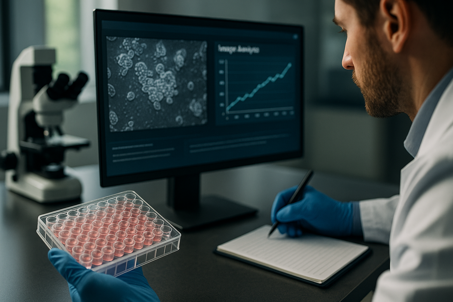
Continuous monitoring with incubator-based systems allows researchers to observe cellular events—such as mitosis, apoptosis, or morphological changes—as they unfold. Such systems are particularly valuable in experiments where dynamic processes are critical to the outcome, such as cell migration assays, wound healing studies, or compound kinetics in drug screens.
Instead of revisiting cells at arbitrary time points, scientists gain a full temporal resolution of cellular events through automated imaging schedules. Combined with quantitative image analysis software, these workflows provide high-content data that are immediately actionable.
- Capture complete cell behavior without disturbing conditions
- Gain real-time feedback on experimental interventions
- Simplify endpoint determination in rate-based assays
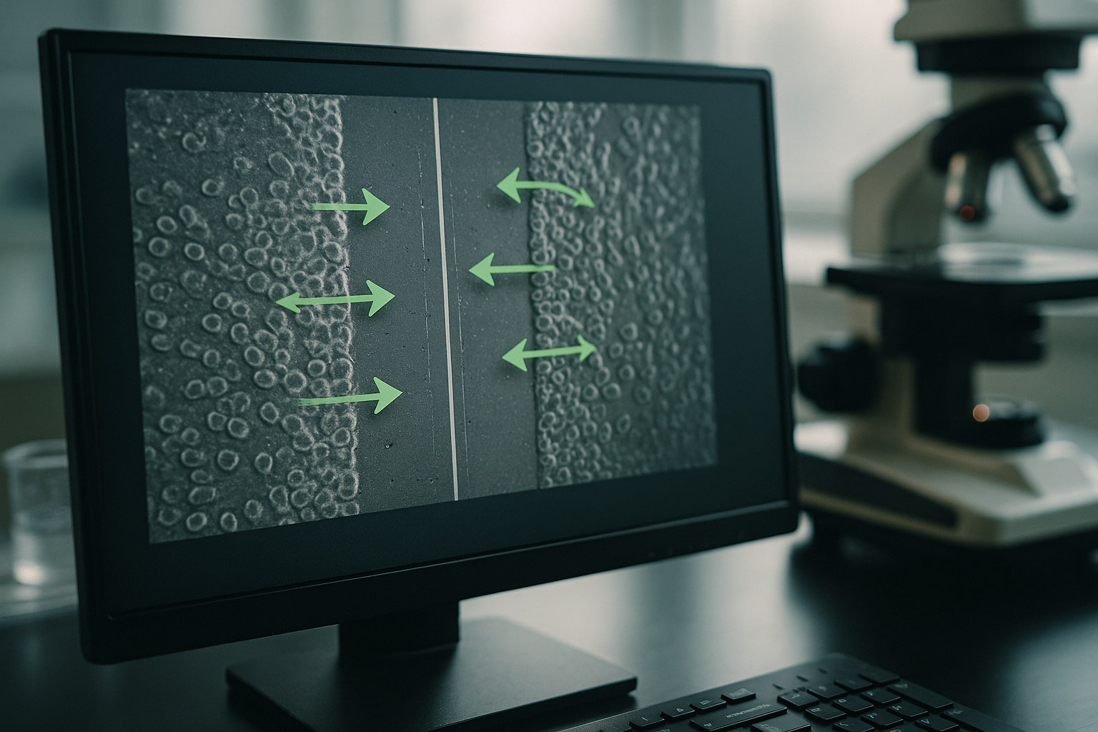
Case Example: 96-Well Migration Assay
In a multicenter wound healing assay using a 96-well scratch format, researchers can program the live-cell imager to capture images every 30 minutes for 72 hours. Devices like the zenCELL owl maintain uniform environmental conditions while collecting consistent, high-resolution data across all wells. Automated image stitching and analysis algorithms quantify wound area closure across the plate, offering kinetic insights into migratory differences among treatment groups.
- Standardize across replicates and treatment groups
- Automated detection of wound areas and coverage timeline
- Reduce variability and manual error in endpoint measurements
Boosting Reproducibility and Data Quality Through Incubator-Based Imaging
Maintaining Physiological Conditions During Imaging
One of the most impactful benefits of live-cell imaging inside the incubator is the maintenance of optimal cell culture conditions throughout the experiment. Devices operable within humidified, CO₂-regulated environments avoid microenvironmental shocks such as temperature drops, pH shifts, or altered gas exchange. These disturbances, even when subtle, can affect cellular metabolism, differentiation, or response to stimuli—leading to misleading results.
- Continuous imaging in an undisturbed cellular environment
- Prevention of artifacts caused by culture stressors
- Improved consistency across experimental replicates
Quantifiable Metrics for Standardization
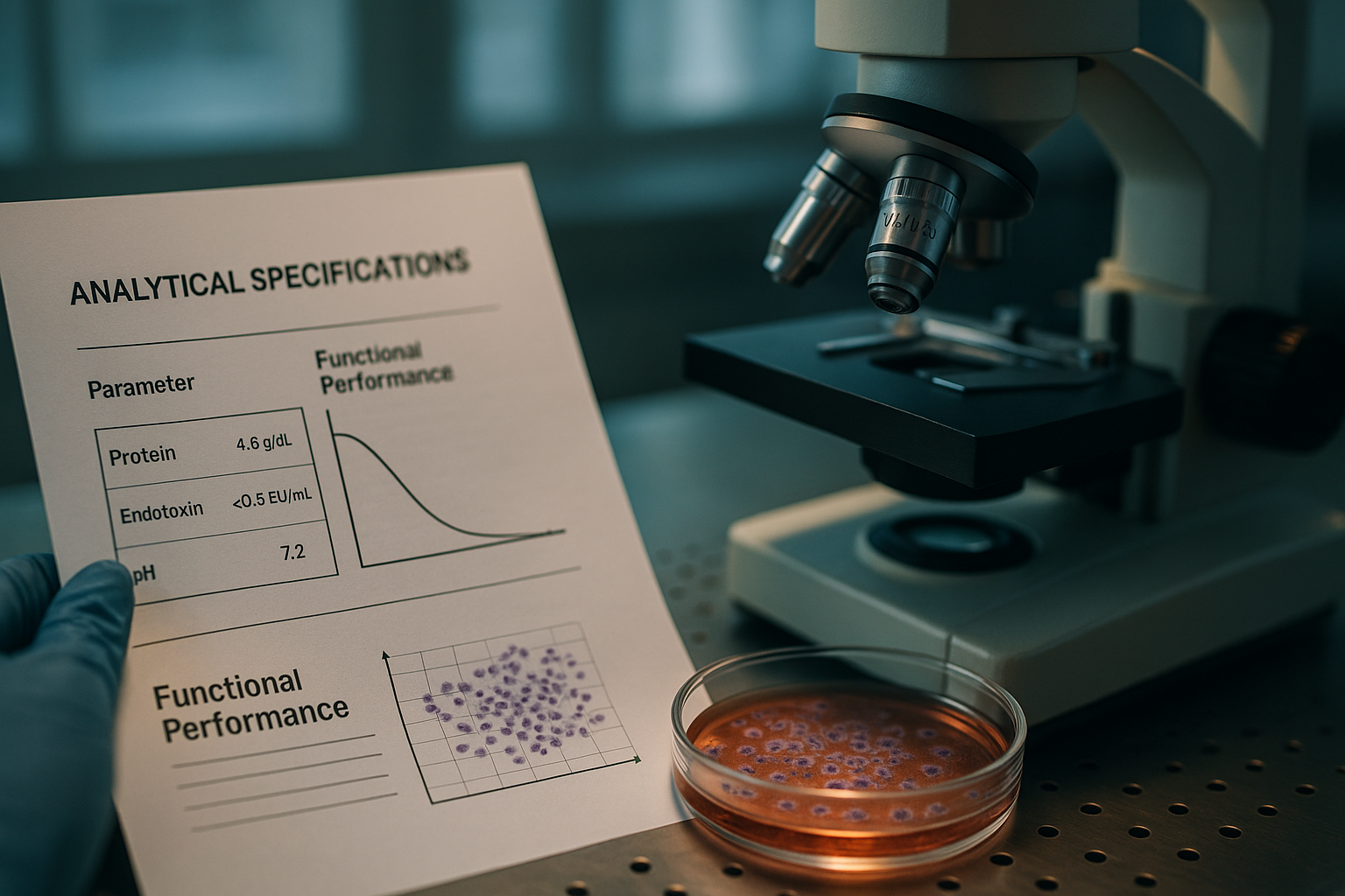
Modern incubator-based imaging systems generate quantitative outputs—such as confluency, cell count, morphology metrics, and migration distance—that can be archived and compared across experiments. This enables better longitudinal studies, inter-laboratory collaboration, and compliance with reproducibility standards set by funding agencies or journals.
- Data-rich outputs facilitate assay validation and protocol optimization
- Support for standardized metrics in regulatory workflows
- Long-term archiving for meta-analysis and peer review
Continue reading to explore more advanced insights and strategies.
Enhancing High-Throughput Screening Efficiency
Accelerating Data Collection in Drug Discovery Pipelines
High-throughput screening (HTS) is an essential process in pharmaceutical research and biotech innovation, requiring fast, reliable data from thousands of samples. Incubator-based live-cell imaging systems streamline HTS by automating image capture across entire multiwell plates without physically relocating the samples. This design allows researchers to perform kinetic and morphological analyses on treatment effects in real time, preserving cell health and boosting data accuracy.
For instance, during compound screening for anti-cancer candidates, a 384-well format can be monitored over several days, assessing proliferation and apoptosis rates using automated confluency metrics and morphological classifiers. The ability to dynamically rank hit candidates by effect onset and duration avoids downstream bottlenecks and speeds lead optimization.
- Use multiwell-compatible imaging platforms to support HTS scalability
Facilitating Longitudinal Cell Line Development
Tracking Morphological Stability Over Time
In cell line development for biologics or genetic engineering, stability monitoring is a critical quality control step. With continuous live-cell imaging, researchers can generate a day-to-day or even cell-division-level record of phenotype changes, eliminating guesswork around optimal passaging timelines, clone selection, or genetic drift.
One application involves monitoring CHO (Chinese hamster ovary) cell lines used in monoclonal antibody production. By imaging these cultures continuously over weeks, lab teams can track proliferation consistency and detect early morphological deviations that compromise yield potential. This enables automated alerting when cultures deviate from expected growth curves, improving culture-to-culture reproducibility.
- Automate clone stability tracking to enhance bioproduction workflows
Integrating With Artificial Intelligence and Image-Based Analytics
Tapping Into Machine Learning for Predictive Insights
The high temporal resolution of incubator-based imaging systems unlocks opportunities to train AI models on cell behavior patterns. Machine learning algorithms can detect subtle changes preceding major events—like apoptosis, differentiation, or detachment—by processing large time-lapse datasets. These tools can uncover patterns invisible to manual observation, aiding in early-response biomarker discovery and cell state classification.
One study applied convolutional neural networks to time-lapse imagery from a zenCELL owl unit to predict toxic compound effects before morphological anomaly onset. By training the model on thousands of images across multiple treatment types, it achieved over 93% predictive accuracy just hours after compound addition—versus 24 hours needed with traditional endpoint assays.
- Expand real-time analytics with AI to accelerate phenotype classification
Improving Adaptive Experimental Designs
Real-Time Data Feedback Enables Mid-Study Adjustments
Live-cell imaging inside the incubator empowers researchers to shift from static designs to responsive experimental strategies. For example, researchers can adjust compound concentrations or time points dynamically in response to observed cellular behavior—optimizing interventions on the fly based on live feedback.
In a stem cell differentiation model, a team at a regenerative medicine lab monitored the emergence of specific morphologies over six days. When early differentiation cues were suboptimal, they altered inducer concentration midway through the experiment. Thanks to live image feeds, outcome trajectories improved measurably without needing to restart the study. Such adaptability is only feasible when continuous data is available in near real time.
- Use real-time monitoring to guide adaptive dose-response curves
Supporting Co-Culture and 3D Model Analysis
Addressing the Complexity of Multicellular and Organoid Systems
Complex cell culture systems, such as co-cultures and 3D organoids, are increasingly used to mimic in vivo conditions. These models introduce new imaging challenges like variable z-depth, non-adherent growth, and asynchronous cell interactions. Incubator-based imaging platforms with adaptive focus and multiple time-point sampling help capture these dynamics without disrupting structural integrity.
A cancer immunotherapy study utilized 3D co-culture spheroids of tumor and immune cells inside a zenCELL owl-compatible bioreactor plate. The system captured migration of cytotoxic T cells into tumor spheroids across 48 hours, enabling researchers to visualize tumor infiltration and quantify spheroid disintegration over time. This level of resolution was critical for validating checkpoint inhibitor efficacy in a physiologically relevant model.
- Apply incubator-based time-lapse imaging to validate complex cell interactions
Streamlining Education and Training in Modern Cell Biology
Remote Access and Cloud Integration Support Virtual Collaboration
As cell biology techniques become more data-centric and collaborative, incubator-based live-cell imaging systems offer a modern solution for research institutions and training facilities. Cloud-connected platforms allow students, collaborators, and remote scientists to access real-time experiment footage, download timelapses, and analyze image data from shared dashboards—no matter their location.
During the COVID-19 pandemic, many educational labs deployed zenCELL owl systems to bridge physical access limitations. At one university, students remotely participated in seven-day proliferation studies, logging into cloud software to annotate cell behavior, perform growth curve analysis, and upload lab reports. This model elevated remote learning while maintaining experimental rigor.
- Leverage remote data access for student training and multi-site collaboration
Reducing Experimental Waste and Resource Use
Non-Invasive Imaging Minimizes Sample Sacrifice
Traditional live-cell methods often require sampling, fixation, or staining that consumes cells per time point. Incubator-based imaging preserves sample viability, enabling full temporal studies from a single culture passage. This reduces the number of replicates needed, cuts down reagent waste, and lowers biosafety burden—especially important in scarce or patient-derived samples.
In oncology research involving patient-derived xenograft (PDX) cells, the ability to perform non-terminal kinetic assays allowed for efficient drug panel screening with minimal sample consumption. This cost-saving approach enhanced experimental density per biopsy and improved ethical use of limited human tissue.
- Adopt label-free, non-invasive imaging to conserve critical sample resources
Compliance With Regulatory and QA Requirements
Traceable, Time-Stamped Data Supports Audit Readiness
Certain laboratory environments—especially GMP and GLP facilities—require detailed experimental traceability. Automated live-cell imaging platforms deliver time-stamped image sequences, standardized metadata, and audit-ready reports integrated with centralized data systems. This makes them particularly well suited for CROs, CMOs, and biotech startups pursuing IND or regulatory filings.
Many platforms, including the zenCELL owl, support exportable datasets containing image timestamps, treatment metadata, and environmental logs. This simplifies integration with lab information management systems (LIMS) and ensures consistent data archiving for long-term compliance or reanalysis in multicenter studies.
- Use timestamped timelapse data to strengthen QA and regulatory submissions
Next, we’ll wrap up with key takeaways, metrics, and a powerful conclusion.
Enabling Scalable Bioprocess Optimization
High-Content Monitoring for Biomanufacturing Advancement
Biomanufacturing pipelines increasingly rely on automated workflows to scale up production without compromising quality. Incubator-based imaging technologies provide continuous visual and quantitative monitoring of culture behavior across multiple vessels in parallel, enabling real-time comparisons of bioprocess conditions such as feed strategy, culture density, and oxygenation. Unlike traditional sampling approaches, integrated imaging systems deliver uninterrupted feedback that supports faster decision cycles and robust optimization.
For example, in a bioreactor scale-up study, researchers used compartmentalized multiwell plates coupled with live-cell imaging to evaluate different nutrient formulations and perfusion rates. The platform’s temporal resolution allowed them to detect culture instability and aggregation early—well before viability dropped—leading to timely process adjustments. This approach enhanced yield consistency while minimizing the risk of batch failure.
- Integrate live imaging into scale-up development to reduce process variability
Advancing Personalized Medicine and Drug Responsiveness Profiling
Using Live-Cell Imaging to Tailor Therapeutic Approaches
As personalized medicine becomes increasingly mainstream, functional assays play a central role in determining patient-specific drug responses. Incubator-based live-cell imaging offers a unique advantage by allowing drug efficacy profiling on rare or patient-derived cells without endpoint biomarkers or destructive assays. The ability to capture individual cell behaviors—such as migration, proliferation, and death—in real time supports more nuanced phenotypic characterization of heterogeneous samples.
Clinical researchers have harnessed this approach to evaluate the effects of drug cocktails on tumor cell dissociation, immune cell motility, and organoid survival. Continuous visualization of how distinct cell subpopulations respond to treatment helps stratify patients based on functional response—not just genomic data. This paradigm shift opens doors to combining cell behavior profiling with AI models to guide precision treatment decisions.
- Utilize dynamic cell behavior data to inform precision therapeutics
Conclusion
Incubator-based live-cell imaging is transforming how researchers across life sciences observe, measure, and understand cellular phenomena. By enabling continuous, non-invasive, and high-resolution data collection directly within culture environments, this technology bridges the gap between traditional static assays and the dynamic nature of living systems. Applications across drug discovery, bioproduction, regenerative medicine, and personalized therapy demonstrate the versatility and far-reaching impact of this approach.
Key takeaways from this exploration emphasize how live-cell imaging inside the incubator accelerates high-throughput screening, supports longitudinal studies, enables adaptive experimentation, and empowers AI-assisted image analysis. The integration of these platforms into research workflows not only enhances biological insight but also reduces experimental waste, ensures regulatory compliance, and fosters collaborative learning. Whether it’s tracking immune cell infiltration in a tumor spheroid, predicting toxicity before it becomes visible, or adjusting differentiation protocols mid-study, incubator-based imaging offers the responsiveness and depth needed for modern cell biology research.
As the demand grows for reproducibility, data richness, and rapid iteration, the ability to collect real-time, traceable image datasets is no longer a luxury—it is a necessity. Scientific innovation depends on tools that are both scalable and insightful. Technologies like the zenCELL owl are paving the way by making high-frequency observation accessible, reliable, and deeply informative.
Institutions and laboratories embracing this shift are not only optimizing their current protocols but positioning themselves for the next wave of scientific discovery. The future of cell culture research lies in continuous monitoring powered by live imaging, data analytics, and intelligent decision-making tools. Now is the time to reimagine how we interact with our cell models and unlock a more efficient, ethical, and insightful era of biological research.
Take the next step—bring your incubator to life by integrating a live-cell imaging system and experience the evolution of cell science in every frame.