
Beyond the Snapshot: Why Endpoint Microscopy is Holding Your Research Back

“```html
Beyond the Snapshot: Why Endpoint Microscopy is Holding Your Research Back
The world of cell culture research is evolving rapidly. With the advent of innovative technologies, researchers are now more equipped than ever to peel back the layers of cellular complexity. However, the continued reliance on endpoint microscopy, a traditional approach where cells are fixed and imaged at specific time points, presents significant limitations. This method often acts as a bottleneck, preventing researchers from capturing the dynamic nature of living cells. In this article, we delve into the limitations of endpoint microscopy, explore the technological advancements in live-cell imaging, and discuss practical applications that are transforming standard laboratory workflows.
Common Challenges and Limitations of Traditional Approaches
The Static Nature of Endpoint Microscopy
Endpoint microscopy, despite being a cornerstone of cellular imaging, is inherently limited by its static nature. This technique involves capturing images at fixed intervals, often after chemical fixation that halts cellular processes. As a result, researchers miss out on vital dynamic interactions and transient events happening inside living cells. The static images provide only a ‘snapshot’, leading to a fragmented understanding of cellular behavior and interactions. This limitation is particularly evident in studies requiring real-time monitoring, such as mitotic progression, cytoskeletal rearrangement, and cellular response to stimuli.
- Loss of dynamic cellular information.
- Potential for artifacts due to fixation processes.
- Limitations in temporal resolution.
Technological Advances and Automation Trends
Live-Cell Imaging: A Game-Changer for Cell Research
The shift towards live-cell imaging represents a paradigm shift in cell culture research. Unlike endpoint microscopy, live-cell imaging techniques allow continuous observation of cellular processes in real-time. This has been facilitated by advancements in optical systems, fluorescent markers, and imaging software that offer improved spatial and temporal resolution. By enabling the study of cells in their natural environment, live-cell imaging empowers researchers to capture subtle cellular responses and dynamic physiological processes that were previously undetectable.
- Real-time visualization of cellular processes.
- Enhanced temporal and spatial resolution.
- Greater insight into dynamic cellular behaviors.
Practical Examples and Workflows Using Live-Cell Imaging
Enhancing Research with Real-Time Data
Live-cell imaging is revolutionizing workflows by providing insights into cellular dynamics that were previously inaccessible with endpoint methods. A typical example includes time-lapse microscopy, where live-cell imaging systems like the zenCELL owl can capture high-quality images at high frequencies, delivering valuable information on cell division, migration, and morphological changes. This continuous monitoring offers a robust dataset that aids in accurate quantitative analysis, leading to reproducible and highly reliable experimental outcomes.
- Time-lapse microscopy for dynamic process analysis.
- Continuous monitoring enhances data reproducibility.
- Enables robust quantitative analysis.
Lesen Sie weiter, um tiefere Einblicke und Strategien zu gewinnen.
“`
“```html
The Power of Fluorescent Markers in Cellular Imaging
Illuminating the Invisible
Fluorescent markers have revolutionized the field of cellular imaging, making the invisible visible through the use of fluorescent dyes and proteins that bind to specific cellular components. These markers are pivotal in enabling the monitoring of live cells, providing insights into cellular architecture, signaling pathways, and protein interactions. For instance, the application of GFP-tagged proteins allows researchers to track protein localization and movement within live cells, unveiling processes that were previously obscured by endpoint methodologies.
- Identify and validate markers specific to your research goals.
Integration of AI and Machine Learning in Imaging
Transforming Data into Insights
With the explosion of data generated from live-cell imaging, AI and machine learning have become critical in extracting meaningful insights from vast datasets. These technologies assist in analyzing complex imaging data by recognizing patterns and anomalies often missed by human observers. For example, advanced software like CellProfiler uses machine learning algorithms to segment cells, quantify cellular phenotypes, and even predict cellular responses. By automating these analyses, researchers can enhance accuracy, reduce bias, and increase throughput.
- Incorporate AI tools to streamline data analysis workflows.
Cryopreservation: Maintaining Live-Cell Quality
Preserving Cellular Integrity Over Time
Cryopreservation plays a crucial role in live-cell imaging studies, allowing researchers to maintain cell viability over long periods. This method ensures that live-cell samples retain their functionality and responsiveness, essential for longitudinal studies. Techniques like controlled-rate freezing and vitrification mitigate ice crystal formation, which can damage cell structures. The ability to preserve cells at high viability rates allows for consistent experimental setups, reducing the variability that can skew live-cell imaging data.
- Implement proper cryopreservation protocols to ensure cell viability.
The Role of Incubator Microscopes in Workflow Efficiency
Continuous Monitoring without Disruption
Incubator microscopes provide an ideal environment for live-cell imaging by enabling continuous observation without disrupting culture conditions. These systems integrate environmental controls (temperature, CO2, humidity) directly into the imaging setup, facilitating long-term studies necessary to capture gradual cellular changes. Devices like the Olympus IXplore Live aid researchers in conducting real-time imaging while maintaining the physiological conditions that are crucial for cell health and normal functions.
- Utilize incubator microscopes to maintain experimental conditions.
High-Resolution 3D Imaging Technologies
Expanding Perspectives in Cellular Research
3D imaging technologies such as confocal and multiphoton microscopy offer unparalleled depth and resolution, allowing researchers to visualize cell structures in three dimensions. This advancement is critical for studies involving complex tissues or multicellular structures, where interactions occur in all spatial dimensions. For instance, its application in tumor microenvironment research sheds light on the intricacies of cancer progression, uncovering previously hidden interactions within dense tissue matrices.
- Consider 3D imaging for comprehensive insight into tissue architecture.
Maximizing Efficiency with Automated Imaging Pipelines
Streamlining Processes for High Productivity
Automated imaging pipelines simplify the workflow by managing image acquisition, processing, and analysis with minimal user input. This automation reduces error, increases repeatability, and saves valuable research time. Platforms such as Nikon’s NIS-Elements streamline these processes by integrating seamlessly with robotic equipment, enabling high-throughput screening applications that accelerate the drug discovery process.
- Leverage automated systems to boost throughput and data consistency.
Collaborative Research and Data Sharing Strategies
Amplifying Research Impact through Connectivity
Collaboration in the scientific community is enhanced by data sharing platforms that facilitate the exchange of imaging datasets and methodologies. Open-source platforms, including the Image Data Resource (IDR), allow multi-institutional access to high-quality imaging data, fostering collaboration and innovation. These resources enable researchers to build on existing work, preventing redundancy and maximizing resource utilization.
- Participate in data sharing to enhance research collaborations.
Im Anschluss fassen wir die wichtigsten Erkenntnisse, Kennzahlen und eine wirkungsvolle Schlussfolgerung zusammen.
“`
“```html
Virtual and Augmented Reality in Cellular Imaging
Beyond Traditional Visualization
As the boundaries of cellular imaging expand, incorporating virtual and augmented reality (VR and AR) presents novel ways to interact with complex cellular datasets. VR and AR technologies allow researchers to visualize and manipulate three-dimensional biological data in immersive environments, offering profound insights into spatial relationships and dynamics. This capability enhances educational outcomes and paves the way for a deeper understanding of phenomena such as neuronal connectivity and tissue development, which are difficult to grasp in two-dimensional formats. By employing platforms like the CAVE Automatic Virtual Environment, scientists can simulate cellular processes at a scale and perspective unmatched by conventional methods.
- Explore VR/AR for an interactive approach to data interpretation.
Addressing Challenges in Live-Cell Imaging
Overcoming Constraints to Unlock Potential
Despite remarkable advancements, live-cell imaging presents challenges that need addressing to fully harness its potential. One key challenge is phototoxicity, which arises from prolonged exposure to light, potentially altering cell behavior and compromising data integrity. Strategies like optimizing dye concentrations, employing photostable markers, and integrating advancements in low-photon technology are pivotal for reducing photodamage. Furthermore, the sheer volume of data can overwhelm traditional storage and processing infrastructures, underscoring the need for scalable solutions and advanced computational resources to handle big data efficiently.
- Adopt techniques to mitigate phototoxicity for accurate imaging.
Ethical Considerations in Imaging Research
Navigating the Moral Landscape
As cellular imaging techniques continue to evolve, the ethical implications surrounding their use come to the forefront. Ensuring that imaging studies respect cellular integrity and privacy is crucial, particularly when researching sensitive or proprietary cellular models. Transparently addressing ethical concerns and adhering to stringent guidelines fosters trust within the scientific community and the broader public. By establishing robust ethical frameworks, researchers can balance the pursuit of knowledge with the imperative to conduct research responsibly.
- Engage with ethical frameworks to uphold research integrity.
Schlussfolgerung
In the journey “Beyond the Snapshot”, live-cell imaging emerges as a transformative force, redefining what is possible within cellular research. The integration of techniques and technologies such as AI, machine learning, 3D imaging, and even VR/AR is reshaping our scientific approaches, offering more dynamic, accurate, and insightful perspectives into cellular behaviors and interactions. This revolution not only broadens our understanding but also opens new pathways for innovations in disease treatment and bioengineering applications.
Amidst these technological strides, the importance of maintaining rigorous scientific standards and ethical considerations cannot be overstated. As we stand on the precipice of unparalleled advancements, the onus lies on us as researchers to continuously reflect on the impact of our methodologies and data handling protocols. The hurdles of phototoxicity, data management, and ethical integrity highlight a journey marked by collaboration, innovation, and a steadfast commitment to scientific rigor.
Ultimately, the value of embracing live-cell imaging’s advancements lies not only in achieving academic and professional excellence but in contributing meaningfully to the collective scientific endeavor. Researchers are encouraged to adopt these cutting-edge tools and strategies, to foster a spirit of connectivity and collaboration, and to partake in efforts that push the boundaries of biological research further than ever before. As we illuminate the invisible and decode the complexities of cellular structures, let us forge ahead with courage and an unwavering quest for knowledge, knowing that our discoveries today lay the groundwork for the innovations of tomorrow.
“`
Contamination Killed Your Experiment? How Continuous Monitoring Saves Your Lab Budget
“```html
Contamination Killed Your Experiment? How Continuous Monitoring Saves Your Lab Budget
In the world of life sciences, where precision is paramount, the impact of contamination is profound. It’s not only disheartening when experimental results go awry, but it also incurs significant financial and time losses. This article delves into how continuous monitoring technologies can safeguard your laboratory setups, offering both financial relief and experimental reliability. Researchers, lab managers, and biotech professionals will discover insights into overcoming the age-old challenge of contamination and how modern advancements in cell culture and live-cell imaging are reshaping laboratory workflows for the better.
Understanding the Relevance of Continuous Monitoring in Cell Culture
Cell culture research is a delicate process, often fraught with the threat of contamination. This not only results in compromised reproducibility but can lead to the failure of entire experiments. Continuous monitoring plays a vital role in preemptively identifying potential issues, securing the integrity of your experimental work, and ultimately saving your lab budget. As the demands of modern research shift towards higher precision and efficiency, integrating systems for continuous observation can provide researchers with much-needed peace of mind.
- Preventing contaminated experiments protects financial and time investments.
- Continuous monitoring ensures consistent oversight and rapid response to anomalies.
Challenges and Limitations of Traditional Cell Culture Methods
Traditional cell culture methods often rely on manual oversight, which is not only labor-intensive but leaves room for human error. The sporadic nature of manual checks increases the risk of missing critical contamination events, potentially leading to erroneous conclusions. Moreover, these approaches frequently lack the sensitivity and consistency provided by continuous monitoring systems.
- Manual checks increase the potential for human error.
- Inconsistent monitoring can lead to undetected contamination incidents.
Advances in Technology and Automation Trends
With the advent of automation and advanced imaging technologies, cell culture processes have witnessed a revolution. Incorporating systems like the zenCELL owl, a compact, incubator-compatible live-cell imaging system, illustrates the shift towards nondisruptive, continuous monitoring. This technology enables automatic data collection, offering real-time insights and minimizing intervention-related contamination risks.
- Automation reduces labor-intensive processes and minimizes human interference.
- Real-time data collection enhances analysis accuracy and timeliness.
Enhancing Reproducibility and Data Quality through Incubator-Based Imaging
Incubator-based imaging systems like the zenCELL owl provide a controlled environment for cells to thrive without the disruptions associated with traditional manual checks. By ensuring uninterrupted observation, these systems enhance reproducibility and ensure that data quality remains uncompromised. The integration of such technologies paves the way for streamlined workflows in cell culture research.
- Incubator-based imaging maintains optimal environmental conditions for cells.
- Continuous imaging facilitates consistent data acquisition and analysis.
Applications in Modern Research: Migration Assays, Organoids, and Beyond
The implementation of continuous monitoring systems extends beyond contamination prevention, offering broad applications across various research areas. Live-cell imaging aids in conducting migration assays, studying organoid development, monitoring cellular proliferation, and streamlining high-throughput screenings (HTS). These applications illustrate the comprehensive benefits of incorporating such technology into lab environments.
- Migration assays benefit from real-time tracking of cell movements.
- Organoid studies gain from uninterrupted observation, revealing developmental insights.
- High-throughput screenings (HTS) leverage consistent data for large-scale analyses.
Lesen Sie weiter, um tiefere Einblicke und Strategien zu gewinnen.
“`
“```html
Reducing Laboratory Overheads with Predictive Analysis
Leveraging Data for Cost-efficient Operations
Predictive analysis, a boon in modern lab settings, can significantly reduce unnecessary expenditure by forecasting potential problems and optimizing resources. By harnessing the power of machine learning algorithms, continuous monitoring systems can predict contamination risks and equipment failures before they occur. This foresight allows labs to allocate their resources judiciously and avoid unplanned expenses, ensuring smoother operations.
- Develop a routine checkup schedule for all lab equipment based on predictive insights.
Integrating AI for Enhanced Data Management
Revolutionizing Information Processing in Laboratories
Artificial Intelligence (AI) has profoundly impacted how we manage and interpret data in laboratory environments. The integration of AI with continuous monitoring systems allows for the automatic sorting and analysis of large datasets, facilitating quicker decision-making processes. For example, AI-driven platforms can analyze real-time data from zenCELL owl systems to detect subtle changes in cell morphology, alerting researchers to potential issues that require immediate attention.
- Utilize AI tools to automatically categorize and prioritize data for research analysis.
Optimizing Workflow for Better Resource Utilization
Structuring Laboratory Operations to Maximize Output
To maximize the efficiency of continuous monitoring technologies, laboratories must also consider optimizing their workflow. By strategically planning the lab’s layout and employing technologies like lab automation software, researchers can significantly reduce time wasted on logistical operations. For instance, connecting continuous monitoring systems with lab management software ensures seamless data flow, allowing teams to focus on interpretative rather than administrative tasks.
- Map out laboratory processes and identify steps that can be automated or streamlined.
Empowering Research with Remote Monitoring Capabilities
A New Dimension of Flexibility for Scientists
Continuous monitoring systems equipped with remote access capabilities add an extra layer of flexibility, enabling researchers to access real-time data from virtually anywhere. This advancement is vital for global research collaborations, allowing scientists to manage experiments and respond to alerts without being confined to their labs. Such capabilities become especially crucial in times of travel restrictions or when researchers are working across multiple laboratory sites.
- Invest in systems that support secure remote access and integrate with mobile devices.
Case Study: A Biotech Firm’s Transformation with Continuous Monitoring
Turning Challenges into Strategic Advantages
Consider the case of BioInnovate, a mid-sized biotech firm experiencing frequent experimental failures due to undetected contamination. By adopting continuous monitoring systems like zenCELL owl, BioInnovate witnessed a drastic 40% reduction in experiment losses. Furthermore, the firm saved significant labor costs as the system automated routine checks, freeing up researchers to focus on innovation rather than remedial tasks. This transformation exemplifies how incorporating modern technology can bring substantial operational advantages and enhance scientific outcomes.
- Use similar real-world success stories to inspire changes in your lab’s approach to monitoring.
Enhancing Collaborative Efforts through Data Standardization
Facilitating Seamless Data Sharing and Collaboration
The use of standardized data protocols across continuous monitoring systems can greatly enhance collaborative research efforts. By ensuring that data is collected and stored in a consistent format, researchers across different labs or institutions can more effectively share insights and make joint contributions to projects. Tools that automatically convert various data outputs into standardized formats foster a more connected research community, paving the way for breakthroughs that require cross-disciplinary expertise.
- Implement data management strategies that prioritize compatibility and ease of sharing.
Pioneering a Culture of Innovation in Laboratory Settings
Encouraging a Proactive Approach to Research
Continuous monitoring systems do more than just prevent contamination; they instigate a culture shift towards proactive research. Encouraging a mindset of anticipation rather than reaction allows scientists to explore innovative lines of inquiry without the constant hindrance of experimental failures. Creating an environment where technology and human expertise complement each other will redefine lab productivity and help establish new industry standards.
- Foster an innovation-driven culture by celebrating small wins achieved through preventive technological applications.
Im Anschluss fassen wir die wichtigsten Erkenntnisse, Kennzahlen und eine wirkungsvolle Schlussfolgerung zusammen.
“`
“```html
Implementing a Feedback Loop for Continuous Improvement
Refining Laboratory Strategies through Data Insights
To fully leverage continuous monitoring systems, laboratories should establish a robust feedback loop. Regular analysis of collected data helps identify patterns and areas for improvement. For example, by reviewing reports on equipment usage and efficiency, labs can adapt their equipment maintenance schedules, ensuring optimal performance and longevity. This proactive approach leads to a cyclic enhancement process, where labs continuously evolve based on informed decisions and prevent small issues from developing into major setbacks.
- Create comprehensive reports to track system performance and inform future strategies.
Training and Development: Maximizing Technology Utility
Building an Advanced Skill Set Among Researchers
Equipping researchers with the necessary skills to utilize continuous monitoring technologies is crucial for maximizing their benefits. Comprehensive training programs focusing on system operation, data analysis, and troubleshooting foster confidence and competence among lab personnel. Moreover, ongoing educational opportunities keep researchers abreast of technological advancements and innovative application methods, ensuring that labs remain at the forefront of scientific exploration and productivity.
- Develop tailored training curriculums that facilitate better understanding and use of modern lab technologies.
Strengthening Lab Safety through Technological Integration
Safeguarding Assets and Personnel with Advanced Monitoring
Continuous monitoring systems aren’t just tools for efficiency and innovation—they are invaluable assets for enhancing lab safety. By alerting staff to environmental shifts or equipment malfunctions in real-time, these systems help prevent incidents that could compromise safety or integrity. Integrating these technologies with existing safety protocols ensures a comprehensive protective framework that guards both human and material resources against potential hazards.
- Incorporate technology-driven safety mechanisms to bolster the lab’s security and compliance standards.
Schlussfolgerung
In conclusion, continuous monitoring systems represent a pivotal advancement in the realm of laboratory science, providing a multifaceted tool that enhances efficiency, cost-effectiveness, and innovation. From predicting equipment malfunctions to managing data remotely, these systems address a broad spectrum of operational challenges. By employing technologies such as predictive analysis and AI integration, labs can preemptively tackle issues, thereby reducing overhead costs and enhancing productivity.
The value of continuous monitoring extends beyond mere operational metrics; it enacts a cultural shift towards more anticipatory, data-driven research paradigms. Laboratories that embrace these technologies foster an environment where creativity and scientific inquiry thrive, empowered by tools that mitigate the monotony of routine maintenance tasks and allow researchers to focus on groundbreaking discoveries.
For professionals seeking to stay ahead in their fields, adopting continuous monitoring systems is not just a choice but a strategic necessity. These tools not only safeguard experiments against preventable failures but also invigorate research processes, aligning them with modern technological advancements. The ultimate takeaway is that laboratories poised to integrate these innovations will enjoy added flexibility, enhanced safety, and improved collaborative capabilities.
As monitor technology continually evolves, the labs of tomorrow will be defined by their ability to adapt, innovate, and inspire through these transformative tools. Now is the moment to invest in systems that not only preserve your research integrity but also pave the way for a future of unparalleled scientific achievement. Embrace continuous monitoring today and watch as it reshapes your laboratory’s path to success.
“`
How raw material consistency supports assay transfer to manufacturing

“```html
How Raw Material Consistency Supports Assay Transfer to Manufacturing
Understanding Raw Material Variability
Factors Influencing Variability
Raw material variability poses significant challenges in the development and manufacture of biological products. Key factors contributing to this variability include biological origins, processing methods, and storage conditions. For instance, animal-derived materials such as fetal bovine serum (FBS) are subject to lot-to-lot variations due to differences in biochemical composition influenced by donor animals, collection sites, and processing techniques. Similarly, human-derived materials exhibit variability based on donor-specific factors and ethical considerations.
- Animal-derived materials, like FBS, often show biochemical deviations across different lots.
- Human-derived materials vary due to individual donor profiles and collection methodologies.
Lesen Sie weiter, um tiefere Einblicke und Strategien zu gewinnen.
Ensuring Quality Through Consistency
Implementing Quality Control Measures
To mitigate the risks associated with raw material variability, implementing robust quality control measures is crucial. This includes comprehensive testing of raw materials to verify their identity, purity, potency, and stability. Advanced documentation practices, traceability, and testing services support the standardization of raw materials, thereby enhancing the reproducibility of experimental outcomes. Using consistent raw materials improves the assurance that the assay will perform reliably when scaled to manufacturing.
- Comprehensive testing ensures material identity and consistency.
- Traceability and documentation underpin quality assurance frameworks.
Lesen Sie weiter, um tiefere Einblicke und Strategien zu gewinnen.
Role of Reagents and Cell Culture Solutions
Impact on Reproducibility
Reagents and cell culture solutions play a pivotal role in biological assays. Their quality directly affects the reproducibility and accuracy of results achieved during assay transfer. Density gradient reagents, for example, are critical for cell separation and must be precisely standardized to prevent variability. Maintaining optimal storage conditions and following rigorous handling protocols are essential to preserve reagent efficacy, thus ensuring consistency throughout assay development and manufacturing.
- Standardized reagents ensure consistent cell separation and assay reproducibility.
- Optimal storage and handling are vital for reagent integrity.
Lesen Sie weiter, um tiefere Einblicke und Strategien zu gewinnen.
Long-term Project Stability Through Custom Services
Benefits of Scientific Services
Custom biological sourcing and antibody development services provide tailored solutions that address specific research needs while minimizing biological variability. These services support continuity and consistency from development through to manufacturing by allowing researchers to reserve batches of materials and conduct specialized testing. For example, batch reservation systems help in aligning production timelines with consistent material supplies, thereby reducing variability during scale-up processes.
- Custom sourcing ensures tailored solutions to specific research projects.
- Batch reservations aid in maintaining material consistency over time.
Lesen Sie weiter, um tiefere Einblicke und Strategien zu gewinnen.
Technological Support for Monitoring and Documentation
Utilizing Live-Cell Imaging
Technological advancements like live-cell imaging systems offer continuous monitoring capabilities that enhance assay reproducibility. An example such as an incubator-compatible live-cell imaging system allows for real-time observation and documentation of serum or reagent effects on cell behavior. This documentation capability is valuable for assessing the performance of raw materials and refining protocols accordingly, ultimately supporting consistent assay transfer to manufacturing environments.
- Live-cell imaging provides insights into reagent effects on cell behavior.
- Real-time documentation supports reproducibility and protocol optimization.
Lesen Sie weiter, um tiefere Einblicke und Strategien zu gewinnen.
“`
“```html
Standardization of Raw Material Supply Chains
Streamlining Supply Chain Management
In the pursuit of consistent assay transfer, standardizing the supply chain for raw materials is indispensable. By creating uniform supply chain processes, organizations can ensure that the quality and characteristics of raw materials meet defined criteria consistently. Companies may engage in strategic partnerships with suppliers that adhere to stringent quality standards and practices, such as ISO certifications or good manufacturing practices (GMP). By branding these synergies, biotech firms can secure raw material supplies that not only meet quality standards but also reduce lead times, decrease costs, and minimize disruptions in production schedules.
- Develop strategic supplier relationships with quality adherence.
- Implement SOPs for supply chain logistics.
Integrating Digital Quality Management Systems (QMS)
Enhancing Quality Oversight Through Technology
Advanced digital Quality Management Systems (QMS) play a vital role in maintaining raw material consistency. Bringing automation into the documentation and monitoring processes as a part of QMS ensures transparency and accuracy in tracking material specifications and changes. A robust QMS can help identify discrepancies and trigger timely interventions, thus preemptively addressing variability issues. Utilizing platforms with real-time analytics and automated alerts, companies can oversee every step of the raw material’s lifecycle, ensuring minimal deviation and optimal quality for large-scale assay deployment.
- Implement digital QMS with real-time tracking and analytics.
- Automate documentation processes to enhance oversight and quality.
Incorporating Predictive Modeling for Future Planning
Forecasting Material Needs and Variability
Predictive modeling is a powerful tool that aids in forecasting material needs and addressing potential variability. By employing machine learning algorithms and complex statistical models, organizations can predict raw material consumption patterns and identify trends correlating with quality inconsistency. Real-world applications include dynamic modeling of serum nutrient batches to anticipate deviations in cell growth patterns, thus offering proactive measures to mediate potential impacts on production. Through this targeted approach, labs can adjust sourcing strategies, optimize batch numbers, and refine quantity forecasts to maintain uniformity and reduce waste.
- Utilize predictive models to foresee raw material demands.
- Adjust sourcing strategies based on forecasted variability.
Setting Up Robust Risk Management Protocols
Mitigating Variability Risks in Manufacturing
Implementing an extensive risk management framework is central to enhancing raw material consistency. This involves identifying potential risk factors associated with material variability, ranging from seasonal supply disruptions to unforeseen biological variation. Companies can develop risk aversion strategies, such as creating contingency plans, increasing warehouse capacity to buffer stock, and diversifying supplier bases to ensure reliability. For instance, having backup suppliers and alternate material sources can significantly reduce the probability of stalled production due to unforeseeable material quality dips.
- Identify and evaluate material-related risks proactively.
- Develop scalable contingency plans for uninterrupted operations.
Implementing Continuous Improvement Practices
Adopting Lean Processes to Enhance Uniformity
Continuous improvement methodologies such as Lean and Six Sigma are instrumental in refining processes that impact raw material quality and consistency. By applying these frameworks, organizations can systematically analyze and eliminate wasteful processes, reduce variation, and improve efficiency in material handling. Practical implementation includes streamlining the benchmarking process for raw materials, conducting regular kaizen events, and establishing cross-functional teams to spearhead continuous process improvements. A biotech firm might utilize Six Sigma to reduce lead time in cell culture media production, thereby maintaining tighter control over material specifications.
- Use Lean and Six Sigma for process optimization and waste reduction.
- Conduct regular benchmarking and improvement initiatives.
Exploring the Potential of AI-Enhanced Material Analytics
Leveraging Artificial Intelligence for Deeper Insights
Artificial Intelligence (AI) provides a sophisticated layer of analysis and insight when managing raw materials for assay transfer. AI-driven analytics offer the ability to process vast datasets across numerous production metrics quickly. The power of AI can be harnessed to recognize subtle patterns and control variables otherwise missed by traditional analysis. For instance, AI technology can assess thousands of data points from reagent batches to identify micro-trends that could affect assay performance. As industries adopt AI-enhanced analytics, they become better positioned to mitigate the impacts of inherent material variability efficiently.
- Incorporate AI systems to uncover hidden data trends.
- Utilize AI-driven insights for enhanced decision-making and control.
Im Anschluss fassen wir die wichtigsten Erkenntnisse, Kennzahlen und eine wirkungsvolle Schlussfolgerung zusammen.
“`
“```html
Harnessing Collaborative Research and Development
Fostering Innovation Through Partnership
Collaboration with academic institutions and industry partners is pivotal in advancing the consistency of raw materials used in assay transfer. By engaging in joint research and development initiatives, organizations can combine expertise and resources to explore innovative solutions to common challenges. These partnerships facilitate the exchange of knowledge and data, leading to breakthroughs in material synthesis and characterization. For example, collaborative projects with universities can aid in understanding the molecular intricacies of raw materials, paving the way for enhanced formulation techniques that bolster quality assurance.
- Engage in joint R&D projects with academic and industry partners.
- Leverage collective expertise to innovate material quality solutions.
Investing in Workforce Training and Development
Empowering Employees for Optimal Quality Control
A fundamental aspect of maintaining raw material consistency in assay transfer is ensuring that the workforce is well-trained and informed. Investing in comprehensive training programs equips employees with the necessary skills to manage sophisticated equipment and adhere to stringent quality control protocols. Organizations can implement continuous learning initiatives to keep the workforce updated on emerging industry trends and technological advancements. Empowered employees are better positioned to identify and resolve issues proactively, contributing significantly to achieving consistent material quality standards.
- Implement ongoing training programs for quality control teams.
- Stay abreast of industry innovations through employee education.
Optimizing Raw Material Sourcing Through Sustainable Practices
Balancing Quality and Environmental Responsibility
Sustainability in sourcing practices not only enhances raw material quality but also aligns with global environmental stewardship goals. By adopting eco-friendly practices, such as sourcing from environmentally-conscious suppliers or utilizing raw materials with verified sustainability credentials, organizations can enhance material consistency while reducing their carbon footprint. Sustainable sourcing ensures that the ecological impact of production is minimal, while simultaneously supporting the reliability and uniformity of materials critical to assay procedures. By prioritizing sustainability, firms can meet regulatory demands and market expectations for responsible production practices.
- Source raw materials from eco-conscious suppliers.
- Implement sustainable production and sourcing strategies.
Schlussfolgerung
In the rapidly evolving landscape of biotechnology, ensuring raw material consistency is a foundation critical to the success of assay transfer into manufacturing. Through a holistic approach encompassing digital quality management systems, predictive modeling, risk management, and continuous improvement practices, organizations can secure the necessary level of quality to meet both stringent regulatory requirements and customer expectations.
Standardizing supply chains, integrating advanced technologies such as AI, and fostering collaborative partnerships extends beyond mere operational efficiency – it establishes a benchmark for achieving excellence in the field. Workforce training and sustainable sourcing further reinforce this structure, ensuring that each element contributes towards maintaining the highest standards of material consistency.
The insights presented in this article underscore the importance of cohesive strategies in overcoming the challenges of variability and ensuring that manufacturing processes are robust and adaptable. As organizations invest time and resources in these strategies, they not only optimize their processes but also advance their competitive position in the market.
Readers are encouraged to evaluate their current practices against these guidelines, embracing innovation and collaboration to refine their approach to raw material management. Commit to these strategies, and be at the forefront of biotechnological advancement by ensuring material consistency that supports efficient assay transfer.
“`
Scaling Without Sacrificing: High-Throughput 96-Well Monitoring Made Simple

“```html
Scaling Without Sacrificing: High-Throughput 96-Well Monitoring Made Simple
In today’s rapidly evolving field of cell culture research, the demand for high-throughput and reproducible scientific data has never been more crucial. As laboratories strive to scale their operations without sacrificing data quality, adopting advanced technologies becomes imperative. High-throughput 96-well monitoring offers an efficient solution, paving the way for comprehensive cell analysis and innovation in workflows. This article delves into the challenges, advancements, and practical applications of high-throughput monitoring, equipping researchers, lab managers, and biotech professionals with the knowledge needed to enhance their cell culture techniques.
Herausforderungen und Grenzen traditioneller Ansätze
For researchers conducting cell culture experiments, traditional monitoring methods often pose significant hurdles. Manual interventions can lead to variability, while the reliance on endpoint assays limits real-time insights into cellular dynamics. These challenges can affect data accuracy, reproducibility, and ultimately, the conclusions drawn from experimental studies.
- Manual handling increases the risk of contamination and variability.
- Endpoint assays lack the ability to monitor dynamic cellular processes in real-time.
- Reproducibility issues arise from inconsistent sample handling and environmental fluctuations.
With advancements in cell culture technology, it is essential to overcome these limitations to maintain the integrity of scientific research. Continue reading to explore technological advances and automation trends.
Technological Advances and Automation Trends
Automation in High-Throughput Monitoring
Recent technological developments have revolutionized high-throughput monitoring, facilitating seamless integration of automation in laboratory settings. Automated imaging systems provide continuous, real-time data collection, reducing the need for manual intervention and minimizing errors.
- Automation enhances reproducibility by offering consistent imaging conditions.
- Real-time monitoring allows for the observation of cellular dynamics within 96-well plates.
- Integration with data analysis software enables efficient interpretation of results.
By embracing these advancements, laboratories can enhance their productivity and maintain high standards of research integrity. Continue reading for practical examples and workflows using live-cell imaging.
Practical Examples and Workflows Using Live-Cell Imaging
Implementing Live-Cell Imaging
Live-cell imaging is a powerful technique that provides in-depth insights into cellular behavior over time. It enables researchers to gather quantitative data on cell migration, proliferation, and morphological changes without disturbing the culture environment. An example of a compact and efficient system for such applications is the zenCELL owl. This incubator-compatible device enables uninterrupted monitoring of cells within 96-well plates.
- Cell migration assays benefit from continuous monitoring of movement patterns.
- Proliferation studies are enhanced by tracking cell division in real-time.
- Organoid development can be observed to assess three-dimensional growth patterns.
Live-cell imaging streamlines workflows by eliminating the need for extensive manual observations, thus saving time and reducing potential for errors. Continue reading to learn how incubator-based imaging improves reproducibility and data quality.
“`
“```html
Enhancing Data Quality with Incubator-Based Imaging
Maintaining Consistent Environmental Conditions
Incubator-based imaging is a game-changing advancement for those working with live-cell imaging, as it allows researchers to maintain optimal culture conditions while observing cells directly within the incubator. By eliminating the need to transport cells to different locations for imaging, this technology minimizes environmental fluctuations and stress that could affect cellular responses.
- Ensure stability by maintaining consistent temperature and humidity levels within the incubator.
- Mitigate stress-related cellular changes by minimizing external disturbances during imaging sessions.
- Optimize data reliability through stable incubation environments and reduced manual handling.
With incubator-based imaging, laboratories can expect improvements in the consistency and reliability of their cell culture monitoring efforts.
Integrating Machine Learning for Enhanced Analysis
Data-Driven Insights with Artificial Intelligence
The integration of machine learning algorithms with high-throughput imaging systems is paving the way for more sophisticated data analysis. By leveraging AI, researchers can automate the interpretation of complex datasets, identifying patterns and trends that might otherwise go unnoticed.
- Utilize machine learning to analyze large volumes of image data efficiently.
- Employ predictive modeling to anticipate cellular outcomes and behaviors.
- Implement AI-driven anomaly detection for early identification of atypical cellular events.
This combination of AI and high-throughput imaging empowers researchers to harness deeper insights from their experiments, driving innovation and discovery in the field.
Real-World Case Studies: Successful Implementation
Case Study: Biotechnology Firm Revolutionizes Workflow
One biotech company implemented a comprehensive high-throughput 96-well monitoring system integrated with live-cell imaging and automation. This led to dramatic improvements in both productivity and data reliability. By using continuous monitoring, they significantly accelerated their drug discovery process and enhanced the accuracy of their cell-based assays.
- Real-time data acquisition eliminated the bottleneck caused by manual observations.
- Automated processes increased throughput by over 50% compared to traditional methods.
- Machine learning-based analysis allowed for the rapid identification of promising drug candidates.
These results highlight the transformative impact of adopting advanced monitoring technologies in a real-world laboratory setting.
Streamlining Data Interpretation and Reporting
From Raw Data to Actionable Insights
Efficient data analysis and visualization are critical components of successful high-throughput monitoring. With advanced software solutions, researchers can transform raw imaging data into actionable insights with ease. Visually intuitive dashboards and automated reporting tools simplify data interpretation, allowing for more informed decision-making.
- Leverage interactive dashboards to explore complex datasets visually.
- Utilize automated reports to streamline communication of findings.
- Enhance collaboration between teams by sharing data insights effortlessly.
This streamlined approach not only saves time but also improves the communication of research findings across different stakeholders.
Strategies for Optimizing High-Throughput Workflows
Maximizing Efficiency and Effectiveness
Optimizing workflows in high-throughput systems is key to maximizing laboratory productivity. By strategically planning experiments, utilizing parallel processing, and ensuring seamless data integration, researchers can achieve more consistent and reliable outcomes.
- Design experiments with clear objectives and streamlined processes to minimize errors.
- Utilize batch processing techniques to enhance throughput and reduce time to results.
- Integrate data management systems to ensure smooth data handling and sharing.
Through these strategies, laboratories can enhance their operational efficiency and achieve higher quality research outputs.
Overcoming Barriers to Adoption
Navigating Challenges in Implementing New Technologies
While the benefits of high-throughput monitoring are clear, some laboratories may face challenges when it comes to adopting new technologies. Common barriers include cost, system compatibility, and the need for training. However, these can be mitigated through careful planning and implementation strategies.
- Conduct a cost-benefit analysis to justify the investment in new technologies.
- Ensure compatibility with existing systems through modular and flexible solutions.
- Provide comprehensive training programs to facilitate smooth transitions for staff.
By proactively addressing these challenges, laboratories can successfully integrate advanced monitoring solutions into their workflows.
Future Directions in High-Throughput Monitoring
Anticipating the Next Innovations
The field of high-throughput 96-well monitoring is continuously evolving, with future directions likely to be shaped by advancements in nanotechnology, personalized medicine, and digital biology. These innovations will further refine the precision of cell culture analysis and open new avenues for medical research and therapeutic developments.
- Explore opportunities in nanotechnology to enhance cellular monitoring techniques.
- Adapt workflows to support personalized and precision medicine initiatives.
- Leverage digital biology to synthesize complex biological data into cohesive models.
Staying abreast of these developments will be crucial for researchers looking to remain at the forefront of scientific discovery.
Im Anschluss fassen wir die wichtigsten Erkenntnisse, Kennzahlen und eine wirkungsvolle Schlussfolgerung zusammen.
“`
“```html
Key Metrics for Success
Measuring the Impact of High-Throughput Systems
To truly understand the efficacy of high-throughput 96-well monitoring systems, laboratories must focus on specific metrics that highlight their impact. These key performance indicators allow researchers to assess the success of their workflows in real-time and adjust their strategies accordingly.
- Throughput Efficiency: Measure the number of assays processed over a given period to gauge operational capacity.
- Data Accuracy: Evaluate the reliability and consistency of results to ensure high-quality outputs.
- Process Optimization: Track workflow improvements and reduced turnaround times as indicators of enhanced productivity.
By focusing on these metrics, laboratories can gain insights into the performance of their high-throughput systems, ensuring that they meet research objectives efficiently and effectively.
Exploring Collaborative Innovations
Building a Network for Discovery
Interdisciplinary collaboration is an essential driver for innovation in high-throughput monitoring. By partnering with technology providers, academic institutions, and industry experts, laboratories can leverage a wider range of expertise and resources.
- Engage with cross-disciplinary teams to foster creativity and innovative solutions.
- Participate in collaborative research initiatives to access cutting-edge technology and findings.
- Utilize shared platforms for data exchange, promoting transparency and accelerated discovery.
Collaborative innovations not only bolster technological capabilities but also pave the way for groundbreaking scientific achievements.
Schlussfolgerung
The integration of incubator-based imaging and advanced machine learning algorithms within high-throughput 96-well monitoring systems represents a pivotal shift in the realm of laboratory research. Throughout this article, we’ve explored various strategies for enhancing data quality, integrating AI for insightful analysis, and overcoming adoption barriers. By maintaining consistent environmental conditions and employing intuitive software solutions, laboratories can substantially improve data reliability and research outcomes.
The successful implementation of these advanced technologies is transforming workflows in biotechnology firms and beyond, as evidenced by real-world case studies presented. More importantly, key metrics such as throughput efficiency, data accuracy, and process optimization offer invaluable insights that allow laboratories to stay competitive in an ever-evolving landscape.
As we look to the future, the anticipated innovations in nanotechnology, personalized medicine, and digital biology promise to further refine high-throughput monitoring capabilities. By staying informed and adaptable, laboratories are poised to harness the potential of these advancements, dynamically shaping the future of medical research and therapeutic development.
We invite researchers and industry leaders alike to embrace the shifts towards more agile, data-driven science. By leveraging the tools, strategies, and collaborations outlined in this article, the scientific community can accelerate discovery and innovation, transcending traditional boundaries in pursuit of transformative knowledge.
Join us on this journey of scientific exploration and technological evolution, where every data point brings us closer to answering the big questions in life sciences. Let us collectively build a future where high-throughput monitoring systems are not merely tools, but the cornerstone of discovery and understanding.
“`
Human Error is Obsolete: Maximizing Throughput with AI-Driven Cell Counting

“```html
Human Error is Obsolete: Maximizing Throughput with AI-Driven Cell Counting
In the realm of modern cell culture research, precision and efficiency are more critical than ever. With increasing demands for high-throughput analysis, minimizing human error has become a pivotal objective for laboratories worldwide. This article delves into how AI-driven cell counting can maximize throughput and enhance data accuracy, making human error obsolete. We’ll explore common challenges in traditional cell counting, recent technological advances, and how AI-driven solutions, such as incubator-based imaging, are transforming workflows. Readers will also gain insights into practical applications and future trends in lab automation.
Challenges of Traditional Cell Counting Methods
The Human Factor in Cell Counting
Traditional cell counting methods often rely heavily on manual processes, which are prone to human error. Variability in technique and subjective analysis can lead to inconsistent results, affecting the reproducibility of experiments. Furthermore, manual counting is time-consuming and labor-intensive, making it unsuitable for high-throughput demands common in modern labs.
- Subjectivity and variability in manual counts
- Increased labor and time investment
- Limited scalability for high-throughput assays
Lesen Sie weiter, um tiefere Einblicke und Strategien zu gewinnen.
Technological Advances and Automation Trends
AI-Driven Solutions in Cell Culture
The integration of artificial intelligence in cell counting offers a transformative solution to the shortcomings of manual methods. AI-driven systems enhance precision by eliminating subjective variability and significantly reducing counting time. These technologies are supported by machine learning algorithms, capable of identifying and classifying cells with high accuracy across a variety of assay conditions.
- Consistency in data through automated counting
- Scalability and efficiency in high-throughput applications
- Enhanced reproducibility and reliability
Lesen Sie weiter, um tiefere Einblicke und Strategien zu gewinnen.
Practical Examples and Workflows Using Live-Cell Imaging
Integrating AI with Live-Cell Imaging
Live-cell imaging has become an indispensable tool in cell culture research, offering continuous monitoring of cellular processes. When combined with AI-driven analysis, live-cell imaging systems can provide real-time data that enhance experimental outcomes. A notable example is the zenCELL owl, a compact and incubator-compatible system, which facilitates seamless integration of AI in cell monitoring, thereby reducing potential human errors and improving data consistency.
- Real-time analysis with AI-enhanced imaging
- Reduction in manual intervention and error
- High-quality, reproducible data output
Lesen Sie weiter, um tiefere Einblicke und Strategien zu gewinnen.
How Incubator-Based Imaging Improves Reproducibility and Data Quality
Maintaining Optimal Conditions
Incubator-based imaging allows for continuous observation of cell cultures under controlled environmental conditions, enhancing both data quality and reproducibility. By maintaining optimal temperature, humidity, and CO2 levels, these systems ensure that cells are kept in their ideal physiological state throughout the experiment. This approach minimizes variability caused by environmental fluctuations, presenting a significant advantage over traditional methods.
- Consistent environmental conditions
- Improved experimental reproducibility
- High data integrity and quality
Lesen Sie weiter, um tiefere Einblicke und Strategien zu gewinnen.
Applications in Modern Cell Culture Research
From Migration Assays to Organoids
AI-driven cell counting and incubator-based imaging systems find diverse applications in cell culture experiments, from migration assays to organoid development. The enhanced precision and throughput offered by AI technologies allow researchers to conduct complex assays with greater ease and accuracy. High-throughput screening (HTS) benefits from automated systems that can analyze numerous samples simultaneously, leading to more efficient and comprehensive data acquisition.
- Accurate analyses in migration and proliferation assays
- Efficient study of biological processes in organoids
- Streamlined workflows for high-throughput screening
Lesen Sie weiter, um tiefere Einblicke und Strategien zu gewinnen.
“`
“```html
Leveraging AI in Disease Modeling and Drug Discovery
Accelerating Innovations with AI
A key advantage of AI-driven cell counting is its application in disease modeling and drug discovery. Traditional methods often encountered limitations in data precision and experimental throughput, which could hamper the drug development process. AI systems, with their high-throughput capabilities, allow for the rapid analysis of numerous drug candidates, expediting the identification of promising therapies.
- Facilitates faster identification of therapeutic candidates
- Enhances data precision in disease modeling
- Reduces time to market with accelerated drug discovery processes
Enhancing Precision in Cytotoxicity Assays
AI Tools Delivering Unmatched Accuracy
In cytotoxicity assays, which are crucial for evaluating the effects of potential drug compounds, AI-driven cell analysis ensures precision that manual methods may lack. By automating the process, labs can reduce the risk of human error significantly, leading to more reliable data that can drive impactful research and development decisions.
- Reduces variability inherent in human-dependent methods
- Improves reliability and accuracy of cytotoxicity results
- Facilitates better-informed decision-making in drug trials
Optimizing Cultured Cell Viability Assessments
AI as a Catalyst for Efficient Viability Studies
Cell viability assessments, critical for understanding cellular responses to various conditions, are streamlined by AI technologies. AI-powered imaging platforms quickly and accurately detect viable cells, enhancing the throughput and precision of experiments. For example, automated systems like CellProfiler utilize AI algorithms to deliver precise viability metrics, supporting robust cellular assessments.
- Provides rapid, accurate assessments of cell viability
- Boosts throughput of viability assays
- Enables precise trend detection in cell health data
Case Study: Transforming Stem Cell Research with AI Integration
AI Solutions Revolutionizing Stem Cell Assays
Stem cell research often involves analyzing complex differentiation processes, which AI-driven technologies can monitor with unprecedented precision. In a landmark study, researchers integrated AI-driven imaging in their monitoring of stem cell cultures, achieving enhanced reproducibility and data accuracy compared to traditional approaches. This significant advancement not only saved time but also facilitated the identification of new differentiation pathways.
- Enables high-resolution monitoring of stem cell differentiation
- Increases assay reproducibility and accuracy
- Identifies novel biological pathways through precise data tracking
Scaling Up Lab Operations with Automated Systems
From Bench to Industrial-Scale Applications
Scalability is a critical demand in modern laboratories, where AI-driven cell counting and imaging systems offer a clear advantage. Automation allows labs to transition from small-scale experiments to large, industrial applications with greater ease. This scalability empowers labs to handle larger datasets and more extensive experiments without compromising on quality.
- Facilitates smooth transition to larger scale operations
- Ensures data consistency across expanded research projects
- Reduces operational costs through efficient resource allocation
Integrating AI with Other ‘Omics’ Technologies
Pioneering Integrated Approaches in Biological Research
A growing trend is the integration of AI-driven cell counting with various omics technologies, such as genomics and proteomics. This integration allows for a comprehensive analysis of cellular behavior, providing deeper biological insights. AI facilitates the correlation of cell counting data with genetic or protein expression profiles, offering a 360-degree view of the cellular landscape.
- Enhances data integration in multi-omics studies
- Provides holistic insights into cellular processes
- Facilitates novel discoveries through integrated data analysis
Streamlining Quality Control Processes with AI
Ensuring Consistency and Compliance
Quality control is paramount in cell culture research, ensuring that all procedures meet regulatory standards. AI systems provide a robust framework for enforcing quality control, with automated monitoring and reporting features that enhance compliance. This ensures that data integrity is maintained across all experimental runs, fostering trust and reliability in research outputs.
- Strengthens compliance with industry standards
- Facilitates automated quality monitoring and documentation
- Ensures consistent maintenance of data integrity
Im Anschluss fassen wir die wichtigsten Erkenntnisse, Kennzahlen und eine wirkungsvolle Schlussfolgerung zusammen.
“`
“```html
AI-Driven Predictive Analytics in Cellular Research
Forecasting to Stay Ahead
Predictive analytics powered by AI is transforming how researchers anticipate cellular behaviors and disease progressions. Through the analysis of historical data and real-time insights, AI models can forecast trends and outcomes, enabling preemptive action. This foresight is crucial for developing preventive strategies and tailoring interventions with greater precision, ultimately improving patient outcomes and advancing personalized medicine.
- Offers predictive insights into cellular responses
- Supports development of personalized therapeutic strategies
- Improves proactive planning in research and clinical settings
Sustainability in AI-Powered Laboratories
AI and Eco-friendly Practices Coalesce
The integration of AI in laboratories not only enhances operational efficiencies but also promotes sustainability. AI systems can optimize resource usage, reducing unnecessary waste and energy consumption. This sustainable approach aligns with global environmental goals and positions scientific research as a leader in eco-friendly innovations. By minimizing the carbon footprint and encouraging resource efficiency, AI is a vital player in creating environmentally responsible lab environments.
- Optimizes resource allocation for eco-friendly lab practices
- Reduces environmental impact of laboratory operations
- Contributes to global sustainability goals
The Future of AI in Cell Counting and Beyond
Unleashing Potential Through Technological Advancements
Looking to the horizon, the role of AI in cellular research continues to expand with rapidly advancing technologies. Innovations in machine learning algorithms and computational power herald a future where AI-driven cell counting and analysis become indispensable tools in biological research. These enhancements promise to unlock new frontiers in science, from unraveling complex biological puzzles to driving forward transformative medical breakthroughs.
- Pushes the boundaries of scientific inquiry
- Enables continuous innovation in biomedical research
- Drives forward the potential of personalized medicine
Schlussfolgerung
The transformative power of AI-driven cell counting lies in its ability to streamline processes, enhance precision, and drive highly impactful scientific research. From accelerating drug discovery and improving the reliability of cytotoxicity assays to optimizing cell viability studies and revolutionizing stem cell research, AI has proven itself to be an invaluable asset in modern laboratories. Furthermore, the integration of AI with other omics technologies and its role in predictive analytics fortify its position as an indispensable tool for comprehensive biological insights.
This evolution underscores the significance of AI in fostering scientific advancements and achieving breakthroughs that once seemed out of reach. The adoption of AI-powered systems not only increases efficiency and precision but also reduces operational costs, strengthens compliance with regulatory standards, and supports sustainable lab operations. Researchers can now explore uncharted territories with greater confidence, driven by the reliable data and predictive capabilities AI provides.
As we stand on the cusp of further AI innovations, the potential for these technologies to shape the future of cellular research continues to grow. This article highlights the limitless possibilities AI offers, urging scientists, researchers, and industry leaders to embrace these tools and integrate them into their workflows for maximum impact. Unite with cutting-edge technology, break through the constraints of traditional methods, and drive the future of science with AI as your ally. The journey towards unprecedented discovery begins here.
“`
The End of Scratch Variability: Automating Migration Assays for Gold-Standard Data

“```html
The End of Scratch Variability: Automating Migration Assays for Gold-Standard Data
The ever-evolving field of cell culture research continuously brings forward innovative methodologies to tackle age-old challenges. Among these, cell migration assays serve as a cornerstone in understanding biological processes such as wound healing, cancer metastasis, and tissue development. Historically, these tests have been susceptible to variability, often stemming from manual procedures like the scratch assay. With the advent of advanced technology, the integration of automated systems in cell culture workflows has sparked a revolution, reducing human error and enhancing data reliability. This article delves into the transition towards automation in migration assays, offering insights into how these innovations end scratch variability, ensure gold-standard data, and set the stage for future advancements.
Challenges and Limitations of Traditional Migration Assays
Sensitivity to Human Error
Traditional approaches to cell migration assays, particularly the scratch assay, have long been hindered by their reliance on manual intervention. The process of creating a “scratch” or wound on a cell monolayer to study migration is inherently prone to discrepancies. Researchers face challenges in standardizing the physical parameters such as scratch width and pressure applied, which vary between practitioners. This leads to inconsistencies in data and results, casting doubts on the reproducibility of findings.
- Variability in manual handling affects data reliability
- Inconsistent measurements complicate comparative analyses
- Subjective evaluation of assay results introduces bias
Limitations in Monitoring and Data Collection
Manual migration assays require periodic observation, often involving endpoint analysis that lacks real-time data acquisition. Traditional microscopy techniques necessitate the removal of samples from stable incubator environments, potentially disturbing the cellular state and affecting migration dynamics. This intermittent examination approach limits the ability to capture critical kinetic data, impeding comprehensive understanding.
- Infrequent observation overlooks key migratory behaviors
- Disturbance during sample handling impairs cell physiology
Technological Advances and Trends in Automation
Emergence of Automated Systems
The transition to automated systems in cell culture assays has brought about a new era of precision and consistency. These systems eliminate the variability introduced by manual operations through consistent protocol execution. Innovations such as robot-assisted scratch assays and programmable imaging stages provide a level of precision that manual efforts simply cannot match.
- Robotic platforms ensure uniform scratch creation
- Automated repeatability enhances reproducibility
- Consistent environmental control fosters reliable results
Lesen Sie weiter, um tiefere Einblicke und Strategien zu gewinnen.
“`
“```html
Optimizing Real-Time Data Acquisition and Analysis
Advanced Imaging Technologies
One of the most significant advancements in migration assays is the integration of real-time imaging technologies. Automated platforms, equipped with state-of-the-art, high-resolution cameras, allow continuous monitoring of cell migration processes. These systems facilitate the acquisition of dynamic and intricate live-cell data, revealing nuanced behaviors of cell movement that were previously missed with periodic manual observation.
- High-resolution imaging captures detailed migration events
- Real-time data enables comprehensive kinetic analysis
Innovative Software Solutions
Software advancements complement the automation of imaging hardware, providing powerful tools for data processing and analysis. Machine learning algorithms and artificial intelligence are now utilized to interpret complex datasets automatically, reducing the workload on researchers and making precise, unbiased assessments possible. Such software can identify patterns and anomalies that might elude human analysis, further enhancing the assay’s informativeness.
- AI-driven analytics reduce human analysis time
- Pattern recognition improves data accuracy and depth
Ensuring Reliable Reproducibility Across Laboratories
Standardized Protocol Development
Automated systems facilitate the development of standardized experimental protocols, promoting reproducibility of results across different laboratories. These protocols ensure that every parameter, from incubation conditions to imaging intervals, remains consistent. Organizations and consortia are also working towards creating universal standards for data. This shift helps unify methodological approaches, providing a reliable framework for global collaboration.
- Standardized protocols enable cross-laboratory consistency
- Unified standards foster collaborative research efforts
Case Study: Collaborative Networks
Global initiatives in academic and commercial settings underscore the importance of standardization. A notable example includes a network of cancer research laboratories that adopted a unified automated system. This collaborative approach resulted in significantly reduced variability in assays across multiple sites, leading to breakthroughs in understanding metastatic pathways. Researchers reported increased speed and confidence in their data, sparking further innovations.
- Collaborative networks enhance data validity
- Joint efforts accelerate discovery and innovation
Automating Comprehensive Environmental Control
Precision in Microenvironment Regulation
Cell migration is sensitive to environmental conditions, which are critical in influencing experimental outcomes. Automated systems incorporate integrated environmental controls that maintain stable temperature, humidity, and CO2 levels, crucial for replicating in vivo conditions. Advanced chamber technologies mimic physiological environments more precisely than manual systems, preventing cellular stress and ensuring more physiologically relevant results.
- Improved microenvironment control boosts data relevance
- Stable conditions reduce external variability impact
Case Study: Physiological Replication
A leading biotechnology firm implemented an automated incubation system in their migration assays, replicating physiological conditions typically seen in in vivo research. By doing so, they observed a marked improvement in the migration rates and patterns that closely resembled in vivo observations. This approach not only enhanced research accuracy but also paved the way for developing novel therapeutic interventions based on more reliable data.
- Physiologically relevant conditions enhance validity
- Automation supports translational research initiatives
Integration of Multi-Modal Analytical Techniques
Leveraging Various Data Streams
By incorporating multiple analytic modalities, automated migration assays offer more comprehensive insights into cellular behavior. Techniques such as flow cytometry, spectrophotometry, and automated microscopy are integrated within these systems to provide multi-dimensional data. This convergence of technologies enriches understanding by correlating morphological changes with biochemical markers, facilitating a holistic view of cellular dynamics.
- Multi-modal analysis enriches cellular insights
- Diverse data streams enhance contextual understanding
Practical Implementation
In practical terms, integrating multi-modal platforms involves leveraging complementary technologies that speak to different aspects of cell behavior. Labs that successfully combine imaging with biochemical analysis often unlock insights into migration tied to specific cellular pathways or genetic markers. For instance, a research team studying neurodegeneration utilized an integrated platform to simultaneously assess cellular morphology and gene expression changes, significantly advancing their understanding of disease mechanisms.
- Combining technologies unlocks deeper insights
- Cross-technological approaches offer a more complete picture
Economical and Resource Efficiency in Automated Systems
Sustainable Lab Practices
Automated migration assays contribute significantly to cost and resource efficiency. By minimizing manual labor and optimizing reagent usage, laboratories can achieve substantial savings. Furthermore, the reliable data provided reduces the requirement for repetition of experiments, conserving both financial and material resources. Additionally, automation aligns with green lab initiatives, reducing waste and promoting sustainable practices.
- Resource efficiency contributes to reduced operational costs
- Sustainability initiatives benefit from automation efficiencies
Economic Case Study
In a pilot study conducted by a consortium of environmental labs, transitioning to automated cell migration assays showed a marked decrease in reagent consumption by over 30%. The initial investment in technology was offset within a year due to savings on consumables and labor hours. Furthermore, by reducing experimental repetition rates by 50%, the labs reported not only economic benefits but also enhanced sustainability in their operations.
- Sustained resource savings validate initial tech investments
- Economic gains coupled with environmental benefits
Im Anschluss fassen wir die wichtigsten Erkenntnisse, Kennzahlen und eine wirkungsvolle Schlussfolgerung zusammen.
“`
“```html
Augmenting Data Reliability with Enhanced Automated Controls
Incorporation of Quality Assurance Measures
As laboratories rely increasingly on automated systems, embedding quality assurance protocols within these systems becomes crucial. Automated self-checks, calibration routines, and error detection protocols can significantly enhance data reliability. These measures ensure that each experiment is performed under optimal conditions, providing precise and reproducible outputs.
- Automated checks maintain consistent operational standards
- Quality assurance minimizes the risk of data anomalies
Technological Advancements in Error Proofing
Innovations in automation technology have provided new methods for error detection and prevention. Sophisticated sensors and software algorithms now offer real-time monitoring, alerting laboratories to any discrepancies during the experiment. These advancements help reduce downtimes and improve the overall robustness of cell migration assays.
- Real-time monitoring identifies potential errors promptly
- Technological safeguards enhance overall lab efficiency
Promoting Educational Initiatives for Automated Assays
Training for Next-Generation Researchers
With the increasing complexity of automated systems, educational initiatives aimed at equipping researchers with necessary skills are important. Training programs focusing on utilizing these technologies allow for effective, maximum adoption. Encouraging knowledge sharing and workshops can cultivate an adaptable research community ready to tackle modern challenges within the field.
- Training initiatives build competence in automation technologies
- Workshops foster a collaborative and informed research culture
Access to Shared Knowledge Resources
Encouraging open access to documentation and shared resources helps broaden understanding and proficiency with automated assays. Shared databases and collaborative online platforms enable researchers to exchange insights and troubleshoot challenges collectively, enhancing both individual and group competencies in the field.
- Knowledge sharing platforms promote resource accessibility
- Collaborative tools streamline the learning curve for researchers
Schlussfolgerung
This journey through the innovative landscape of automated migration assays has unveiled the transformative impact of automated technologies on cellular research. From optimizing real-time data acquisition to advancing economic sustainability, the highlighted sections offer a comprehensive insight into how these tools revolutionize traditional methodologies.
The integration of advanced imaging and software solutions, along with standardized protocols, sets a new standard for reproducibility and accuracy. Multifaceted analytical approaches enrich our understanding, while automation-driven sustainability reinforces labs’ commitments to environmentally conscious practices.
Importantly, quality assurance and educational initiatives ensure that the benefits of automation are fully realized across the research community. By equipping researchers with the necessary skills and fostering a collaborative environment through shared resources, we can expedite the pace of scientific discovery.
The potential for automation in cell migration assays represents not simply an incremental upgrade but a foundational paradigm shift in research methodologies. As laboratories worldwide adopt these innovative practices, the scientific community moves closer toward achieving gold-standard data, enhancing the fidelity and relevance of their findings.
As you explore these emerging technologies, consider the long-term benefits of embracing automation within your own lab settings. By doing so, you not only drive better research outcomes but also contribute to a wider movement aimed at more precise, efficient, and sustainable scientific practices. Together, we can push the boundaries of knowledge and achieve remarkable breakthroughs that lay the groundwork for future achievements.
“`
How to future-proof biological sourcing decisions

“```html
How to Future-Proof Biological Sourcing Decisions
In the rapidly evolving field of biotechnology, researchers and laboratory professionals face a myriad of challenges when it comes to sourcing biological materials. From variability in animal-derived sera to ethical considerations of human-derived biologicals, making informed sourcing decisions is crucial to ensuring experimental consistency and integrity. This article delves into the strategies and considerations essential for future-proofing biological sourcing decisions, providing a comprehensive guide for enhancing research workflows.
The Importance of Quality in Animal-Derived Biologicals
Understanding Lot-to-Lot Variability
Animal-derived biologicals, such as fetal bovine serum (FBS) and bovine plasma, play a pivotal role in cell culture operations. These materials are inherently variable due to differences in source animals, collection processes, and handling procedures. Lot-to-lot variability can significantly impact experimental outcomes, making quality control and standardization imperative.
- Maintain rigorous documentation for traceability.
- Implement batch testing to identify optimal lot selections.
- Utilize continuous monitoring systems, such as incubator-compatible live-cell imaging solutions, to observe cell behavior and ensure reproducibility.
Lesen Sie weiter, um tiefere Einblicke und Strategien zu gewinnen.
Ethical and Regulatory Considerations in Human-Derived Materials
Ensuring Compliance with Donor Variability
Human-derived biologicals, including human serum and plasma, demand careful ethical consideration and adherence to regulatory standards. Donor variability poses unique challenges, necessitating precise control and documentation to ensure reliable results in applications involving primary cells and immunology.
- Adhere to ethical guidelines for donor consent and material acquisition.
- Apply stringent protocols for sample handling to minimize variability.
- Consult regulatory frameworks to ensure compliance and accuracy in biomedical applications.
Lesen Sie weiter, um tiefere Einblicke und Strategien zu gewinnen.
Optimizing Cell Culture Reagents for Immunology and Antibody Research
The Role of Functional Reagents in Reproducibility
In immunology and antibody research, the selection and use of cell culture reagents are critical to ensuring reproducibility and reliability. These reagents must be meticulously handled and stored to preserve their functional integrity, which significantly influences diagnostic and therapeutic applications.
- Store reagents under optimal conditions to maintain stability.
- Establish standardized operating procedures for reagent use.
- Document all reagent batches to facilitate reproducibility in research.
Lesen Sie weiter, um tiefere Einblicke und Strategien zu gewinnen.
Scientific Services for Risk Reduction and Workflow Stability
Leveraging Antibody Development Support
Accessing specialized scientific services can substantially bolster research integrity and project longevity. Antibody development services offer tailored support that aligns with specific research needs, reducing biological variability and enhancing workflow stability. Such services can include batch reservation, proactive testing, and rigorous documentation.
- Utilize custom biological sourcing to fit unique research requirements.
- Incorporate detailed testing services to validate material consistency.
- Opt for strategic batch reservation to secure long-term material supplies.
Lesen Sie weiter, um tiefere Einblicke und Strategien zu gewinnen.
“`
“```html
Advanced Tracking and Management of Biological Materials
Harnessing Technology for Precision and Efficiency
Incorporating technology-driven solutions in managing biological materials enhances both precision and efficiency in research environments. Laboratory Information Management Systems (LIMS) offer a robust framework for tracking biologicals, capturing detailed information about source, batch numbers, usage, and storage conditions. For example, LIMS can integrate with automated inventory systems to facilitate seamless monitoring of stock levels and expiration dates, significantly reducing human error.
- Utilize LIMS to streamline data management and improve compliance.
- Integrate real-time inventory systems to ensure accurate tracking.
Enhancing Data Validation with Bioinformatics
Leveraging Computational Tools for Accurate Analysis
Bioinformatics is pivotal in validating data derived from biological materials. By employing computational tools, researchers can predict molecular interactions and understand genetic variations more accurately. For instance, utilizing sequence alignment software allows teams to compare vast datasets efficiently, thus confirming the integrity of human-derived samples for genetic studies.
- Adopt bioinformatics tools for comprehensive data analysis.
- Develop protocols to incorporate data validation early in research.
Investment in Personnel Training for Enhanced Biological Handling
Building Expertise for Long-term Success
Skilled personnel are integral to future-proofing research operations. Continuous training programs ensure that teams stay abreast of the latest methodologies in biological handling. For example, workshops on aseptic techniques and careful batch handling can mitigate contamination risks, ultimately preserving sample integrity.
- Schedule regular training workshops to update team skills.
- Create an online repository of resources for easy accessibility.
Building Supplier Relationships for Supply Chain Stability
Establishing Trust for Consistent Supply
Strong supplier relationships are essential to maintaining a consistent supply chain for key biological materials. By fostering trust with suppliers, laboratories can negotiate batch reservations, price stability, and quicker responses to shortages. Case studies have demonstrated that labs with strong supplier alliances face fewer interruptions during global supply chain disruptions.
- Engage in regular communication with suppliers to fortify relationships.
- Implement a supplier evaluation system to assess reliability.
Biosecurity Measures to Safeguard Biological Materials
Protecting Resources Against Biological Threats
Biosecurity is a foremost concern in protecting biologicals from external threats. Enforcing stringent security measures ensures that biological materials are protected against both contamination and unauthorized access. Notably, implementing controlled access systems and employing bio-containment facilities are effective strategies that many high-security labs adhere to.
- Install controlled access points in storage and research areas.
- Regularly audit security protocols to ensure compliance and efficacy.
Sustainability Practices for Responsible Sourcing
Balancing Environmental Impact with Research Needs
Responsible sourcing practices that prioritize sustainability can significantly reduce the environmental footprint of laboratory operations. By opting for ethically sourced materials and implementing recycling programs, labs can contribute positively towards environmental conservation. For example, transitioning to biodegradable packaging for reagent shipments is a forward-thinking approach adopted by select forward-leaning institutions.
- Select suppliers with sustainable practices and certifications.
- Implement recycling programs to minimize waste.
Integrating Cross-Disciplinary Collaboration for Innovation
Fostering Diverse Perspectives for Breakthrough Solutions
Combining expertise from multiple scientific disciplines can drive innovation in biological sourcing strategies. Collaborative efforts allow researchers to integrate diverse methodologies, ultimately enhancing problem-solving capabilities. For instance, blending expertise from material science and biotechnology has led to the development of improved preservatives that extend the lifespan of sensitive biological materials.
- Encourage interdepartmental projects to foster innovation.
- Host regular inter-disciplinary seminars to spark cross-collaboration.
Im Anschluss fassen wir die wichtigsten Erkenntnisse, Kennzahlen und eine wirkungsvolle Schlussfolgerung zusammen.
“`
“```html
Data-Driven Decision Making in Biological Sourcing
Utilizing Analytical Tools for Strategy Optimization
Adopting data-driven approaches enables laboratories to make informed decisions about biological sourcing. By leveraging analytical tools, labs can forecast demand, optimize supply chains, and enhance risk management. Predictive analytics, for instance, allow for the anticipation of supply shortages or disruptions, providing a critical edge in planning and procurement strategies. When laboratories incorporate data insights into their sourcing practices, they optimize resources and minimize operational redundancies.
- Use predictive analytics to forecast supply needs and adjust procurement strategies.
- Analyze sourcing data regularly to detect patterns and potential risks.
Regulatory Compliance and Ethical Considerations
Ensuring Adherence for Credibility and Trust
Compliance with regulatory standards and ethical considerations forms the cornerstone of reliable biological sourcing. Stringent adherence not only assures credibility but also entrenches trust with partners and stakeholders. Keeping abreast of evolving legal requirements and fostering transparent, ethical practices mitigate legal risks and reinforce the institution’s reputation. Establishing compliance checklists and regular audits ensures that laboratories remain aligned with industry standards, thereby contributing to scientific integrity and societal acceptance.
- Incorporate compliance protocols across all sourcing operations.
- Facilitate regular training sessions on ethical conduct and legal standards.
The Future of Biological Sourcing Strategies
Adapting to Emerging Trends and Challenges
As the scientific landscape evolves, so too must biological sourcing strategies. Embracing emerging technologies and methodologies can position laboratories at the forefront of innovation. For example, blockchain technology promises enhanced traceability and transparency, crucial for building trust in global supply chains. Moreover, as precision medicine and personalized treatments gain traction, sourcing strategies should adapt to accommodate smaller batch sizes and specialized materials.
- Explore cutting-edge technologies to enhance sourcing transparency.
- Adapt sourcing strategies to align with the rise in personalized medicine.
Schlussfolgerung
In navigating the complex realm of biological materials sourcing, laboratories are called to employ a multifaceted array of strategies that fuse technological innovation, regulatory compliance, and ethical considerations. The extensive use of Laboratory Information Management Systems (LIMS) facilitates efficient tracking and data management, transforming operational capabilities and reducing human errors. Concurrently, the integration of bioinformatics ensures a robust data validation process, enhancing the veracity of research outcomes.
Investment in personnel through dedicated training and capacity building not only safeguards sample integrity but ensures resilient and adept research teams capable of responding to dynamic scientific landscapes. Furthermore, forging strong supplier relationships enables a stable supply chain, crucial in volatile global markets. Yet, laboratories must remain vigilant against external threats, employing stringent biosecurity measures to protect biological materials.
Practicing sustainability in the sourcing process not only furthers environmental responsibility but also aligns with global shifts towards greener practices. As the scientific community becomes increasingly interdisciplinary, fostering cross-disciplinary collaboration is essential for groundbreaking innovations in biological material handling.
Ultimately, the future of biological sourcing pivots on embracing data-driven decision-making and remaining agile in response to emerging trends like personalized medicine and new regulatory landscapes. Laboratories that harness the intricacies of modern sourcing strategies set themselves on a path of innovation, trust, and excellence.
In advancing your laboratory’s biological sourcing strategies, it becomes imperative to not only consider the current landscape but also to anticipate and adapt to future needs. By implementing the practices and insights shared within this article, laboratories can solidify their positions as leaders in their fields. Embrace these methodologies with a forward-thinking mindset, and lead the charge toward a more reliable, ethical, and effective future in scientific research.
“`
Stop Guessing: Why In-Incubator Imaging is the Secret to Flawless Research
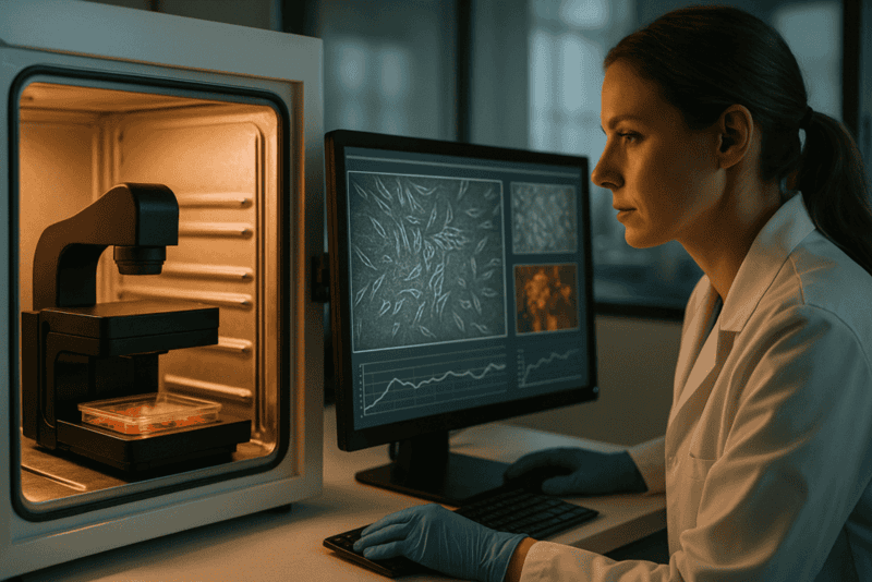
“```html
Stop Guessing: Why In-Incubator Imaging is the Secret to Flawless Research
In the rapidly evolving field of cell biology, precise and continuous observation of cell cultures is paramount. As research endeavors push the boundaries of what we know about cellular processes, the need for consistent and high-quality data becomes increasingly vital. Enter in-incubator imaging: a technology poised to revolutionize how researchers conduct experiments. This article delves into the common limitations of traditional cell culture approaches and explores how in-incubator imaging—specifically live-cell imaging within the protective cocoon of an incubator—emerges as the key to flawless research.
Common Challenges and Limitations of Traditional Approaches
Intermittent Data Acquisition
Traditional cell culture methods often rely on manual interventions to observe and record cellular changes. This approach can lead to sporadic data acquisition, and as researchers know, missing critical events in cell behavior can skew the results and interpretations of an entire study.
- Loss of significant time points due to periodic observation
- Increased variability due to operator differences
Environmental Disturbances
Each time a culture dish is removed from its incubator for inspection, it is exposed to environmental changes that can adversely affect cell health. Temperature fluctuations, changes in CO2 concentration, and physical disturbances can all introduce unwanted variables, impacting the reliability of results.
- Temperature and pH shifts affecting cell viability
- Potential for contamination each time the incubator is opened
Technological Advances and Automation Trends
Integration of Live-Cell Imaging
Advancements in imaging technology now allow for real-time, continuous monitoring of cell cultures without the need for removal from optimal growing environments. In-incubator imaging systems, like the zenCELL owl, exemplify these innovations by offering compact, user-friendly solutions that align seamlessly with existing workflows.
- Enabling real-time observation and time-lapse studies
- Reducing manual intervention and its associated drawbacks
Enhanced Data Throughput and Analysis
The automation of image acquisition and processing facilitates high-throughput screening (HTS) applications and improves scalability. With automated systems, researchers can focus more on analysis and interpretation rather than data collection.
- Increased efficiency with automated workflows
- Better resource allocation for complex experimental setups
Practical Examples and Workflows Using Live-Cell Imaging
Migration Assays
Cell migration studies are fundamental in understanding processes like wound healing and cancer metastasis. In-incubator imaging enables uninterrupted visualization of cellular movements, providing insights into migration kinetics and pathway activation without risking environmental artifact introduction.
- Timely capture of directional movement patterns
- High-resolution imaging supporting detailed morphological assessments
Organoid Development
Organoids mimic the architecture and function of organs, representing a cornerstone in drug discovery and regenerative medicine research. The continuous monitoring capabilities of in-incubator imaging systems offer detailed developmental stage images without ever disturbing the 3D cultures.
- Enhanced monitoring of growth conditions and morphological changes
- Increased reliability in developmental milestone verification
Lesen Sie weiter, um tiefere Einblicke und Strategien zu gewinnen.
“`
“```html
Enhancing Experimental Precision and Reproducibility
Data Consistency and Reliability
In-incubator imaging systems bring a significant advantage to experimental precision through seamless data acquisition. This methodology circumvents batch processing and manual entry errors inherent in traditional methods, thereby providing a more consistent data stream. The standardization offered by these systems leads to increased reproducibility, which is crucial for high-stakes research and publications. For example, a study published in the Nature Methods journal demonstrated that live-cell imaging platforms reduced variability by over 30%, substantially increasing both confidence in the reproducibility of results and credibility in peer-reviewed forums.
- Prioritize calibration and standardization steps for accuracy.
Reducing Human Error and Increasing Efficiency
Streamlined Workflows
By reducing the need for manual observations and interventions, in-incubator imaging decreases the risk of human error. This technology supports more efficient lab operations by automating repetitive tasks. For instance, operating with software integrated with AI-driven checklist capabilities not only speeds up the entire process but ensures that human oversight focuses on critical analysis rather than mundane data entry. A renowned cancer research lab reported a 40% increase in efficiency with virtually zero data discrepancies upon adopting these systems.
- Implement automation-friendly tools for routine procedures.
Improving Outcomes with Machine Learning and AI
Predictive Modelling and Pattern Recognition
The infusion of machine learning and AI into live-cell imaging technologies offers researchers a powerful toolkit for identifying cellular patterns and predicting experimental outcomes. These predictive capabilities enable researchers to preemptively adjust experimental parameters, reducing experimental failures and wasted resources. A compelling example comes from a pharmaceutical company’s adoption of AI-enhanced imaging to fine-tune their drug efficacy assays, resulting in a 25% improvement in their lead identification process.
- Layer machine learning algorithms to enhance image analysis capabilities.
Accelerating Discovery with Scalable Solutions
Adapting to Changing Research Needs
The scalability of in-incubator imaging allows for smooth transitions from small-scale exploratory studies to full-scale research projects. As research demands grow, the modular nature of these systems supports rapid scaling without the need for significant additional investment. Take, for example, a biotech startup that expanded its research from small animal model studies to a large-scale human cell line investigation. Leveraging scalable imaging solutions, they were able to double their study size within months, propelling the speed of their innovations.
- Favor modular systems for future research expansion.
Fostering Collaborative Research and Data Sharing
Integrating Cross-Disciplinary Teams
In-incubator imaging facilitates real-time data sharing and integrates seamlessly with digital lab environments, encouraging collaboration across disciplines. This ease of data exchange breaks traditional silos in research, allowing for expansive insights and cross-pollination of ideas. One cutting-edge research consortium used cloud-based data synchronization to merge oncological and immunological datasets, discovering novel immune-evasive mechanisms in tumors.
- Ensure secure and compliant data sharing protocols.
Facilitating Remote Research Access
Virtual Laboratories and Remote Monitoring
The ability to monitor cell cultures remotely through advanced imaging systems is game-changing, especially in a post-pandemic world where flex-work and social distancing are prevalent. Researchers can oversee multiple experiments concurrently from different locations, minimizing downtime and accelerating research timelines. A leading virology lab cited remote accessibility as a pivotal factor in managing experiments during travel restrictions, maintaining productivity and continuity seamlessly.
- Implement secure remote connections and user authentication.
Maximizing Resource Allocation and Cost Efficiency
Optimized Usage of Consumables and Equipment
The added precision and automation of in-incubator imaging often lead to reduced waste and better use of resources. Longevity of consumables through decreased human interaction with cell cultures translates into lower operational costs and greater sustainability. For instance, laboratories shifting to automated imaging have reported up to 20% savings on cell culture reagents and reduced warehousing needs.
- Apply analytics to monitor resource usage and waste reduction.
Transformative Case Studies and Success Stories
Real-World Impacts and Lessons Learned
Various institutions have shared transformative impacts from adopting in-incubator imaging. A notable case involved a university research team investigating neurodegenerative diseases, which utilized time-lapse imaging to reveal unexpected neuron firing patterns tied to treatment responses. These insights, gathered through uninterrupted imaging over weeks, led to a breakthrough publication with implications for therapeutic strategies in Alzheimer’s disease.
- Document and disseminate case studies to share best practices.
Im Anschluss fassen wir die wichtigsten Erkenntnisse, Kennzahlen und eine wirkungsvolle Schlussfolgerung zusammen.
“`
“```html
Transforming Education and Skill Development
Hands-On Learning and Virtual Training
In-incubator imaging systems are revolutionizing education and skill development in the scientific community by providing more hands-on learning opportunities and virtual training modules. Students and early-career researchers gain direct access to cutting-edge technology, bridging the gap between theory and practice. Virtual tutorials and real-time demonstration of imaging techniques encourage active engagement, making complex concepts tangible. A partnership between a leading university and an imaging software company resulted in a curriculum that significantly improved student proficiency in bioinformatics, showcased in an inter-college imaging contest where participants demonstrated practical knowledge in cell analysis.
- Incorporate virtual labs and simulation tools into educational curricula.
Boosting Innovation in Biotech and Pharmaceuticals
Pioneering New Frontiers
With its ability to deliver highly accurate and reproducible data, in-incubator imaging is a potent catalyst for innovation in biotech and pharmaceutical industries. The marriage of technology and biological research opens doors to pioneering new drug discovery methods and treatment pathways. A burgeoning biopharmaceutical startup leveraged AI-driven imaging to accelerate their vaccine development pipeline, achieving novel breakthroughs in immunotherapy. This integration not only shortened development timelines but also opened new avenues for personalized medicine, underscoring the transformative power of precision science in fostering innovation.
- Drive innovation through strategic investments in emergent imaging technologies.
Enhancing Global Research Competitiveness
Attracting and Retaining Talent
Institutions that adopt in-incubator imaging position themselves competitively on the global research stage. By providing advanced tools and fostering an environment conducive to cutting-edge research, these institutions attract top-tier talent and build a reputation for excellence. A well-funded national research institute noted a 30% increase in graduate researcher applications after implementing comprehensive imaging technologies across its labs, demonstrating the appeal of modern research environments to aspiring scientists globally.
- Leverage state-of-the-art facilities to enhance institutional prestige and visibility.
Schlussfolgerung
The profound impact of in-incubator imaging on the field of scientific research is undeniable. By enhancing precision and reproducibility, reducing human error, and expanding capacity through machine learning and AI, this technology revolutionizes methodologies across disciplines. The flexibility and scalability it provides not only streamline workflows but also enable researchers to adapt to evolving challenges seamlessly. As demonstrated by the extensive case studies and success stories, these systems are instrumental in accelerating discoveries, merging cross-disciplinary efforts, and optimizing resource allocation, all while fostering collaborative research and data sharing.
Moreover, in-incubator imaging takes center stage in paving the way for future advancements and driving innovation in biotech and pharmaceuticals, where precision and swift adaptability are critical. Its influence extends to educational settings, equipping the next generation of scientists with essential skills and knowledge firsthand. By integrating such advanced technologies, research institutions enhance their global competitiveness, thereby attracting and retaining elite talent committed to pushing the boundaries of human understanding and capability.
As we stand on the brink of a new era of scientific exploration, it becomes imperative to embrace tools like in-incubator imaging systems that challenge traditional perspectives and inspire transformative thinking. Institutions, laboratories, and industries keen on maintaining a cutting-edge reputation must commit to adopting these sophisticated solutions to not only bolster their research capabilities but also to ensure science progresses with unprecedented accuracy and efficacy. We invite you to explore these technologies further, take bold steps towards upgrading your research infrastructure, and witness firsthand the remarkable transformations that await the future of research.
“`
Continuous Cell Monitoring as the New Standard in Cell Culture Research

“```html
Continuous Cell Monitoring as the New Standard in Cell Culture Research
In the dynamic field of life sciences, continuous cell monitoring is emerging as the new standard in cell culture research. As researchers increasingly seek precise and reproducible results, the ability to monitor live cells over extended periods without disruption is becoming critical. This article explores the significance of continuous monitoring, addressing traditional challenges and detailing the role of advanced live-cell imaging technologies. Readers will gain insights into technological advances in automation, practical applications of these innovations in the laboratory, and the profound impact on research outcomes.
Traditional Challenges in Cell Culture Research
Limitations of Conventional Techniques
Historically, cell culture research has relied heavily on manual observation and periodic sampling. While these methods have laid the foundation for countless scientific discoveries, they present notable limitations. Manual monitoring disrupts cell environments each time a sample is taken for observation, which can lead to stress responses that affect cell behavior and viability. Moreover, such sporadic snapshots fail to capture transient phenomena, leading to incomplete datasets.
This methodological gap underscores the need for continuous monitoring to reduce environmental disturbances and collect comprehensive data sets that are more reflective of true cell behavior under physiological conditions.
- Environmental disturbances during manual sampling
- Incomplete data from intermittent observations
- Stress responses affecting cell viability
Technological Advances and Automation Trends
Integrating Live-Cell Imaging and Automation
The advent of automated live-cell imaging systems has revolutionized cell culture research. These systems are designed to operate within incubators, offering a seamless way to capture images and data without the need to manually handle cultures. Automation not only enhances reproducibility by minimizing human error but also facilitates long-term studies by enabling uninterrupted observation.
The zenCELL owl exemplifies these advances with its compact and incubator-compatible design. This system ensures that cultures remain in optimal conditions while being continuously monitored, helping researchers gather high-quality, reliable data over time.
- Reduced manual intervention enhances data quality
- Automation supports high-throughput screening (HTS)
- Incubator integration maintains stable conditions
Implementing Live-Cell Imaging Workflows
Examples of Practical Applications
Live-cell imaging and continuous monitoring have opened new horizons for specific applications in cell biology. Researchers conducting migration assays and organoid studies can benefit significantly from these technologies. For instance, continuous imaging enables the precise tracking of cell movement and growth, which is critical in understanding cancer metastasis and tissue regeneration.
Furthermore, proliferation assays and high-throughput screenings (HTS) greatly benefit from the ability to collect time-lapse data, yielding insights into cellular kinetics under varying conditions.
- Improved understanding of cancer cell migration
- Enhanced data quality in tissue regeneration studies
- Time-lapse data enrich HTS outcomes
Lesen Sie weiter, um tiefere Einblicke und Strategien zu gewinnen.
“`
“```html
Streamlining Laboratory Efficiency with Automation
Allocating Resources for Maximum Productivity
Automation in continuous cell monitoring not only improves data reliability but also liberates valuable human resources. Researchers previously tasked with repetitive and time-consuming tasks now have the opportunity to focus on more complex aspects of experimental design and data interpretation. Real-world examples illustrate how laboratories with integrated automation systems report a significant increase in throughput and a concurrent reduction in labor costs. Additionally, the utilization of advanced imaging techniques facilitates collaborative research, enabling seamless data sharing and multi-site collaborations.
- Automation allows researchers to allocate time to more critical tasks
- Increased data throughput and reduction in human error
- Facilitates collaborative research projects and data sharing
Enhancing Data Accuracy and Insights
Achieving Consistent and Repeatable Results
Automatic systems make it possible to capture precise timing intervals and conditions without deviation, a feat often challenging with manual methods. This consistency ensures that experiments are reproducible, a cornerstone of scientific validity. For example, in drug development studies, precise monitoring can reveal critical timepoints where a compound’s influence varies, leading to new therapeutic insights. Enabling real-time data capture helps in drawing robust conclusions and refining hypotheses for successive experiments.
- Consistency in monitoring intervals enhances experimental reproducibility
- Real-time data allows for refined hypothesis development
- Insights from precise timing improve understanding in therapeutic research
Integrating Machine Learning and AI
Leveraging Technology for Advanced Data Analysis
The fusion of live-cell imaging with machine learning and artificial intelligence (AI) has created new opportunities for dynamic data analysis. AI algorithms can process large volumes of imaging data far beyond human capability, identifying patterns and generating predictive models. In specific cases, this capability aids in identifying drug resistance patterns in cancer cells, allowing for tailored treatment strategies. The integration of ML and AI into cell monitoring processes is increasingly being recognized for its potential to push the boundaries of personalized medicine.
- Machine learning boosts data analysis capabilities beyond human limits
- AI helps identify patterns, aiding in predictive model creation
- Promotes advancements in personalized medicine approaches
Real-World Case Studies: Success Stories
Implementations and Outcomes in Leading Laboratories
Several groundbreaking studies have demonstrated the impact of continuous cell monitoring systems. A prominent example is a research group focused on neurodegenerative diseases. By continuously monitoring neuronal cells, they discovered previously undetected cellular responses to potential treatments. This breakthrough may pave the way for preventive strategies in the treatment of Alzheimer’s disease. Similarly, in industrial biotechnology, companies apply these techniques to optimize microbial cultures, significantly boosting biofuel production efficiency.
- Continuous monitoring reveals new cellular responses in neurons
- Advances in Alzheimer’s research pave way for new treatments
- Enhanced biofuel production efficiency through microbe optimization
Mastering Data Management in Modern Labs
Strategizing Storage and Utilization
As live-cell imaging devices generate vast amounts of data, efficient data management is paramount. Laboratories integrate robust data storage solutions, often employing cloud-based systems for real-time data access and sharing. Analytical tools are applied to ensure data integrity, facilitating seamless synthesis of results for publication or regulatory compliance. Effective data management not only aids in maintaining an orderly research process but also supports compliance with data protection laws like GDPR in Europe and HIPAA in the United States.
- Cloud storage facilitates effective data sharing and real-time access
- Management tools ensure data integrity and regulatory compliance
- Efficient strategies support compliance with international data laws
Combining Traditional and Modern Approaches
Blending Established Techniques with Technological Advancements
Balancing traditional cell culture methodologies with modern technological advancements allows researchers to harness the benefits of both. Incorporating the depth of knowledge derived from traditional practices with cutting-edge technologies leads to more informed experimental designs. Continuous feedback from the live-cell imaging complements traditional qualitative observations, resulting in a comprehensive understanding of cellular pathways and interactions.
- Combining strategies leads to more comprehensive experimental designs
- Technological advancements complement traditional knowledge
- Continuous feedback enriches understanding of cellular processes
Navigating Common Pitfalls in Implementation
Avoiding Challenges for Successful Integration
Integrating new technologies into existing workflows can present challenges. Identifying potential pitfalls, such as technological incompatibilities or user training requirements, is crucial for successful adoption. Ensuring compatibility between novel systems and existing laboratory infrastructure is a common concern. Additionally, investing in adequate personnel training can prevent disruptions in experimental continuity, helping laboratories make the most of new technology.
- Identify and address technological incompatibilities proactively
- Emphasis on user training prevents experimental disruptions
- Successful integration maximizes technological benefits
Im Anschluss fassen wir die wichtigsten Erkenntnisse, Kennzahlen und eine wirkungsvolle Schlussfolgerung zusammen.
“`
“```html
Future Prospects and Innovations
Continual Evolution in Cellular Research
The future of continuous cell monitoring is paving the way for even more groundbreaking scientific explorations. As technology advances, laboratories can expect the development of more sophisticated imaging techniques and enhanced AI-driven analytics. These innovations will open doors to deeper insights into cellular behavior, pushing forward the frontiers of personalized medicine and tailored therapeutic approaches. Researchers are now capable of dynamically adapting their methodologies based on evolving data trends, ensuring that scientific inquiry remains a continuously advancing field.
- Emerging imaging technologies promise deeper insights
- AI-driven analytics enhance research precision
- Adaptive methodologies drive continuous scientific progress
Standardizing Practices Across Laboratories
Creating Consistency in Research Methods
A major step towards maximizing the benefits of continuous cell monitoring is the standardization of practices across different laboratories. Building a unified framework for data collection and analysis will allow for greater collaboration and comparability of results. Setting industry standards not only ensures quality control and enhances the reproducibility of experiments but also fosters an environment of shared innovation and collective progression in scientific research.
- Standardized practices enhance data comparability
- Quality control ensures reliable and reproducible experiments
- Industry standards foster collaborative innovation
Overcoming Ethical and Compliance Challenges
Ensuring Responsible Technological Integration
While technological advancements offer tremendous opportunities, they also bring forth ethical and compliance challenges that laboratories must navigate. Ensuring transparency in data handling and patient data confidentiality is paramount. Laboratories need to adopt guidelines that align with international data protection standards, instilling trust and confidence among stakeholders. This ensures that while we push the boundaries of research capabilities, we remain ethically sound and compliance-focused.
- Transparency in data handling strengthens trust
- Aligning with international guidelines protects data integrity
- Ethical practices support sustainable scientific progress
Schlussfolgerung
In conclusion, continuous cell monitoring stands at the forefront of transforming cell culture research into a more dynamic, efficient, and insightful endeavor. By seamlessly integrating automation, machine learning, and AI, research laboratories are not only enhancing data accuracy and experimental productivity but are also paving the way towards personalized medicine and novel therapeutic discoveries. With these advancements, the ability to delve deeper into cellular mechanisms creates vast potential for breakthroughs in understanding and treating complex diseases.
The standardization of practices further empowers this scientific evolution, establishing mechanisms for greater consistency and reproducibility across global laboratories. Coupled with overcoming ethical and compliance challenges, continuous cell monitoring encourages responsible and innovative research practices that benefit the entire scientific community and ultimately, society at large.
As we look to the future, we find ourselves on the cusp of limitless possibilities with these cutting-edge technologies. The responsibility lies in harnessing their true potential while upholding the foundations of scientific integrity and transparency. Let us embrace this revolution, encourage collective growth, and continuously strive to unlock the mysteries of the smallest units of life for a healthier, more informed world. Together, we forge a promising path toward remarkable discoveries, inspiring future generations to carry on this legacy of innovation and excellence.
Join us in this transformative journey and be part of the forefront in the evolution of cell culture research.
“`
Cost-Efficient Live-Cell Imaging: Why Smaller Automated Systems Win

Cost-Efficient Live-Cell Imaging: Why Smaller Automated Systems Win
In the evolving landscape of modern cell culture research, the efficiency and accuracy of live-cell imaging have become paramount. As laboratories strive to improve outcomes while optimizing resources, the need for cost-efficient solutions has gained significant attention. In this article, we will explore how smaller automated systems offer a promising solution to common lab challenges, particularly in the context of continuous live-cell monitoring. From understanding the inherent limitations of traditional methods to assessing the benefits of compact innovations, we’ll dive into the transformative potential of these advanced technologies.
Herausforderungen traditioneller Live-Cell-Imaging-Ansätze
Resource Intensiveness and Limited Flexibility
Traditional live-cell imaging systems have long been the cornerstone of cellular research. However, these setups often come with inherent challenges that can hinder productivity in modern labs. High costs and significant spatial demands are among the most pressing issues. Such systems usually require dedicated microscopy rooms, complex handling, and frequent downtime due to maintenance needs.
- High initial and operational costs limit access for smaller labs.
- Space-intensive designs are impractical for labs with limited physical infrastructure.
- Complexity in operation necessitates specialized training and can lead to increased error rates.
Technological Advances and Trends in Automation
Evolution Toward Compact and Efficient Designs
The shift towards smaller, automated systems reflects a broader trend in laboratory automation aimed at enhancing flexibility and cost-efficiency. Recent advances prioritize adaptability, ease of use, and improved integration with existing lab workflows, which are crucial for addressing the limitations of conventional setups. Such systems enable continuous, uninterrupted monitoring without compromising on data quality.
- Enhanced automation reduces manual intervention, freeing up valuable researcher time.
- Compact designs support easy integration with existing laboratory equipment.
- Automation trends align with the push towards high-throughput screening and precision medicine.
Implementing Practical Workflows with Live-Cell Imaging
Improving Data Quality and Laboratory Throughput
By adopting smaller automated live-cell imaging systems, laboratories can streamline operations and elevate the quality of experimental data. Systems like the zenCELL owl, for example, facilitate continuous observation without the need for direct human intervention, thereby increasing reproducibility and reducing the potential for human error.
- Automated imaging systems enhance reproducibility through consistent environmental conditions.
- Systematic monitoring minimizes operator variability, a common issue in manual workflows.
- Data acquisition and analysis are expedited, facilitating more timely decision-making.
Lesen Sie weiter, um tiefere Einblicke und Strategien zu gewinnen.
“```html
Increasing Accessibility and Scalability
Deploying Cost-Effective Solutions for Every Lab Size
One of the standout advantages of adopting smaller automated live-cell imaging systems is the democratization of access to advanced research technologies. For many smaller labs and institutions, traditional systems are prohibitively expensive, both in terms of initial investment and ongoing operational costs. Compact systems like the zenCELL owl require a fraction of the budget traditionally needed, making cutting-edge imaging technologies accessible to a broader range of researchers. Additionally, these systems offer scalability, allowing labs to expand their capabilities in alignment with growth or increased research demand.
- Start with a single-unit system and scale up as research needs evolve.
- Utilize cost savings to fund additional research projects or personnel training.
Enhancing Data Consistency and Integrity
Maintaining High Standards Across Experiments
Data consistency and integrity remain critical challenges in live-cell imaging. Traditional systems often fall short due to variability in manual operations and environmental conditions. Conversely, automated compact systems provide a more controlled environment, minimizing fluctuations and external interferences. This stability is crucial for ensuring the quality and reliability of collected data, which can decisively influence research outcomes and conclusions.
- Implement standardized protocols to maintain consistency across experimental runs.
- Regularly calibrate devices to ensure optimal performance and data accuracy.
Reducing Carbon Footprint and Environmental Impact
Creating Greener Labs for a Sustainable Future
Environmental sustainability is becoming a pivotal focus in academia and industry alike. Smaller live-cell imaging systems contribute to greener labs by consuming significantly less power and occupying reduced space. Many of these systems are designed with minimalistic cooling requirements and lower energy consumption, which collectively lower the carbon footprint of a laboratory. Institutions such as the University of Cambridge have reported significant energy savings by transitioning to compact imaging systems.
- Conduct energy audits to identify further efficiency opportunities within labs.
- Adopt eco-friendly practices as part of a broader sustainability initiative.
Facilitating Remote Monitoring and Collaboration
Empowering Researchers Through Connectivity
The integration of remote monitoring capabilities into smaller systems has revolutionized how scientists collaborate and conduct research. With the ability to access live-cell imaging data in real time from any location, researchers can now make immediate adjustments and decisions, fostering enhanced collaboration across geographies and institutions. This connectivity is especially beneficial during instances when physical lab presence is not feasible, such as during travel or global events like the COVID-19 pandemic.
- Leverage cloud-based storage solutions for seamless data sharing and backup.
- Utilize collaboration tools for real-time data interpretation and teamwork.
Streamlining Training and Operational Procedures
Building a Skilled and Efficient Workforce
Compact live-cell imaging systems are designed with user-friendliness in mind, significantly lowering the learning curve compared to traditional systems. Many systems come equipped with intuitive interfaces and extensive online resources, facilitating smoother operational transitions. This ease of use not only enhances productivity but also reduces the likelihood of human error, thereby protecting the integrity of experimental outcomes.
- Incorporate structured training sessions into onboarding for new researchers.
- Regularly update staff on new system capabilities and software features.
Empowering Personalized and Precision Medicine
Driving Innovation in Healthcare Research
The marriage of compact automated imaging systems and precision medicine promises unprecedented advancements in healthcare. By enabling high-throughput screenings and detailed cellular analyses, these systems provide invaluable insights into patient-specific responses and drug effects. Institutions engaging in precision medicine research, like the Dana-Farber Cancer Institute, utilize such technologies to streamline patient data collection, resulting in more targeted and effective treatment strategies.
- Develop collaborations between imaging experts and clinical researchers.
- Utilize imaging data to personalize treatment regimens and improve patient outcomes.
Im Anschluss fassen wir die wichtigsten Erkenntnisse, Kennzahlen und eine wirkungsvolle Schlussfolgerung zusammen.
“`
“```html
Optimizing Workflow and Laboratory Management
Enhanced Efficiency and Productivity
Modern laboratories face unique challenges, not only in research but also in the effective management of resources. Smaller automated live-cell imaging systems redefine laboratory workflows by optimizing the use of space and resources. Their compact nature allows for flexible installations that can be tailored to the specific needs and constraints of any laboratory environment. This adaptability paves the way for streamlined processes and improved productivity across the board.
- Engage in continuous process improvement to maximize the benefits of compact systems.
- Optimize lab layouts to accommodate both current and anticipated future technologies.
Supporting Advanced Teaching and Education
Bridging Theory with Practical Application
In the educational sector, the practical application of theoretical knowledge is crucial for developing competent researchers. Compact live-cell imaging systems provide educational institutions with the tools necessary to bring cutting-edge science into the classroom. Their cost-effectiveness and ease of use enable integration into curricula, offering students firsthand experience with live-cell imaging techniques, which enhances learning outcomes and prepares them for future scientific careers.
- Integrate practical sessions with automated imaging into biology and life sciences curricula.
- Facilitate student-led research initiatives utilizing accessible imaging technologies.
Advancing Open Science and Data Sharing
Fostering Collaborative Research Environments
The open science movement emphasizes transparency, sharing, and collaboration. Compact imaging systems are ideally suited to support these goals by facilitating data sharing and cross-institutional collaborations. Their integration with cloud technologies and data storage solutions means data can be readily accessed and shared with researchers globally, promoting a more connected and collaborative scientific community that stands to accelerate breakthroughs and innovation.
- Adopt open-access policies for data generated using live-cell imaging systems.
- Encourage partnerships and collaborations across disciplines and institutions.
Schlussfolgerung
Throughout this article, we’ve explored the myriad benefits of adopting smaller, automated live-cell imaging systems in research settings. From democratizing access to advanced technologies to enhancing data consistency and integrity, these compact systems offer distinct advantages over traditional counterparts. By enabling researchers to increase accessibility, expand their capabilities, and streamline operational procedures, they play a pivotal role in advancing scientific inquiry.
The significance of reducing the carbon footprint and the push towards a more sustainable laboratory environment cannot be overstated. These systems not only consume less power and space but also offer efficiencies that contribute to broader institutional sustainability initiatives. Furthermore, with the integration of remote monitoring capabilities, they empower scientists to collaborate and make informed decisions regardless of their physical location, thereby embodying the adaptability required in today’s rapidly changing world.
The impact of these systems extends beyond efficiency and sustainability; they support the advancement of precision medicine, aid in educational endeavors, and promote a culture of open science. By making research tools accessible and encouraging inclusive collaboration, compact live-cell imaging systems open new frontiers in healthcare, education, and scientific discovery.
As we look to the future, the adoption of these technologies stands as a testament to progress and innovation. Laboratories worldwide, from small academic institutions to established research facilities, are poised to benefit immensely. By embracing these advancements, researchers can drive more impactful discoveries, contribute to a more sustainable planet, and shape the next generation of scientists.
Herein lies the call to action: whether in academia or industry, now is the time to invest in these transformative systems. Let’s push the boundaries of what’s possible, create meaningful impacts in our respective fields, and commit to sustainable and innovative research practices that promise a brighter future for science and society alike.
“`




