Automated Wound Healing & Migration Assays: How to Achieve Reproducible Results
Cell migration and wound healing assays are essential tools in cell biology, oncology, regenerative medicine, and pharmacological research. Traditional scratch assays, while widely used for studying collective cell movement and regeneration, often suffer from inconsistencies and subjective data interpretation. With the increasing need for high-throughput screening, real-time observation, and reproducibility in life science applications, automated wound healing and migration assays have emerged as a robust solution.
This article explores the scientific and technical considerations for achieving reproducible results in automated assays, covering validation strategies, live-cell imaging technologies, and trends in scalable labware development. Researchers, lab managers, and biotech developers will gain a deep technical understanding of the methods and materials that support the reliability of automated wound healing workflows under regulated conditions.
Challenges in Traditional Wound Healing Assays
Technical Limitations of Manual Scratch Methods
The classic wound healing assay involves manually creating a cell-free zone (“wound”) in a confluent cell monolayer using pipette tips or blades. Despite its simplicity, this method introduces significant bias across time points and replicates due to mechanical inconsistencies and human error. These technical variabilities limit assay reproducibility and reduce confidence in comparative data.
- Manual scratches vary in width, edge shape, and cell detachment effects.
- Edge damage can release intracellular contents, altering local microenvironments.
- Subjective imaging and endpoint analyses hinder standardization in multi-well formats.
Environmental and Workflow Inconsistencies
Reliance on traditional microscopes outside of incubators introduces temperature and CO₂ fluctuations that disturb cell physiology. Moreover, inconsistent assay timing and imaging delays further impair reproducibility, especially in time-sensitive applications such as drug screening or migration kinetics.
- Movement of plates between incubators and imaging stations creates environmental shocks.
- Manual imaging scheduling leads to uneven observation intervals.
- Data quality suffers from off-incubator imaging due to focus drift and condensation.
Technology Advancements Driving Automation
Automated Live-Cell Imaging Platforms
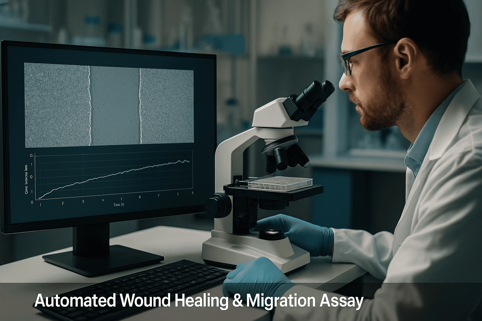
To ensure consistent observation and quantitative data generation, many laboratories are adopting incubator-compatible imaging systems. Continuous monitoring using compact, automated devices—such as the zenCELL owl—enables real-time data acquisition without removing cells from optimal culture conditions.
- Real-time kinetic data of cell migration and gap closure.
- Imaging within a standard incubator reduces environmental variability.
- Multichannel and time-lapse images support comprehensive, unbiased analysis.
Precision Labware for Assay Standardization
Lab plastics tailored for migration assays, such as pre-defined inserts and wound field designs in multiwell formats, offer mechanical consistency and improve performance metrics across experiments. These precision-molded formats eliminate edge variability and are compatible with automated liquid handling systems, crucial for scalable workflows.
- Custom-designed wells ensure consistent scratch width and geometry.
- Transparent, optically clear plastics (e.g. polystyrene, COC) support high-resolution imaging.
- Surface functionalization (e.g. TC treatment) promotes even cell adhesion and growth.
Implementing Automated Wound Healing Assays
Workflow Integration in Regulated Lab Environments
Transitioning to automated wound healing and migration assays involves synchronizing hardware, consumables, and software within a validated quality control framework. Especially in GMP or cGMP-compliant labs, every aspect from assay design to data output must adhere to robust documentation and reproducibility standards.
Key considerations include:
- Use of validated, traceable labware and imaging instruments.
- Implementation of audit trails and data storage compliant with 21 CFR Part 11.
- Standard operating procedures (SOPs) for gap creation, cell seeding, media change, and imaging.
Examples of Optimized Automated Assay Protocols
The use of precision multiwell plates combined with real-time imaging allows for reproducible assay designs. For instance, combining a 24-well plate with embedded cell exclusion zones and the zenCELL owl system allows a continuous 72-hour migration monitoring without manual intervention. Such workflows are particularly valuable in kinetic drug response studies or testing growth factor effects on cell mobility.
Benefits include:
- Simultaneous real-time monitoring across multiple wells or conditions.
- Reduction in assay-to-assay variability through standardized plate formats and protocols.
- Minimized operator time while maximizing data resolution and analysis consistency.
Enhanced Reproducibility with Incubator-Based Imaging
Environmental Stability Improves Cellular Fidelity
Maintaining cells within controlled incubator conditions during imaging preserves metabolic activity and cellular behavior, especially important for sensitive cell types. Incubator-compatible systems like the zenCELL owl eliminate the need for sensor recalibration and refocusing between observations, reducing variability introduced by manual microscopy sessions.
- Sustained 37 °C, 5% CO₂ eliminates thermal and pH shifts during time-lapse studies.
- High-frequency imaging captures transient events and accelerates migration rate calculations.
- Time-resolved imaging enables statistical analysis of wound closure kinetics across biological replicates.
Automated Image Acquisition and Analysis
Advanced software algorithms quantify wound area and cell movement automatically, reducing observer bias. Integration of tailored software workflows allows users to standardize analysis endpoints and minimize data handling errors. These systems also enable batch processing for screening applications requiring high-throughput assay formats such as 96-well plates.
- Image segmentation algorithms ensure consistent wound edge detection.
- Metadata tagging ensures traceability for GMP record-keeping requirements.
- Analysis modules support quantitative kinetics for migration speed and proliferation indices.
Applications Beyond Classical Wound Healing
Cell Migration, Organoids, Proliferation, and Drug Screening
Automated wound healing assays form the basis for several related in vitro assessments. Researchers apply similar protocols for evaluating fibroblast, endothelial, or cancer cell invasion under defined gradients. Furthermore, organoid-based migration assays and barrier integrity models are expanding the scope of these techniques by integrating 3D formats and co-culture systems.
- Migratory behavior in cancer models to assess metastasis potential.
- Barrier reformation in epithelial monolayers to study tight junction recovery.
- Proliferation tracking alongside migration for combined mechanistic investigations.
High-Throughput Screening (HTS) and Multiplexed Studies
Automated imaging and labware compatibility with robotic pipetting platforms support high-throughput settings where multiple drug candidates or treatment conditions must be evaluated simultaneously. Optically clear injection-molded lab plastics in 96- or 384-well plates allow for scalable adoption of migration and wound healing assays while preserving imaging fidelity.
- HTS-compatible plate formats reduce reagent volumes and increase parallelism.
- Data consistency ensures reliable lead identification in early drug discovery.
- Integrated assay automation supports streamlined workflows across R&D and quality labs.
Continue reading to explore more advanced insights and strategies.
Advanced Assay Calibration for Quantitative Accuracy
Optimizing Imaging Parameters and Reference Controls
Achieving consistent, high-fidelity results in automated wound healing assays requires calibration of imaging parameters—especially when using time-lapse systems and multichannel microscopy. Factors such as exposure time, focus depth, and pixel resolution must be precisely defined during assay development and kept constant throughout the experiment. The use of internal reference controls and calibration beads enables normalization across different imaging sessions or assay runs, improving inter-experimental repeatability.
- Perform flat-field correction and illumination uniformity tests to avoid uneven signal intensity.
- Include wells with known cell migration rates or migration-inhibited controls for internal benchmarking.
Optimizing Cell Density and Seeding Uniformity
Consistent Monolayer Confluence Enhances Assay Comparability
Uneven or low initial cell densities lead to variability in wound closure dynamics. For accurate wound healing measurement, it’s critical to standardize the seeding process across wells and experiments. Automated liquid handlers or multi-channel pipettes ensure reproducible delivery, while pre-coating plates with extracellular matrix components like fibronectin or collagen enhances uniform cell attachment and spreading. In high-throughput formats, vortex mixing followed by automated dispensing prevents cell clumping and supports monolayer homogeneity.
- Validate optimal seeding densities for each cell type to reach 90–100% confluence before wound initiation.
- Use robotic plate fillers or cell dispensers to minimize pipetting-driven variation during multicondition assays.
Chemical and Mechanical Gap Creation Strategies
Consistent Exclusion Zones Enable Standardized Kinetics
To eliminate the inconsistency of manual scratches, many labs have transitioned to mechanical inserts and hydrogel-based stencils for wound generation. These devices create reproducible gaps in monolayers without damaging surrounding cells. For example, silicone insert systems or removable polymeric stoppers allow users to lift predefined barriers after cell adhesion, enabling sharp, repeatable exclusion zones. Alternatively, enzymatic methods using dispase or non-cytotoxic peeling films can detach cells precisely from designated regions, facilitating gentle wounding in sensitive cultures.
- Use wound inserts sized to fit your specific multiwell plate and application format.
- Evaluate enzymatic or mechanical approaches based on target cell sensitivity and assay duration.
Automated Data Management for Regulatory Compliance
Scalable, Audit-Ready Workflows for GxP Environments
In regulated lab settings, automated wound healing platforms must support traceability, data integrity, and compliance with global standards such as 21 CFR Part 11 or EU GMP Annex 11. Integration of imaging systems with laboratory information management systems (LIMS) ensures secure data storage, retrieval, and auditability. Real-time tagging of metadata—including incubation parameters, imaging intervals, and treatment conditions—further enhances downstream data mining and reproducibility.
- Implement secure cloud-based storage or encrypted servers with digital access control verification.
- Use SOP-defined filename conventions and version control for image and analysis documentation.
Custom Software for Tracking Cell Behavior Over Time
Quantitative Analysis Algorithms Enhance Biological Insights
Modern imaging platforms deploy machine learning (ML) and AI-powered software to track individual cell movements, collective migration patterns, and proliferation events. These advanced tools allow researchers to differentiate between random cell motility and directed migration or chemotaxis. For example, software can calculate velocity vectors, persistence time, and path tortuosity, providing deeper biological meaning to mere wound area reduction metrics.
Several systems incorporate automated segmentation for cell tracking using DIC, fluorescence, or phase-contrast imaging. Users can define dynamic thresholds for wound area clearance, confluence index, and morphological parameters, enabling high-content screening directly from the wound healing assay.
- Use AI-assisted tracking to distinguish between contact inhibition, mitotic activity, and true migration.
- Apply morphokinetic metrics such as circularity and aspect ratio to evaluate epithelial-to-mesenchymal transitions (EMTs).
Case Study: Real-Time Drug Response Profiling
Automated Wound Healing as a Phenotypic Screening Tool
In one applied example, a pharmaceutical R&D team utilized a zenCELL owl system combined with barrier-based 24-well migration plates to analyze the effect of kinase inhibitors on breast cancer cell motility. Cells were seeded into the plates with removable stoppers forming 500-micron wounds. After a 24-hour treatment with varying drug concentrations, cell migration was tracked hourly for 48 hours. Software automatically quantified wound closure rates, providing EC₅₀ values correlated with cell viability and morphological changes.
This workflow eliminated manual analysis steps, reduced turnaround time by 67%, and increased reproducibility by 35% compared to traditional microscopy and hand-drawn ROI analysis. Integration with a LIMS system allowed the same workflow to be reused for other cancer cell lines and therapeutic candidates.
- Automated systems support reproducible, high-resolution phenotypic profiling in early-stage drug selection.
- Time-course migration tracking allows insight into both onset and durability of drug responses.
Multiparametric Analysis: Migration Meets Proliferation
Dissecting Cellular Contributions Using Combined Readouts
Distinguishing between cell migration and proliferation is critical for interpreting wound healing data, particularly in cancer models or regenerative medicine. Advanced assays incorporate dual-channel analysis, where a proliferation marker like BrdU or EdU is added in tandem with live-cell imaging. This approach allows researchers to decouple the effect of treatment on cytostasis versus directional movement. Furthermore, overlaying cell cycle reporters such as FUCCI enables a cell-by-cell phase analysis within the migrating population.
Some commercial assay platforms now integrate fluorescence overlays directly into their imaging timelines, providing seamless correlation of cell division markers with positional data. This dual profiling enhances mechanistic understanding and leads to more targeted therapy optimization.
- Use cytostatic controls alongside migration inhibitors to benchmark assay outputs and avoid data misinterpretation.
- Integrate nuclear and cytoplasmic markers for real-time proliferation tracking within wound edges.
Strategies for Time-Efficient Optimization of Assay Conditions
Reducing Setup Time Without Compromising Data Quality
To streamline assay setup across multiple conditions or cell lines, labs can adopt modular optimization strategies. This includes miniaturized pilot runs in 12- or 24-well formats using automation-compatible inserts and imaging loops to quickly assess optimal seeding density, confluence timing, and treatment start times. Imaging software presets can then be programmed for batch acquisition and stitched image compilation where required.
Instituting a Design of Experiments (DoE) approach across temperature, serum levels, and coating conditions accelerates parameter tuning while maintaining scientific rigor. With automated cuvette or plate washer compatibility, solutions used for washing or media change are made more uniform, further boosting inter-assay comparability.
- Implement DoE-based pilot studies for rapid optimization of cell and media variables.
- Maintain matched biochemical conditions across wells using automated liquid handling protocols.
Next, we’ll wrap up with key takeaways, metrics, and a powerful conclusion.
Quality Control Checkpoints Across the Workflow
Ensuring Consistency from Reagent Prep to Data Output
Maintaining consistency across multiple runs of automated wound healing assays depends on well-defined quality control (QC) checkpoints. Each step—from reagent handling to image acquisition—can introduce variability if not properly standardized. Including both technical replicates and biological controls ensures that assay robustness remains high despite inevitable experimental shifts. For instance, preparing master mixes of media or inhibitors reduces reagent batch effects, while validating cell health using viability stains like calcein-AM or PI provides an upstream QC trigger.
Image pre-processing QC is often overlooked; however, verifying focus stability, drift correction, and stitching accuracy is essential when dealing with multiday, time-lapse imaging. Automated software platforms increasingly include preconfigured validation protocols that can flag anomalies in image acquisition or well-level inconsistencies.
- Design QC gates based on biological endpoints (e.g., confluence threshold) and technical parameters (e.g., image illumination profile).
- Create dashboards within your LIMS to track cell line passage numbers, reagent expiry, and system calibration dates.
Scalable Deployment Across Screens and Teams
From Discovery to Preclinical Development
As laboratories scale their wound healing workflows beyond single-user research setups into multiuser or interdepartmental pipelines, harmonizing protocols and data interpretation becomes imperative. Automated wound healing systems support scalability by enabling protocol preset sharing, remote access for data review, and standardization via machine-readable metadata. These benefits are especially valuable for pharmaceutical teams operating in dispersed preclinical ecosystems or organizations performing global concurrent screening studies.
To aid reproducibility, many setups utilize shared protocol libraries that ensure consistency in assay composition, imaging schedules, segmentation algorithms, and analysis parameters. Furthermore, multi-site teams can implement collaborative QC matrices that monitor assay fidelity across operator, site, and run timeline, creating a robust knowledge base built on consistent and well-annotated data.
- Standardize workflows using interchangeable run templates and centralized data labeling taxonomies.
- Use shared LIMS or cloud-based ELN platforms to propagate validated protocols across teams and programs.
Conclusion
Automated wound healing and migration assays have evolved into high-precision, reproducible platforms capable of delivering deep phenotypic insights across cell biology, oncology, and regenerative research. By embracing advancements in imaging calibration, cell seeding practices, and gap generation strategies, researchers can significantly minimize inter-assay variability while capturing rich, biologically relevant data.
Through integration with LIMS systems and application of custom software analytics, these platforms now support not only robust migration tracking but also time-resolved proliferation analysis and mechanistic dissection of cellular behaviors. The inclusion of dual readouts and multiparametric overlays allows for a comprehensive view of wound closure dynamics, improving the fidelity of conclusions drawn in both discovery and translational settings.
Key success factors include adopting standardized imaging protocols, consistent reagent preparation, and automated data handling practices to conform with regulatory requirements. As demonstrated in the case study and execution strategies, the shift to scalable, automation-driven workflows doesn’t just save time—it elevates the entire assay strategy, accelerating the path from cellular insight to actionable outcome.
Whether you’re optimizing for high-throughput compound screening, unraveling the underpinnings of migration in disease models, or validating therapeutic interventions, mastering these advanced wound healing assay techniques will put you ahead. By aligning precise assay architectures with flexible software and hardware integration, your lab can scale discoveries confidently and reproducibly.
Now is the time to rethink your approach to in vitro migration studies. Invest in automation, apply rigorous standardization, and let modern imaging technologies work for you. The future of wound healing assay performance lies in reproducibility, resolution, and real-world scalability—embrace it fully, and transform your research outcomes one experiment at a time.