Trends in Impedance Measurement for Cell Culture
Impedance-based analysis is transforming how researchers monitor and quantify cellular behavior in real time. With increasing demand for non-invasive, label-free monitoring across biomedical research, drug discovery, and biotechnology development, electrical impedance spectroscopy (EIS) is receiving renewed attention. This article investigates the latest trends in impedance measurement for cell culture, explores the limitations of traditional methods, and outlines how integration with automated, incubator-based systems enhances reproducibility, throughput, and data richness.
Why Impedance Measurement Matters in Modern Cell Culture
Non-invasive, label-free monitoring for continuous data acquisition
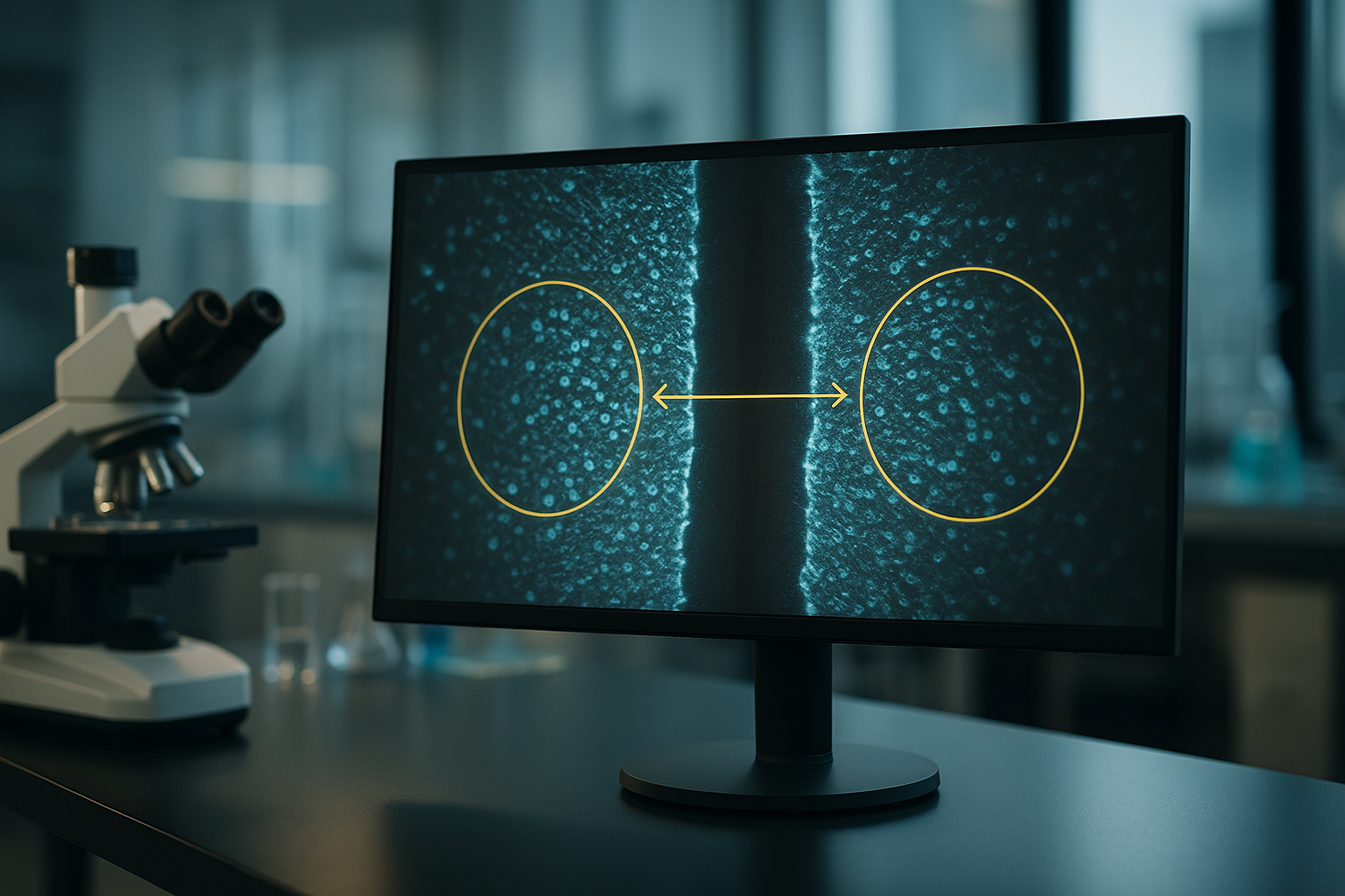
Modern cell biology requires high-resolution, high-content data—with minimal interference to the cell microenvironment. Impedance measurement, particularly electrical impedance spectroscopy (EIS), offers a unique capability: monitoring living cells continuously without staining, washing, or optical systems. This technique is highly sensitive to cell attachment, proliferation, barrier function, and changes in morphology, making it ideal for real-time assessments of cell behavior in vitro.
- Continuous data acquisition over hours or days
- Compatible with various adherent cell types
- Ideal for assessing cell proliferation, migration, and cytotoxicity
- Minimal disruption to cell culture conditions
Increasingly, impedance-based readouts are being integrated into automated, high-throughput platforms, supporting complex assays such as wound healing models, barrier integrity tests (TEER), and 3D culture systems including organoids and spheroids.
Limitations of Conventional Methods in Live Cell Monitoring
Endpoint assays and manual workflows hinder reproducibility
For decades, optical microscopy, colorimetric assays (e.g., MTT, XTT), and fluorescence-based methods have been standard in cell culture laboratories. While effective for many applications, these systems introduce several limitations that impact high-throughput and longitudinal studies:
- Endpoint nature restricts temporal resolution
- Labeling or staining can influence cell physiology
- Manual workflows limit consistency and throughput
- Results often require cell lysis or fixation, ending the experiment
Furthermore, results can vary significantly depending on technician skill, reagent stability, and microscope calibration—factors that limit reproducibility, especially in multi-user or multi-site environments. In regulated sectors such as pharmaceutical development or diagnostics QA/QC, where lot-to-lot comparability and traceability are essential, these inconsistencies can impede assay validation and regulatory submission timelines.
Advances in Impedance-Based Technologies and Automation
From benchtop readers to integrated, incubator-compatible imaging systems
Contemporary impedance measurement technologies now support label-free, real-time monitoring with outputs that can be automated, digitized, and integrated into cloud-based workflows. Integrated systems such as incubator-compatible readers combine data acquisition and environmental control, reducing fluctuations that typically influence sensitive measurements.
An example is the zenCELL owl, a compact system designed to fit within standard incubators and to deliver continuous impedance-based cell monitoring under consistent temperature and humidity conditions. Such systems address key pain points in live-cell analysis by reducing the need to remove plates from CO₂ incubators, maintaining stable conditions and minimizing mechanical disturbances.
Core technical advances fueling the adoption of impedance systems include:
- Miniaturization of readout electronics, enabling multiwell integration (e.g., 24-, 96-, 384-well formats)
- Improved electrode manufacturing techniques for reproducible, low-noise signal acquisition
- Digital data handling, supporting scalable cloud storage and real-time analytics
- Compatibility with automation platforms for liquid handling and high-throughput screening
These developments have significantly advanced impedance applications beyond basic research, making them increasingly relevant in diagnostics development, biosensor validation, and pharmaceutical screening workflows.
Using Impedance Measurement with High-Content Workflows
Linking morphology, confluency, and viability to quantitative data
Modern cell culture research often integrates impedance measurements with live-cell imaging, enabling researchers to interpret complex cell behaviors more holistically. In such systems, impedance provides continuous quantification of cell attachment, proliferation, and confluency, while imaging captures morphological changes, organoid structure, and intercellular interactions.
Workflows combining impedance with high-content imaging support nuanced analysis in areas including:
- Cell differentiation and maturation (e.g., iPSC systems)
- Barrier function evaluation in endothelial or epithelial cell models
- Migration and wound healing assays through dynamic impedance mapping
- Drug sensitivity screening under physiologically relevant conditions
In HTS (high-throughput screening) settings, impedance readouts offer normalization capabilities for cell number variability and reduce the need for post-assay viability staining, expediting turnaround and minimizing material costs. By digitizing and timestamping each data point, these systems also enhance traceability during assay development and validation, a key requirement in GMP-compliant laboratory environments.
Benefits of Incubator-Based Impedance Systems
Improved reproducibility, sterility, and environmental consistency
Impedance systems integrated directly into incubators—rather than operated externally—offer crucial advantages for laboratories aiming to reduce variability and standardize workflows. As cell behavior is highly sensitive to environmental changes, even minor temperature fluctuations or mechanical disturbances can affect assay outcomes. By enabling true in situ monitoring, incubator-based systems provide:
- Stable CO₂, humidity, and temperature conditions throughout the experiment
- Reduced risk of contamination from plate handling or transport
- Higher data fidelity over extended culture periods
- Compatible setup with automated imaging and liquid handling systems
For facilities operating under Good Laboratory Practice (GLP) or transitioning into GMP workflows, these systems also offer advantages in traceability, as each monitored parameter is logged and time-stamped, enabling retrospective analysis and supporting audit readiness.
Key Applications of Impedance Measurement in Life Science Laboratories
Translational use cases across drug discovery and diagnostics
Impedance-based technologies support a wide range of biological analyses across preclinical research, translational biology, and quality control. Notable application fields include:
- Cell proliferation and cytotoxicity: Continuous monitoring of cell viability in response to compounds, without manual endpoint assays
- Barrier integrity and TEER: Real-time assessments of tight junction formation in epithelial and endothelial cell monolayers
- Migration and wound-healing assays: Dynamic impedance mapping following mechanical or chemical injury to the cell monolayer
- 3D culture models: Organoid growth assessed via impedance combined with microscopic imaging to track structural maturation
- Infectivity and pathogen assays: Host-pathogen interactions modeled through disruption in impedance profiles following viral or bacterial exposure
Use in diagnostic assay development is also growing, particularly in validating cellular responses to specific biomarkers or gene-editing strategies (e.g., CRISPR/Cas9). Because impedance systems offer quantifiable, label-free readouts, they are well-suited to early-stage screening as well as GMP-regulated validation phases, provided that system calibration and documentation standards are maintained.
Continue reading to explore more advanced insights and strategies.
Optimizing Experimental Design with Impedance Parameters
Choosing the right frequency range and electrode setup for target assays
One of the most critical parameters influencing impedance measurements is the frequency range used for detection. Different frequencies probe specific electrical properties of cells and their surrounding matrix. Low frequencies (up to ~10 kHz) primarily assess extracellular ionic currents and barrier functions, while high frequencies (above 100 kHz) gauge intracellular dielectric properties. Therefore, selecting the appropriate impedance spectrum can tailor the analysis to specific biological behaviors—whether measuring tight junction formation during endothelial cell monolayer maturation or evaluating cytoplasmic changes during apoptosis.
In addition, electrode configuration—in terms of spacing, geometry, and coating—affects sensitivity and resolution. For instance, interdigitated electrodes with narrow gaps maximize surface area contact for adherent cells, enhancing signal quality. High-throughput systems often embed multiple electrode types within plates to support simultaneous analysis across conditions.
- Match frequency range to target readout: low (as low as 100 Hz) for barrier integrity, mid (10–100 kHz) for adhesion, high (>100 kHz) for intracellular changes.
Integrating Real-Time Impedance Data with AI-Based Analysis
Leveraging machine learning to detect subtle phenotypic shifts
With the proliferation of real-time impedance datasets, researchers are increasingly using machine learning (ML) algorithms to classify cell behavior patterns, detect anomalies, and predict outcomes. Modern impedance platforms often generate tens of thousands of data points per experiment, ideal for supervised learning approaches in phenotyping or toxicity prediction. Training ML models on labeled impedance profiles—for example, correlating characteristic patterns with apoptosis, senescence, or proliferation—can reveal subvisual physiological changes before morphology shifts are visibly apparent in imaging workflows.
One example is using convolutional neural networks (CNNs) to segment impedance data streams by pre-labeled profiles of cancer cell lines exposed to chemotherapeutic agents. This allows early identification of responder vs. non-responder populations in personalized oncology models.
- Use time-series clustering and ML classifiers to differentiate subtle phenotypes in high-throughput impedance datasets.
Case Study: Real-Time Drug Screening with Integrated Impedance Systems
High-throughput pharmacology in cancer cell lines using automated platforms
A pharmaceutical startup investigating kinase inhibitors adopted incubator-based impedance systems to accelerate their oncology pipeline. Using an integrated 96-well platform, they screened over 200 compounds across 10 cancer cell lines in a single week. The impedance system continuously monitored cytotoxicity and cell confluency in real time, eliminating the need for endpoint staining or plates withdrawal. Key advantages included early detection of acute toxicity, real-time EC50 curve generation, and reduced reagent costs.
Furthermore, integration with an automated liquid handler streamlined drug dilution and dispensing, producing fully reproducible conditions between replicates and across batches. Data export directly into cloud-based dashboards enabled pharmacokinetics teams to analyze curve shifts over time and correlate with imaging-derived morphology changes.
- Deploy impedance systems with automated liquid handling to dramatically reduce screening time while improving accuracy and replicability in compound libraries.
Combining Label-Free Impedance with Fluorescent Imaging
Multimodal workflows enhance mechanistic insight
While impedance gives excellent quantification of cellular status, combining it with fluorescence microscopy can enhance mechanistic investigations by pinpointing intracellular responses. Some impedance platforms support dual-modality analysis by synchronizing measurements with optical readouts in transparent-bottom well plates. This enables researchers to track cell membrane dynamics and nucleus organization alongside adhesion or proliferation indices.
Consider a wound healing assay using keratinocyte monolayers: impedance maps the closure of the wound in real time, while fluorescent tags such as phalloidin (F-actin regulator) reveal cytoskeletal alignment during migration. This dual approach allows a richer understanding of both macro (gap closure) and micro (migration directionality) dynamics.
- Use synchronized impedance and fluorescence imaging to explore both qualitative and quantitative dimensions of cell responses in one assay.
Reducing Reagent Costs and Error Potential with Label-Free Monitoring
Streamlining workflows while enhancing validity and reproducibility
Traditional live-cell assays often involve costly reagents, washes, and staining steps that increase variability and introduce user bias. Impedance-based systems require no labeling, significantly lowering consumables costs and minimizing potential for pipetting errors. The fact that experiments are monitored in real time also reduces the need for repeat runs due to missed time points or reagent instability.
In practical terms, shifting to a label-free impedance workflow saved one biotech firm over $25,000 annually in viability dye purchases during routine toxicity screens. Moreover, the switch freed up personnel from time-intensive tasks related to plate handling and endpoint preparation.
- Replace endpoint assays with impedance for cost-effective, high-throughput screening that minimizes user intervention and assay deviations.
Adoption in GMP and Regulated Workflows
Supporting documentation, traceability, and validation in compliant environments
As impedance platforms move into regulated environments such as biopharma QA/QC, diagnostic validation, and personalized medicine, they must meet standards for documentation and traceability. Leading systems now provide audit trails, exportable metadata, encrypted storage, and user access management—all essential for FDA 21 CFR Part 11 compliance. In biologics manufacturing, for instance, impedance readings are used to monitor cell growth in bioreactor-based systems, ensuring consistent lot-to-lot quality.
At a cell therapy manufacturer, impedance data are used to non-invasively evaluate stem cell expansion and differentiation, replacing destructive manual sampling. Historical datasets are then stored and compared to batch release criteria during regulatory reviews.
- Validate impedance measurement tools within compliant frameworks by using platforms equipped for auditability and GMP-ready reporting features.
Extending Impedance Applications to Co-Cultures and Organoids
Capturing complex biological dynamics in 3D and multi-cell models
With a growing emphasis on physiologically relevant models, impedance is now applied to 3D structures such as spheroids and organoids, as well as co-cultures modeling tissue interfaces. Impedance systems can measure collective adhesion forces, proliferation in dense matrices, or barrier dynamics in systems such as the blood-brain barrier (BBB). In these models, impedance can even help quantify lumen formation or detect necrotic core collapse in maturing spheroids—all without destructive sampling.
Researchers creating lung organoids to model COVID-19 used impedance as a readout of epithelial fusion, barrier tightness, and viral infectivity. Overlaying impedance data onto morphological reconstructions supported a better understanding of viral entry mechanics.
- Apply impedance to co-culture and 3D models to gain insight into multicellular dynamics, integrity, and differentiation in real time.
Cloud Connectivity and Remote Experiment Monitoring
Enabling flexible research environments and global collaboration
Cloud-connected impedance systems allow users to monitor experiments remotely, track data anomalies, or adjust protocols in real time. This capability has become especially relevant in hybrid research labs with offsite staff or global collaborative teams. Researchers can receive alerts about signal spikes, power interruptions, or threshold exceedances, ensuring minimal data loss. Shared dashboards allow real-time collaboration and troubleshooting across institutions.
During the COVID-19 pandemic, multiple academic centers reported that remote access to incubation-based impedance systems kept their drug screening workflows operational even under staffing restrictions. Dashboards enabled investigators to select hits, schedule follow-ups, or modify treatment protocols remotely without accessing the lab bench.
- Use cloud-based systems for real-time oversight and collaboration, ensuring productivity continuity across decentralized research teams.
Next, we’ll wrap up with key takeaways, metrics, and a powerful conclusion.
Future-Proofing Impedance Workflows with Modular Hardware
Scalable designs to support evolving assay demands
As experimental paradigms shift toward multiplexed, multi-organoid, and patient-derived models, impedance systems must be flexible enough to evolve. Modular impedance hardware—such as swappable electrode inserts, plate formats, and channel expansions—ensures compatibility with diverse applications, from cardiac spheroid beating assays to stem cell lineage tracking. Newer platforms now offer plug-and-play electrode arrays for microfluidic integration, allowing seamless incorporation into organ-on-chip setups.
This scalability means a single impedance reader can support both basic research and commercial screening simply by adjusting inserts or software parameters. For example, a startup developing gut-brain axis organoids migrated from planar 2D impedance plates to custom 3D well designs with integrated perfusion and real-time barrier monitoring—all while retaining the same analytic backend.
- Future-proof your lab by selecting impedance systems with modular hardware and cross-compatible accessories to support growing assay complexity.
Enhancing Interpretability with Integrated Metadata and Visual Dashboards
Making complex datasets actionable for diverse stakeholders
While impedance data is rich in temporal resolution, its interpretability depends heavily on context. Integrating metadata—such as cell type, well location, compound ID, exposure duration, and environmental conditions—ensures that patterns observed in impedance profiles can be interpreted and reused meaningfully across teams. Visualization tools now package this data into interactive dashboards, letting biologists explore signals alongside phenotypic annotations, and data scientists train AI models on standardized inputs.
One advanced approach overlays impedance traces with microscopy snapshots and drug identity, allowing real-time drill-down into anomalous wells or diverging phenotypes. For biopharma and translational teams, these dashboards facilitate data reviews without needing to parse raw signal files, enabling faster go/no-go decisions during early-stage development.
- Combine metadata integration and visual analytics to make impedance results accessible, reproducible, and actionable across interdisciplinary teams.
Conclusion
As the life sciences field continues its shift toward high-information, physiologically relevant, and automation-compatible methodologies, impedance measurement stands out as a powerful, label-free modality capable of delivering real-time insights into cellular function. From optimizing electrode configurations to selecting frequency windows that align with biological endpoints, fine-tuning impedance parameters brings unmatched precision to experimental design.
By overlaying impedance maps with fluorescence imaging, or feeding continuous streams of data into machine learning models, researchers gain access to both qualitative and quantitative dimensions of cellular behavior. This multimodal synergy transforms standard assays—like wound healing or cytotoxicity screening—into dynamic platforms for mechanistic discovery and predictive insight. In co-culture and organoid settings, impedance excels by non-invasively tracking 3D dynamics, tissue integrity, and differentiation over time, providing a robust replacement or complement to endpoint-based techniques.
Moreover, the push toward digitized, remote-capable workflows has made cloud-connected impedance systems indispensable. Teams spanning continents can now collaborate in real time, adjusting protocols and making decisions without ever stepping into the lab. That flexibility isn’t just efficient—it’s transformative in a world where resilience, speed, and connectivity are essential to scientific progress.
As platforms grow increasingly modular and AI-integrated, and adoption rises across regulated environments like GMP and personalized medicine pipelines, impedance is no longer a niche technique—it is a core analytical pillar of modern cell biology, drug development, and biomanufacturing.
Whether you are optimizing a novel 3D assay, accelerating a drug screen, or building next-generation diagnostic models, impedance-based technologies offer the resolution, scalability, and insight needed to revolutionize your workflows. Now is the time to invest—not only in the hardware, but in the mindset shift toward dynamic, label-free, and data-rich experimentation. The future of cell culture analytics starts with an electric signal—and it’s already here.