Deja de Adivinar: Por Qué la Imagenología Intra-Incubadora es el Secreto para una Investigación Impecable
“`
Deja de Adivinar: Por Qué la Imagenología Intra-Incubadora es el Secreto para una Investigación Impecable
En el campo de la biología celular, de rápida evolución, la observación precisa y continua de los cultivos celulares es primordial. A medida que los esfuerzos de investigación amplían los límites de lo que sabemos sobre los procesos celulares, la necesidad de datos consistentes y de alta calidad se vuelve cada vez más vital. Aquí entra la imagen dentro de la incubadora: una tecnología destinada a revolucionar la forma en que los investigadores llevan a cabo sus experimentos. Este artículo profundiza en las limitaciones comunes de los enfoques tradicionales de cultivo celular y explora cómo la imagen dentro de la incubadora —específicamente la imagen de células vivas dentro del capullo protector de una incubadora— emerge como la clave para una investigación impecable.
Desafíos y limitaciones comunes de los enfoques tradicionales
Adquisición Intermitente de Datos
Los métodos tradicionales de cultivo celular a menudo dependen de intervenciones manuales para observar y registrar los cambios celulares. Este enfoque puede generar una adquisición de datos esporádica y, como saben los investigadores, la omisión de eventos críticos en el comportamiento celular puede sesgar los resultados y las interpretaciones de un estudio completo.
- Pérdida de puntos temporales significativos debido a la observación periódica
- Mayor variabilidad debido a diferencias entre operadores
Perturbaciones ambientales
Cada vez que se retira una placa de cultivo de su incubadora para su inspección, se expone a cambios ambientales que pueden afectar negativamente la salud celular. Las fluctuaciones de temperatura, los cambios en la concentración de CO2 y las perturbaciones físicas pueden introducir variables no deseadas, lo que afecta la fiabilidad de los resultados.
- Cambios de temperatura y pH que afectan la viabilidad celular
- Posibilidad de contaminación cada vez que se abre la incubadora
Avances tecnológicos y tendencias de automatización
Integración de la imagenología de células vivas
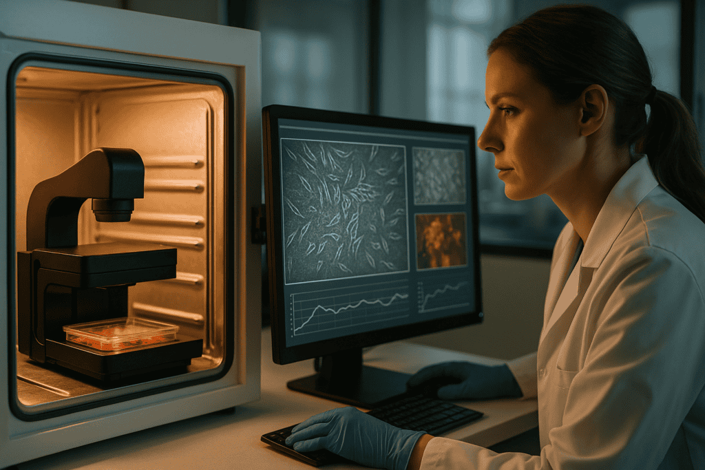
Los avances en la tecnología de imagen permiten ahora la monitorización continua en tiempo real de los cultivos celulares sin necesidad de sacarlos de entornos de crecimiento óptimos. Los sistemas de imagen dentro de incubadoras, como el zenCELL owl, ejemplifican estas innovaciones al ofrecer soluciones compactas y fáciles de usar que se alinean perfectamente con los flujos de trabajo existentes.
- Habilitar la observación en tiempo real y estudios de tiempo-lapse
- Reducción de la intervención manual y sus inconvenientes asociados
Rendimiento de Datos y Análisis Mejorados
La automatización de la adquisición y el procesamiento de imágenes facilita las aplicaciones de cribado de alto rendimiento (HTS) y mejora la escalabilidad. Con sistemas automatizados, los investigadores pueden centrarse más en el análisis y la interpretación en lugar de la recopilación de datos.
- Mayor eficiencia con flujos de trabajo automatizados
- Mejor asignación de recursos para configuraciones experimentales complejas
Ejemplos Prácticos y Flujos de Trabajo Utilizando Imágenes de Células Vivas
Ensayos de migración
Los estudios de migración celular son fundamentales para comprender procesos como la curación de heridas y la metástasis del cáncer. La obtención de imágenes dentro de la incubadora permite la visualización ininterrumpida de los movimientos celulares, proporcionando información sobre la cinética de migración y la activación de vías sin arriesgar la introducción de artefactos ambientales.
- Captura oportuna de patrones de movimiento direccional
- Imagen de alta resolución que soporta evaluaciones morfológicas detalladas
Desarrollo de organoides
Los organoides imitan la arquitectura y la función de los órganos, representando una piedra angular en la investigación para el descubrimiento de fármacos y la medicina regenerativa. Las capacidades de monitorización continua de los sistemas de imagen dentro de incubadoras ofrecen imágenes detalladas de las etapas de desarrollo sin perturbar nunca los cultivos en 3D.
- Monitoreo mejorado de las condiciones de crecimiento y los cambios morfológicos
- Fiabilidad mejorada en la verificación de hitos del desarrollo
Continúe leyendo para explorar información y estrategias más avanzadas.
“`
“`
Mejora de la Precisión y Reproducibilidad Experimental
Consistencia y confiabilidad de los datos
Los sistemas de imagenología dentro de incubadora brindan una ventaja significativa para la precisión experimental a través de la adquisición de datos sin problemas. Esta metodología evita el procesamiento por lotes y los errores de entrada manual inherentes a los métodos tradicionales, proporcionando así un flujo de datos más consistente. La estandarización que ofrecen estos sistemas conduce a una mayor reproducibilidad, lo cual es crucial para investigaciones y publicaciones de alto riesgo. Por ejemplo, un estudio publicado en la Métodos de la naturaleza La revista demostró que las plataformas de obtención de imágenes de células vivas redujeron la variabilidad en más de un 301 % (TP3T), lo que aumentó considerablemente tanto la confianza en la reproducibilidad de los resultados como la credibilidad en los foros revisados por pares.
- Prioriza los pasos de calibración y estandarización para la precisión.
Reducir el error humano y aumentar la eficiencia
Flujos de trabajo optimizados
Al reducir la necesidad de observaciones e intervenciones manuales, la obtención de imágenes dentro de la incubadora disminuye el riesgo de error humano. Esta tecnología contribuye a que las operaciones de laboratorio sean más eficientes al automatizar las tareas repetitivas. Por ejemplo, el uso de software integrado con funciones de listas de comprobación basadas en inteligencia artificial no solo agiliza todo el proceso, sino que garantiza que la supervisión humana se centre en el análisis crítico en lugar de en la simple introducción de datos. Un reconocido laboratorio de investigación oncológica informó de un aumento del 40% en la eficiencia, con prácticamente cero discrepancias en los datos, tras la adopción de estos sistemas.
- Implementa herramientas amigables con la automatización para procedimientos rutinarios.
Mejorando los resultados con aprendizaje automático e inteligencia artificial
Modelado predictivo y reconocimiento de patrones
La incorporación del aprendizaje automático y la inteligencia artificial a las tecnologías de imagenología de células vivas ofrece a los investigadores un potente conjunto de herramientas para identificar patrones celulares y predecir los resultados experimentales. Estas capacidades predictivas permiten a los investigadores ajustar de forma preventiva los parámetros experimentales, lo que reduce los fallos en los experimentos y el desperdicio de recursos. Un ejemplo convincente es el de una empresa farmacéutica que ha adoptado la imagenología mejorada con IA para ajustar con precisión sus ensayos de eficacia de fármacos, lo que ha dado lugar a una mejora del 251 % en su proceso de identificación de compuestos líderes.
- Superponer algoritmos de aprendizaje automático para mejorar las capacidades de análisis de imágenes.
Acelerando el Descubrimiento con Soluciones Escalables
Adaptándose a las necesidades cambiantes de la investigación
La escalabilidad de la imagen dentro de la incubadora permite transiciones fluidas desde estudios exploratorios a pequeña escala hasta proyectos de investigación a gran escala. A medida que crecen las demandas de investigación, la naturaleza modular de estos sistemas admite una escalada rápida sin la necesidad de una inversión adicional significativa. Tomemos como ejemplo una startup de biotecnología que expandió su investigación de estudios en modelos de animales pequeños a una investigación a gran escala de líneas celulares humanas. Aprovechando soluciones de imagen escalables, pudieron duplicar el tamaño de su estudio en meses, impulsando la velocidad de sus innovaciones.
- Favorecer sistemas modulares para la futura expansión de la investigación.
Fomentar la investigación colaborativa y el intercambio de datos
Integración de equipos multidisciplinarios
La imagenología dentro de la incubadora facilita el intercambio de datos en tiempo real y se integra perfectamente con los entornos de laboratorio digital, fomentando la colaboración entre disciplinas. Esta facilidad de intercambio de datos rompe los silos tradicionales en la investigación, permitiendo obtener amplias perspectivas y la polinización cruzada de ideas. Un consorcio de investigación de vanguardia utilizó la sincronización de datos basada en la nube para fusionar conjuntos de datos oncológicos e inmunológicos, descubriendo nuevos mecanismos de evasión inmunológica en tumores.
- Asegurar protocolos de intercambio de datos seguros y conformes.
Facilitando el acceso a la investigación remota
Laboratorios Virtuales y Monitoreo Remoto
La capacidad de monitorear cultivos celulares de forma remota a través de sistemas de imagen avanzados está cambiando las reglas del juego, especialmente en un mundo pospandemia donde prevalecen el trabajo flexible y el distanciamiento social. Los investigadores pueden supervisar múltiples experimentos simultáneamente desde diferentes ubicaciones, minimizando el tiempo de inactividad y acelerando los plazos de investigación. Un laboratorio de virología líder citó la accesibilidad remota como un factor fundamental para gestionar experimentos durante las restricciones de viaje, manteniendo la productividad y continuidad sin problemas.
- Implementa conexiones remotas seguras y autenticación de usuarios.
Maximizar la asignación de recursos y la eficiencia de costos
Uso optimizado de consumibles y equipo
La mayor precisión y automatización de la obtención de imágenes dentro de la incubadora suelen traducirse en una reducción de los residuos y un mejor aprovechamiento de los recursos. La mayor durabilidad de los consumibles, gracias a la menor intervención humana en los cultivos celulares, se traduce en menores costes operativos y una mayor sostenibilidad. Por ejemplo, los laboratorios que han adoptado la obtención de imágenes automatizada han informado de un ahorro de hasta un 20 % en reactivos para cultivos celulares y una reducción de las necesidades de almacenamiento.
- Aplicar análisis para monitorear el uso de recursos y reducir el desperdicio.
Estudios de Caso Transformadores e Historias de Éxito
Impactos en el mundo real y lecciones aprendidas
Varias instituciones han compartido impactos transformadores de la adopción de la imagenología dentro de la incubadora. Un caso notable involucró a un equipo de investigación universitaria que investigaba enfermedades neurodegenerativas, el cual utilizó imágenes de lapso de tiempo para revelar patrones de disparo neuronal inesperados relacionados con las respuestas al tratamiento. Estas perspectivas, recopiladas a través de imágenes ininterrumpidas durante semanas, condujeron a una publicación innovadora con implicaciones para las estrategias terapéuticas en la enfermedad de Alzheimer.
- Documentar y difundir estudios de caso para compartir las mejores prácticas.
A continuación, concluiremos con los puntos clave, métricas y una conclusión contundente.
“`
“`
Transformación de la Educación y el Desarrollo de Habilidades
Aprendizaje práctico y formación virtual
Los sistemas de imagenología dentro de la incubadora están revolucionando la educación y el desarrollo de habilidades en la comunidad científica al brindar más oportunidades de aprendizaje práctico y módulos de capacitación virtual. Los estudiantes y los investigadores en las primeras etapas de su carrera obtienen acceso directo a tecnología de vanguardia, cerrando la brecha entre la teoría y la práctica. Los tutoriales virtuales y la demostración en tiempo real de técnicas de imagenología fomentan la participación activa, haciendo tangibles los conceptos complejos. Una asociación entre una universidad líder y una empresa de software de imagenología resultó en un plan de estudios que mejoró significativamente la competencia de los estudiantes en bioinformática, lo cual se demostró en un concurso de imagenología interuniversitaria donde los participantes exhibieron conocimientos prácticos en análisis celular.
- Incorporar laboratorios virtuales y herramientas de simulación en los planes de estudio educativos.
Impulsando la innovación en biotecnología y farmacéutica
Pioneros en Nuevas Fronteras
Con su capacidad para entregar datos altamente precisos y reproducibles, la imagenología dentro de incubadoras es un potente catalizador para la innovación en las industrias biotecnológica y farmacéutica. La unión de la tecnología y la investigación biológica abre puertas a la creación de nuevos métodos de descubrimiento de fármacos y vías de tratamiento. Una emergente startup biofarmacéutica aprovechó la imagenología impulsada por IA para acelerar su proceso de desarrollo de vacunas, logrando avances novedosos en inmunoterapia. Esta integración no solo acortó los plazos de desarrollo, sino que también abrió nuevas vías para la medicina personalizada, subrayando el poder transformador de la ciencia de precisión en el fomento de la innovación.
- Impulsa la innovación a través de inversiones estratégicas en tecnologías emergentes de imagen.
Mejora de la competitividad global en investigación
Atraer y Retener Talento
Las instituciones que incorporan la obtención de imágenes en el propio laboratorio se posicionan de forma competitiva en el panorama mundial de la investigación. Al proporcionar herramientas avanzadas y fomentar un entorno propicio para la investigación de vanguardia, estas instituciones atraen a los mejores talentos y se forjan una reputación de excelencia. Un instituto de investigación nacional bien financiado observó un aumento del 30% en las solicitudes de investigadores de posgrado tras implementar tecnologías de imagen integrales en todos sus laboratorios, lo que demuestra el atractivo de los entornos de investigación modernos para los aspirantes a científicos de todo el mundo.
- Aprovechar instalaciones de vanguardia para mejorar el prestigio y la visibilidad institucional.
Conclusión
El profundo impacto de la imagenología en incubadora en el campo de la investigación científica es innegable. Al mejorar la precisión y la reproducibilidad, reducir el error humano y ampliar la capacidad a través del aprendizaje automático y la inteligencia artificial, esta tecnología revoluciona las metodologías en diversas disciplinas. La flexibilidad y escalabilidad que proporciona no solo agilizan los flujos de trabajo, sino que también permiten a los investigadores adaptarse sin problemas a los desafíos en evolución. Como lo demuestran los extensos estudios de caso e historias de éxito, estos sistemas son fundamentales para acelerar los descubrimientos, fusionar los esfuerzos interdisciplinarios y optimizar la asignación de recursos, todo ello al tiempo que fomentan la investigación colaborativa y el intercambio de datos.
Además, la imagenología dentro de incubadoras cobra protagonismo para allanar el camino a futuros avances e impulsar la innovación en biotecnología y productos farmacéuticos, donde la precisión y la rápida adaptabilidad son fundamentales. Su influencia se extiende a los entornos educativos, equipando a la próxima generación de científicos con habilidades y conocimientos esenciales de primera mano. Al integrar tecnologías tan avanzadas, las instituciones de investigación mejoran su competitividad global, atrayendo y reteniendo así a talentos de élite comprometidos con ampliar los límites de la comprensión y la capacidad humanas.
Al encontrarnos en el umbral de una nueva era de exploración científica, se vuelve imperativo adoptar herramientas como los sistemas de imagenología dentro de incubadoras que desafían las perspectivas tradicionales e inspiran un pensamiento transformador. Las instituciones, laboratorios e industrias interesadas en mantener una reputación de vanguardia deben comprometerse a adoptar estas sofisticadas soluciones no solo para potenciar sus capacidades de investigación, sino también para garantizar que la ciencia progrese con una precisión y eficacia sin precedentes. Lo invitamos a explorar estas tecnologías, dar pasos audaces hacia la actualización de su infraestructura de investigación y ser testigo de primera mano de las notables transformaciones que aguardan al futuro de la investigación.
“`
