From Images to Impact: Continuous Data for High-Ranking Publications & QA
“`
From Images to Impact: Continuous Data for High-Ranking Publications & QA
In the fast-evolving landscape of cell culture research, the ability to capture high-quality continuous data has become pivotal. This development isn’t just about enhancing visual documentation but transforming these images into significant scientific impact, contributing to high-ranking publications and rigorous quality assurance (QA). As researchers, lab managers, and biotech professionals increasingly turn to advanced technologies, understanding the role of continuous data in modern workflows is crucial. This article delves into the existing challenges, offers insights into technological advances, and provides examples of practical workflows using live-cell imaging. Readers will gain valuable knowledge on how to leverage incubator-based imaging systems to improve data quality and reproducibility.
Common Challenges and Limitations of Traditional Approaches
Why Traditional Methods Fall Short
Traditional cell culture techniques have been foundational in biological research; however, they often come with significant drawbacks that can impede progress. Manual observation of cell growth and behaviors risks introducing human error, leading to biased data interpretations. These methods also lack the ability to capture continuous data, which is crucial for understanding dynamic cellular processes.
- High potential for human error in manual observations
- Inability to capture real-time data for dynamic processes
- Variable conditions that affect reproducibility across experiments
The absence of continuous data collection results in fragmented insights, making it challenging to rank highly in publications that prioritize comprehensive datasets. Moreover, traditional methods struggle to meet the increasing demands for data quality and reproducibility, critical components of successful QA.
Continúe leyendo para explorar información y estrategias más avanzadas.
Technological Advances and Automation Trends
The Shift Towards Automation in Cell Culture
The move towards automation in cell culture is not merely an industry trend but a necessity for advancing research capabilities. Integrating automated systems can significantly reduce manual errors, enhance reproducibility, and boost data throughput. Technologies such as live-cell imaging systems have transformed how researchers collect and analyze data, offering real-time insights into cellular behavior.
- Automation reduces manual intervention, enhancing data integrity
- Continuous data capture with live-cell imaging provides unparalleled insights
- Automation supports scalability of experiments, improving productivity
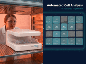
The zenCELL owl is an example of a compact, incubator-compatible live-cell imaging system that facilitates these advancements. Its design supports continuous monitoring, ensuring researchers stay informed of cellular changes in precise detail, thus laying the groundwork for reproducible, high-quality publications.
Continúe leyendo para explorar información y estrategias más avanzadas.
Practical Examples and Workflows Using Live-Cell Imaging
Implementing Live-Cell Imaging for Enhanced Research
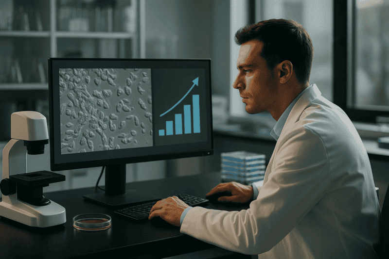
Live-cell imaging has opened new avenues for observing the intricate dynamics of cells over time. By employing advanced live-cell imaging systems, researchers can streamline their workflows, allowing for the seamless integration of continuous data into their research methodologies. Whether tracking cell proliferation, analyzing cell behavior, or conducting migration assays, the continuous data offers a significant advantage.
- Real-time monitoring enhances understanding of cellular dynamics
- Data-rich environments facilitate high-ranking academic publications
- Improved data quality supports robust QA processes
For instance, employing a live-cell imaging system like the zenCELL owl enables continuous, detailed observation of cellular processes within an incubator environment. Researchers gain access to consistent data crucial for comparative studies and long-term experiments.
Continúe leyendo para explorar información y estrategias más avanzadas.
How Incubator-Based Imaging Improves Reproducibility and Data Quality
The Benefits of Integrating Imaging within Incubators
Incorporating imaging systems directly within incubators enhances reproducibility and data quality by maintaining stable environmental conditions crucial for cell cultures. These systems minimize disturbances caused by environmental fluctuations, which can skew data and affect reproducibility.
- Consistent environment reduces variability in experimental outcomes
- Continuous monitoring diminishes the need for intrusive interventions
- High-quality, reproducible data fortifies rigorous QA protocols
This approach is particularly effective when using the zenCELL owl, which provides seamless integration within typical incubator setups. Its capacity to deliver real-time data ensures ongoing oversight, significantly reducing the likelihood of variability between experimental replicates.
Continúe leyendo para explorar información y estrategias más avanzadas.
Applications Such as Migration Assays, Organoids, Proliferation, or HTS
Exploring Diverse Applications in Cell Culture Research
Live-cell imaging finds application in an array of research areas, from migration assays to organoid culture and high-throughput screening (HTS). Each application benefits from the rich, continuous datasets generated, which enhance both the depth and breadth of cellular insights attainable.
- Migration assays: Real-time data reveal cell dynamics and interactions
- Organoid culture: Continuous monitoring supports developmental studies
- Proliferation assays: Accurate growth measurements bolster research findings
- HTS: High data throughput accelerates discovery and validation phases
These applications underscore the transformative impact of technologies like the zenCELL owl, which foster more comprehensive and insightful research outcomes, laying the foundation for innovation in cell culture methodologies.
Continúe leyendo para explorar información y estrategias más avanzadas.
“`
“`
Enhancing Quality Assurance with Advanced Imaging Metrics
Beyond Surface Evaluations: Deep Diving into QA
Quality assurance in cell culture is paramount, as it ensures the reliability and repeatability of experimental results. The integration of incubator-based live-cell imaging systems has revolutionized QA protocols by offering metrics that go beyond mere visual inspections. These advanced systems provide quantifiable insights into cellular behaviors and health, which are crucial for consistent QA checks.
- Adopt imaging metrics such as cell viability, morphology assessment, and growth rates as standard QA parameters.
By implementing these sophisticated metrics, laboratories can significantly enhance their QA processes, leading to reduced variability and heightened confidence in experimental results. For example, tracking morphological changes over time can predict early signs of cell health deterioration, preventing flawed data collection and enhancing study outcomes.
Case Study: Adoption of Live-Cell Imaging in Pharmaceutical Research
A Leap Forward in Drug Discovery
In the pharmaceutical industry, the pace at which drug discovery occurs is critical. The adoption of live-cell imaging has been a game-changer, offering unparalleled insights that are vital for accelerating this process. A notable study within a leading pharmaceutical company demonstrated the efficacy of live-cell imaging systems in streamlining the drug discovery pipeline.
- Implement continuous imaging to monitor drug effects on cellular physiology in real-time, improving discovery timelines.
By using technologies like the zenCELL owl, the research team was able to reduce the time taken to screen compounds by obtaining real-time data on cellular responses, thus enhancing decision-making processes and expediting the preclinical phase.
Data-Driven Decision Making in Cell Culture
Leveraging Data for Strategic Insights
In the realm of cell culture, data-driven decision-making involves utilizing continuous data streams to inform and optimize experimental processes. Modern imaging systems capture data not only for immediate analysis but also for strategizing ongoing and future experiments. This approach is instrumental in refining research methodologies.
- Develop a robust data management strategy to enhance reproducibility and facilitate comprehensive data analysis.
Data collation from varied temporal datasets enhances the ability to predict outcomes, adjust variables dynamically, and implement iterative improvements across experiments, ultimately improving research quality and outputs.
Automating Documentation and Reporting with Imaging Systems
Simplifying Administrative Overheads
The administrative burden of maintaining detailed experimental records can sometimes detract from the primary focus of research activities. The automation of documentation through advanced imaging systems alleviates some of this strain by ensuring that data capture is intrinsic and effortless, keeping researchers concentrated on analysis rather than record-keeping.
- Leverage software solutions tied to live-cell imaging systems to automate the documentation of cellular changes.
Automated documentation minimizes the risk of data loss or inaccuracies in manual entry, enhances compliance with research protocols, and simplifies the generation of reports necessary for publications and regulatory submissions.
Scaling Research Capabilities with Continuous Monitoring
Expanding Horizons through Scalability
Continuous monitoring facilitated by live-cell imaging expands the potential scale of research projects. Experimentation can move from individual to high-throughput scale without compromising data quality, thus accommodating ambitious research objectives and larger sample sizes.
- Integrate scalable imaging solutions to extend experimental scopes and accommodate growing research needs.
With scalable systems like the zenCELL owl, laboratories have successfully managed to increase their throughput, undertaking more extensive and complex studies while maintaining stringent scientific standards.
Empowering Collaborative Research Across Geographies
Seamless Integration in Collaborative Environments
Research collaborations often span multiple locations, demanding seamless data sharing and integration. Live-cell imaging systems empower these collaborations by providing real-time data access across geographies, promoting timely decision making and unified analysis across research teams.
- Use cloud-based data platforms linked with imaging systems to support real-time data sharing among geographically dispersed teams.
This global accessibility removes barriers that historically limited collaborative efforts, paving the way for more synchronized and cohesive research outcomes, crucial for tackling grand scientific challenges.
Predictive Modeling and AI in Cellular Analysis
The Role of Artificial Intelligence in Shaping Future Research
The integration of AI with live-cell imaging systems represents the cutting edge of cellular research. AI-driven algorithms can interpret complex datasets more rapidly and accurately than traditional methods, allowing for predictive modeling and enhanced cellular analysis.
- Incorporate AI tools in your imaging workflows to unlock predictive insights and identify trends that inform future research directions.
Applying AI to live-cell imaging data delivers predictive capabilities that streamline experimental design and refine research hypotheses, positioning researchers at the forefront of innovation.
A continuación, concluiremos con los puntos clave, métricas y una conclusión contundente.
“`
“`
Redefining Standard Protocols with Imaging Metrics
Setting New Benchmarks in Research Standards
As research methodologies advance, the traditional protocols must evolve to incorporate technological advancements for more robust and efficient outputs. The use of imaging metrics in setting new benchmarks for standard protocols ensures high-fidelity data acquisition and interpretation.
- Revise existing QA protocols to integrate systematic imaging data assessments, fostering greater accuracy and repeatability.
Enhanced guidelines ensure that research remains competitive and innovative, capitalizing on end-to-end solutions that maximize both the capture and analysis of critical data points.
Training the Next Generation of Scientists
Fostering Expertise Through Technological Mastery
With scientific research becoming ever more reliant on advanced technology, equipping future researchers with the necessary skills to manage and interpret complex data sets is imperative. Comprehensive training in the use of live-cell imaging systems ensures that new scientists are adept at navigating sophisticated research environments.
- Implement comprehensive training programs that emphasize not only technical proficiency but also strategic thinking in interpreting imaging data.
By investing in education and training, laboratories ensure that they produce technologically literate graduates ready to drive innovation across various research sectors.
Conclusión
As we journey through the age of technological revolution in research, the integration of continuous data monitoring through advanced imaging metrics represents a quantum leap. Key takeaways from our exploration emphasize significant enhancements in quality assurance, data-driven decision-making, and the facilitation of collaborative research efforts. LIVE-cell imaging technologies like the zenCELL owl have emerged as instrumental allies, reducing time frames for drug discovery, fostering better data management strategies, and minimizing administrative overheads.
The article underscores the growing indispensability of implementing scalable and sophisticated imaging systems. These technologies have empowered laboratories to undertake ambitious research, monitor experimental variables in real-time, and leverage predictive insights through artificial intelligence. The adoption of AI-enhanced imaging transforms cellular analysis, paving the path for cutting-edge breakthroughs and revolutionizing the established paradigms of research.
This continuous evolution in research methodologies necessitates a corresponding evolution in training programs and standard protocols. It highlights the importance of preparing the next generation of scientists with the necessary skills to harness these technological advances efficiently and strategically. By redefining benchmarks and integrating comprehensive training, we ensure that our research legacy nurtures innovation and scientific excellence.
At the heart of these advancements lies the power to transcend geographical and technological barriers, fostering unprecedented collaboration and integration across global research efforts. The transformative capabilities of live-cell imaging, combined with state-of-the-art AI technologies, now lead to more informed decision-making, strategic research planning, and ultimately, more impactful publications.
As researchers, stakeholders, and innovators, we stand on the precipice of a new era of scientific inquiry. Let us embrace these tools to enhance our understanding, drive prolific research outputs, and rewrite the fundamentals of scientific exploration. The challenge lies not only in utilizing these technologies but in pioneering pathways that redefine how we perceive and interact with the cellular world. Let this era mark the dawn of refined research methodologies, where our commitment to scientific inquiry fuels a brighter, innovation-driven future. Seize this opportunity to transcend traditional boundaries and redefine the landscape of cellular research.
“`