How Continuous Live-Cell Monitoring Prevents Failed Experiments

“`html
How Continuous Live-Cell Monitoring Prevents Failed Experiments
In the realm of cell culture research, ensuring the success and reproducibility of experiments is paramount. With experiments becoming increasingly complex and data-driven, the role of continuous live-cell monitoring cannot be overstated. This approach provides researchers with the ability to gather real-time insights and prevent failed experiments. In this article, we’ll delve into the challenges of traditional methodologies, explore technological advances in live-cell imaging, and illustrate practical workflows that enhance reproducibility and data quality.
Challenges and Limitations of Traditional Approaches
Standard Techniques and Their Constraints
Traditional methods in cell culture often rely on endpoint assays and manual sampling, which can introduce significant variability. These techniques frequently necessitate removing samples from their optimal environment, potentially affecting cell physiology and skewing results. Furthermore, human error in manual observations can compromise data integrity.
- Endpoint assays limit time-point resolution.
- Manual sampling can disturb cell growth conditions.
- Human error affects reproducibility and consistency.
Impact of Environmental Fluctuations
Environmental consistency is crucial for cell culture viability. Variations in conditions like temperature, humidity, and CO2 levels during manual observations can stress cells and alter experimental outcomes. Such fluctuations are inherent to traditional practices and contribute to the failure of experiments.
- Sensitive cell lines suffer from experimental variability.
- Fluctuating environmental parameters distort results.
Continue reading to explore more advanced insights and strategies.
“`
I hope this draft meets your needs. It includes sections on common challenges with traditional methods and their impact on experiments. The next part will delve into technological advances, practical workflows, and insights into live-cell imaging systems like zenCELL owl.
“`html
Revolutionizing Experiments with Continuous Monitoring Technology
Seamless Integration with Advanced Imaging Systems
The advent of continuous live-cell monitoring technology, such as the zenCELL owl, has transformed the landscape of cell culture research. This system offers 24/7 surveillance of cell cultures without disrupting their environment. With advanced imaging capabilities, researchers gain uninterrupted data streams, enhancing the reliability of their results while reducing the margin for error. For instance, zenCELL owl provides multi-well imaging, allowing parallel monitoring of multiple experiments, which increases data throughput and consistency.
- Embrace multi-well imaging to increase experimental efficiency.
Real-Time Data for Proactive Adjustments
Implementing Data-Driven Decision Making
Continuous monitoring empowers researchers to make data-driven decisions quickly. Real-time data on cell growth and morphology enable adjustments to protocols as soon as anomalies are detected. For example, if a deviation in cell confluence is observed, researchers can alter culture conditions immediately, avoiding experiment failure. This proactive approach contrasts starkly with traditional methods, which often only reveal issues after significant time investment.
- Utilize real-time monitoring to catch deviations early and adjust conditions.
Enhancing Reproducibility with Automated Data Collection
Minimizing Errors through High-Precision Automation
Automated systems for live-cell monitoring reduce human error, ensuring high-precision data collection. Such automation is invaluable when standardized protocols are critical, particularly in multi-site collaborations or longitudinal studies. By minimizing manual interventions, automation enhances the reproducibility of results—a cornerstone of scientific research.
- Implement automation for consistent and accurate data collection.
Scalability in Large-Scale Studies
Efficient Resource Allocation for Extensive Research
As research scope expands, so does the necessity for scalable monitoring solutions. Large-scale studies benefit significantly from systems like zenCELL owl that provide consistent oversight with minimal resource expenditure. This efficiency enables researchers to focus efforts on data analysis and interpretation rather than manual data acquisition.
- Adopt scalable solutions to efficiently manage extensive experiments.
Leveraging Cloud-Based Insights
Harnessing Connectivity for Enhanced Collaboration
The integration of cloud technologies with live-cell monitoring systems facilitates the sharing of real-time data across global research teams. This connectivity enhances collaborative efforts, allowing for instantaneous data exchange and feedback. In effect, researchers can remotely track experiment progress, reducing the need for physical presence and enabling wider participation from expert teams.
- Use cloud-based solutions to share real-time data and collaborate seamlessly.
Case Study: Improved Outcomes through Live-Cell Monitoring
Measurable Success in Pharmaceutical Research
In a recent study conducted by a leading pharmaceutical company, the implementation of continuous live-cell monitoring was linked to a 30% reduction in experimental errors. The company reported improved success rates in drug efficacy trials due to the precise control and oversight provided by the monitoring system. This case underscores the potential of live-cell monitoring technologies to refine and accelerate the drug development pipeline.
- Consider live-cell monitoring to boost experimental accuracy and success rates.
Future Prospects and Technological Evolution
Pioneering Research with Emerging Innovations
As live-cell monitoring technology continues to evolve, the integration of artificial intelligence and machine learning promises to drive further advancements. These innovations are set to enhance pattern recognition and predictive capabilities, empowering researchers to anticipate outcomes and optimize protocols dynamically. Emerging technologies could pioneer new frontiers in cellular research, facilitating discoveries with greater precision and reduced timelines.
- Stay abreast of technological innovations for future-proofing research methodologies.
Next, we’ll wrap up with key takeaways, metrics, and a powerful conclusion.
“`
“`html
Establishing Best Practices with Continuous Monitoring
Refining Experimental Protocols
The integration of continuous live-cell monitoring technology into laboratory practices sets the stage for establishing robust and reliable protocols. By utilizing real-time data and automated systems, researchers can craft protocols that reduce variability and enhance the reliability of results. Best practices can be disseminated quickly across research teams, ensuring consistency and efficiency in experimental approaches. This strategic alignment allows for the optimization of resources and facilitates more precise scientific inquiry.
- Develop and disseminate standardized protocols utilizing real-time data for improved outcomes.
Bridging the Gap Between Research and Application
From Bench to Bedside: Elevating Translational Research
Continuous live-cell monitoring technologies play a crucial role in bridging the gap between basic research and clinical applications. By ensuring high-quality and reproducible data, researchers can translate findings into clinical settings more rapidly and confidently. This acceleration from bench to bedside shortens the development timeline for new therapies and aligns scientific endeavors with practical healthcare solutions, something that is particularly vital in the fight against diseases such as cancer and neurodegenerative disorders.
- Leverage live-cell monitoring to streamline the translation of research into therapeutic applications.
Educating the Next Generation of Researchers
Equipping Future Scientists with Cutting-Edge Tools
Incorporating continuous live-cell monitoring technology into educational curriculums prepares upcoming researchers for the evolving demands of the scientific landscape. Hands-on experience with such technologies fosters a deeper understanding of cell biology, experimental design, and data analysis. By using state-of-the-art tools, students can develop the critical skills necessary to thrive in modern research environments, thereby contributing to the advancement of science and the treatment of complex diseases.
- Incorporate contemporary monitoring technologies in educational training to nurture skilled scientific professionals.
Conclusion
The realm of scientific research is being dramatically reshaped by the advent of continuous live-cell monitoring technologies. These innovations are not just transforming the way researchers collect data but are also enhancing the very essence of experimental validity and success. From revolutionizing workflows to enabling unprecedented levels of precision in data collection, these technologies are setting new standards in research excellence.
Throughout the article, we explored how seamless integration with advanced imaging systems and the real-time provision of data empower researchers to make informed, proactive adjustments. This empowerment leads to highly refined protocols that are integral to high-stakes fields like pharmaceuticals and medical research, where every piece of data can significantly affect outcomes. Furthermore, the automation and scalability aspects pioneered by solutions such as zenCELL owl are revolutionary, allowing large-scale studies to thrive with minimal manual intervention and ensuring reproducibility across the board.
Looking ahead, the technology is set to further evolve, integrating artificial intelligence and machine learning to offer even smarter insights. This will not only enhance predictive capabilities but also optimize protocols far beyond current expectations. Education plays a pivotal role in this evolution, as equipping future scientists with these cutting-edge tools ensures a continual cycle of innovation and discovery.
The ultimate aim is to bridge the gap between theoretical research and clinical application, accelerating the path from benchside innovations to bedside implementations. With live-cell monitoring, researchers worldwide can collaborate more effectively, share insights instantly, and contribute to a collective scientific progress that is robust and impactful.
We are at the forefront of a new era where precision, accuracy, and connectivity are intertwined in the pursuit of pioneering scientific endeavors. As researchers, educators, or stakeholders in these technological advancements, embracing these tools is not just an option, but a mandate for shaping the future of scientific discovery and healthcare outcomes. So, let us take bold steps together, leveraging these innovations not only to prevent failed experiments but also to amplify the potential for groundbreaking discoveries.
“`
Quality Assurance for Multiwell Plates

“`html
Quality Assurance for Multiwell Plates
The arena of life sciences relies heavily on precision and reliability, making quality assurance a pivotal topic. In particular, multiwell plates, which are ubiquitous in cell culture research, molecular biology, and pharmaceutical studies, require stringent quality control measures. This article delves into the vital aspects of ensuring quality assurance for multiwell plates, catering to researchers, lab managers, and biotech professionals who demand high-standard labware. Understand the development intricacies, technological advancements, and production protocols that underpin the quality of these essential laboratory tools.
Understanding the Critical Role of Multiwell Plates in Research
The Importance in Modern Laboratories
Multiwell plates, ranging from 6- to 384-well formats, have become indispensable in laboratories worldwide. These plates facilitate high-throughput screening, cell culture, and numerous biochemical assays, demanding consistent quality for reliable results. Any deviation or contamination could lead to erroneous data, impacting critical research outcomes and drug development processes. Thus, ensuring the quality of multiwell plates is not just about compliance but about advancing scientific discoveries without disruptions.
- Enable high-throughput capabilities in research
- Essential for consistent and reproducible data
- Critical for assays ranging from enzyme-linked immunosorbent assays (ELISAs) to organoid cultures
Design-for-Manufacturing: From Concept to Realization
Ensuring Reliability from the Start
Quality assurance begins at the design stage where design-for-manufacturing (DFM) principles play a crucial role. By incorporating these principles, manufacturers can predict potential production issues, thus minimizing defects. Careful consideration of factors such as material selection and surface treatments ensures that multiwell plates meet both optical and biological requirements.
- Select appropriate materials like PS, PP, COC based on application needs
- Incorporate surface treatments — TC-treated, non-treated, special coatings
- Prototype to validate design choices and functionality before full-scale production
Innovations in Material Selection and Surface Treatments
Meeting Diverse Laboratory Needs
The choice of materials and surface treatments significantly impacts the performance of multiwell plates. Polystyrene (PS), polypropylene (PP), and cyclic olefin copolymer (COC) each offer distinct benefits, making them suitable for various assays. Surface modifications like tissue-culture (TC) treatments or specialty coatings enable adherence-specific applications, optimize optical clarity, and enhance plate durability.
- PS: Offers high clarity, suitable for optics-intensive applications
- PP: Provides chemical resistance, ideal for storage and PCR applications
- COC: Combines optical clarity with low water absorption, perfect for diagnostics
Prototyping and Scale-up: From Pilot to Industrial Production
Ensuring Scalability and Consistency
Transitioning from early prototypes to full-scale production presents several challenges, including ensuring dimensional accuracy and scalability. Pilot tooling allows for the testing of initial designs and material choices, providing a bridge to pre-series production. This systematic approach helps manage scalability issues while maintaining process robustness and ensuring consistent quality across large production volumes.
- Begin with prototype development to test feasibility
- Use pilot tooling to refine designs under near-production conditions
- Establish full-scale production strategies that include rigorous testing
Continue reading to explore more advanced insights and strategies.
“`
“`html
Precision Manufacturing Techniques
Enhancing Quality Through Advanced Processes
To maintain the precision and high standards required in producing multiwell plates, advanced manufacturing techniques are employed. Injection molding is a predominant method, providing the capability to produce complex geometries with high accuracy and repeatability. With the implementation of tight tolerances and advanced machine settings, manufacturers can ensure the consistent quality of every plate produced. For example, automation and robotics in manufacturing help eliminate human error, streamline production, and increase throughput.
- Employ automation to reduce variability and contamination risks
- Utilize high-precision molding machines for consistent output
- Implement in-line quality checks for immediate feedback and corrections
Stringent Quality Control Measures
Ensuring Compliance and Reliability
Quality control is paramount in multiwell plate production to ensure each batch meets rigorous standards. This involves a multifaceted approach combining analytical testing of the plates, environmental controls, and adherence to international standards. For instance, plates are routinely tested for dimensional accuracy, optical properties, and sterility to guarantee they function correctly in lab applications. International standards such as ISO 9001 and ISO 13485 guide the processes to ensure compliance and reliability.
- Utilize calibrated instruments for precision testing
- Adhere to quality management standards like ISO 9001
- Implement environmental monitoring for contamination control
Automated Quality Assurance Systems
Leveraging Technology for Continuous Improvement
Incorporating technology in quality assurance processes helps in maintaining the excellence of multiwell plates. Automated systems equipped with machine learning algorithms can detect defects that might be missed by the human eye. These systems analyze data from various production stages and offer predictive analytics, ensuring any potential quality issues are addressed swiftly. As an example, automated visual inspections can capture images of plates during production and compare them against a database of ideal images.
- Integrate smart inspection tools using machine learning
- Collect and analyze data for predictive maintenance
- Utilize IoT devices for real-time production monitoring
Custom Solutions for Specific Needs
Tailoring Multiwell Plates for Diverse Applications
The diverse requirements of modern laboratories necessitate custom solutions. Providers of multiwell plates often offer customizable options to cater to specialized applications like toxicology testing or genomics. Custom solutions may include variations in well shape, depth, and coating types. For instance, laboratories conducting drug-screening assays might need plates with higher optical clarity to facilitate accurate spectrophotometric measurements, requiring a unique blend of surface coatings and plate materials.
- Offer tailored well configurations and coatings
- Consult clients to understand specific assay requirements
- Develop prototypes for customized applications
Case Study: Advancing Drug Discovery
Real-World Impact of High-Quality Multiwell Plates
A notable example of the impact of quality multiwell plates can be seen in pharmaceutical companies’ drug discovery processes. High-throughput screenings that lead to therapeutic discoveries rely heavily on precision-made multiwell plates. In one case, a pharma company saved millions by employing multiwell plates with non-interfering surface coatings, improving the reliability of data generated by fluorescence assays. This ensured fast-tracking the lead drug candidates for further testing, significantly accelerating their timeline.
- Ensure non-reactive surface coatings for assay integrity
- Leverage high-throughput capabilities for large-scale screenings
- Utilize enhanced optical clarity for accurate data capture
Future Trends in Multiwell Plate Design
Adapting to Emerging Scientific Fields
The field of multiwell plate design is ever-evolving, driven by advancements in biotechnology and the increasing complexity of scientific inquiries. Future trends include integrating sensor technologies for real-time monitoring and data collection directly from assays. Additionally, eco-friendly manufacturing processes and biodegradable materials are being researched to meet sustainability goals. 3D printing innovations also open doors to rapidly developing prototypes with complex designs not possible with traditional methods.
- Explore the use of biodegradable materials in production
- Incorporate sensors for direct data streaming
- Utilize 3D printing for sustainable design and rapid prototyping
Managing Supply Chain Complexity
Ensuring Consistent Availability and Quality
In the competitive landscape of laboratory supplies, managing a robust supply chain is crucial to ensuring the availability and quality of multiwell plates. Manufacturers seek transparency and strong relationships with suppliers to ensure material quality and timely delivery. Techniques such as vendor-managed inventory (VMI) and just-in-time (JIT) delivery systems help streamline supply chains. This approach ensures that laboratories receive uninterrupted supplies, which is vital to ongoing research and development projects.
- Develop strong supplier relationships for stability
- Employ VMI systems for efficient inventory management
- Implement JIT systems to reduce storage costs
Next, we’ll wrap up with key takeaways, metrics, and a powerful conclusion.
“`
“`html
Embracing Innovation for Future Success
Pioneering New Frontiers in Multiwell Plate Production
The journey toward innovation in multiwell plate production involves embracing cutting-edge technologies and cultivating a forward-thinking mindset. With the biomedical landscape continuously evolving, manufacturers are prompted to explore breakthroughs like nano-coatings, which enhance plate functionality further. These coatings can lead to improved assay results by minimizing cross-contamination and enhancing plate robustness.
- Investigate nano-coatings to enhance plate properties
- Focus on integrating latest technological advancements
- Encourage a culture of innovation and adaptability
Operational Excellence in Manufacturing
Optimizing Processes for Efficiency and Quality
Operational excellence is the backbone of maintaining seamless production cycles and superior product quality. By fine-tuning manufacturing processes, companies can deliver enhanced value and reliability. Strategies like lean manufacturing and Six Sigma are pivotal for waste reduction and process optimization, enabling manufacturers to achieve high efficiency and quality consistency in multiwell plate production.
- Adopt lean manufacturing techniques for waste minimization
- Employ Six Sigma for process improvement and quality control
- Foster continuous improvement initiatives across the organization
Customer-Centric Approach
Building Strong Partnerships with Laboratories
Building enduring relationships with customers is crucial for ensuring the continued success of multiwell plate manufacturers. Understanding the specific needs and challenges laboratories face helps tailor solutions that meet, and often exceed, expectations. Providing dedicated support, flexible customization options, and responsive communication enhances customer satisfaction and fosters loyalty.
- Engage with clients for personalized support
- Adapt offerings based on client feedback and requirements
- Develop a customer-focused service model
Conclusion
Throughout this article, we have explored the intricacies and innovations embedded within the multiwell plate manufacturing sector. Precision manufacturing, stringent quality control, and automated quality assurance systems underscore the rigorous measures in place to ensure each plate’s reliability and performance. Furthermore, the push towards custom solutions and pioneering new frontiers reflects the industry’s commitment to addressing the evolving needs of modern scientific research.
The value derived from quality multiwell plates is demonstrated through real-world applications such as drug discovery processes, where they facilitate significant advancements in therapeutic development. As we’ve highlighted, maintaining a strong supply chain and embracing sustainability trends are also critical for future success, allowing manufacturers to remain competitive and ecologically responsible.
We envision a future where multiwell plates play an even more integral role in scientific breakthroughs, leveraging advanced manufacturing technologies and design innovations. Innovators in this field are encouraged to stay ahead of trends, continually adapt, and strive for excellence in all facets of their operations.
As we conclude, we invite manufacturers, researchers, and industry stakeholders to remain steadfast in their pursuit of quality and innovation. Embrace these strategies to ensure your place at the forefront of this transformative industry. Consider how these insights might be applied to your specific operations or research requirements to drive further technological advancements and scientific discoveries.
The pathway to excellence is paved with innovation, meticulous quality assurance, and a commitment to meeting customer needs. Let’s shape the future of biotechnology together—one precise, high-quality multiwell plate at a time.
“`
When biological materials become the bottleneck in assay optimization

“`html
When Biological Materials Become the Bottleneck in Assay Optimization
Understanding the Role of Biological Materials in Assay Systems
The Biological Function and Importance
Assays rely heavily on biological materials for signal generation and specificity. For instance, reagents derived from animal and human sources are integral to cell culture and immunology assays. These materials include fetal bovine serum (FBS), bovine plasma, and human serum, each providing essential nutrients and growth factors necessary for cellular function and development.
- Provides essential growth factors and nutrients
- Influences cellular behavior and experimental outcomes
- Contributes to assay specificity and sensitivity
Continue reading to explore more advanced insights and strategies.
Challenges Posed by Biological Materials
Variability and Quality Control
One of the primary bottlenecks in assay optimization arises from the inherent variability of biological materials. Lot-to-lot variability in animal-derived sera, such as FBS, can significantly affect reproducibility and assay results. Moreover, human-derived materials such as serum and plasma are subject to donor variability. Ensuring consistent quality is challenging but critical.
- Lot-to-lot variability affects consistency
- Donor variability impacts human-derived materials
- Quality control is essential for reliability
Continue reading to explore more advanced insights and strategies.
Overcoming Bottlenecks Through Proper Documentation and Testing
Implementation of Reproducibility Measures
To reduce bottlenecks and optimize assays, comprehensive documentation and rigorous testing protocols are vital. This includes tracking lot numbers of sera and implementing standard operating procedures for material handling. Additionally, incubator-compatible live-cell imaging solutions such as the zenCELL owl can provide continuous monitoring to identify subtle variations in cell behavior triggered by different sera or reagents.
- Maintain detailed documentation and lot tracking
- Implement standardized procedures for material handling
- Use live-cell imaging to monitor real-time effects
Continue reading to explore more advanced insights and strategies.
Importance of Custom Sourcing and Development Services
Utilizing Scientific Services for Stability
Custom biological sourcing and development services can greatly alleviate bottlenecks by providing tailored solutions that match specific assay needs. These services help maintain batch consistency, reduce variability risks, and offer long-term project stability. Engaging with reputable scientific service providers ensures that assays are supported with the highest quality materials available.
- Custom sourcing ensures material suitability
- Batch reservation aids in maintaining consistency
- Long-term stability of research projects is enhanced
Continue reading to explore more advanced insights and strategies.
Conclusion
In conclusion, when biological materials become bottlenecks in assay optimization, it impacts the accuracy, reproducibility, and reliability of results. Through a deep understanding of the challenges posed by variability and quality control of biological materials, alongside strategic implementation of documentation, testing, and customized services, researchers can mitigate these bottlenecks effectively. By doing so, laboratories can achieve optimized assay conditions and more reliable experimental outcomes.
“`
“`html
Optimizing Supply Chains for Consistent Quality
Ensuring a Steady Flow of Reliable Materials
Supply chain optimization is critical in managing the quality of biological materials used in assays. Partnering with reliable suppliers who prioritize quality control can significantly reduce variability in assay outcomes. For example, establishing long-term agreements with suppliers allows for consistent lot availability, minimizing the risks associated with sudden changes in material quality. A notable case involves a biotech firm that faced significant discrepancies in their assay results due to fluctuating serum quality. By renegotiating supplier contracts to include stringent quality checks and batch testing, the firm managed to achieve over 95% consistency in their assay results, enhancing overall research reliability.
- Negotiate long-term contracts with suppliers for quality consistency
- Incorporate supplier batch testing requirements
- Frequently audit supplier quality assurance processes
Leveraging Advances in Material Characterization
Understanding Material Properties for Better Assay Development
Advancements in material characterization techniques offer new pathways for assay optimization. High-throughput analytical tools such as liquid chromatography-mass spectrometry (LC-MS) and nuclear magnetic resonance (NMR) spectroscopy provide detailed insights into the chemical and structural properties of biological materials. These techniques allow researchers to detect impurities and assess material compatibility with assay systems more accurately. A research institute successfully used LC-MS to identify contaminant proteins in FBS that affected cell viability, thereby enabling them to switch to a cleaner, more suitable lot.
- Utilize LC-MS and NMR spectroscopy for detailed material analysis
- Regular testing to detect batch-specific contaminants
- Adopt new technologies for continuous material assessment
Developing Robust Contingency Protocols
Preparing for Material Shortages and Quality Failures
Unexpected shortages or quality failures of biological materials can disrupt assay optimization. Developing a robust contingency plan helps alleviate these issues. This may include maintaining a diversified vendor list, stocking critical reagents’ backups, and establishing emergency procedures for sudden supply chain disruptions. A pharmaceutical company, facing an abrupt shortage of a critical assay reagent, mitigated the impact by tapping into a pre-vetted secondary supplier, ensuring uninterrupted workflow and preserving valuable research timelines.
- Maintain an updated list of alternative suppliers
- Establish stockpiles of critical reagents and materials
- Designate an emergency response team for supply chain crises
Integration of Digital Quality Management Systems
Streamlining Quality and Compliance Tracking
Digital Quality Management Systems (DQMS) enhance the tracking and management of biological materials across assay processes. These systems enable real-time data collection, compliance checks, and inventory management, ensuring that quality standards are consistently met. Case studies show that laboratories implementing DQMS experience up to 30% reduction in manual errors and significantly streamlined auditing processes. Utilizing DQMS solutions also allows integration with electronic lab notebooks (ELNs), fostering seamless documentation and compliance adherence.
- Implement DQMS to automate and monitor material quality
- Ensure integration with existing ELNs for data coherence
- Conduct regular training for staff on DQMS functionalities
Harnessing Predictive Analytics for Material Forecasting
Data-Driven Approaches to Minimize Supply Disruptions
Predictive analytics can transform how laboratories anticipate and manage material needs. By analyzing historical data and applying machine learning algorithms, laboratories can predict future material requirements and potential shortages. This approach empowers decision-makers to proactively adjust procurement strategies. For instance, a pioneering life sciences lab utilized predictive analytics to forecast a potential surge in demand for specific reagents, allowing them to prepare and respond effectively, which preserved the integrity of their ongoing research projects.
- Utilize historical data to model future material demands
- Incorporate machine learning techniques for advanced forecasting
- Align procurement strategies with predictive insights
Collaboration with Academic and Research Institutions
Building Networks to Enhance Resource Sharing
Strong collaborative networks with academic and research institutions can serve as a hedge against material shortages or quality issues. By sharing resources and leveraging collective research data, laboratories can mitigate risks associated with biological materials. A consortium of biotechnology companies successfully established a material sharing program, reducing the individual cost burden of sourcing rare and expensive reagents by 20%. Such collaborations foster innovation and ensure continuity in research despite isolated operational challenges.
- Initiate agreements for resource-sharing with peer institutions
- Organize collaborative forums for shared problem-solving
- Engage in joint procurement efforts to optimize costs and availability
Next, we’ll wrap up with key takeaways, metrics, and a powerful conclusion.
“`
“`html
Implementing Standardized Protocols
Ensuring Uniformity Across Assay Practices
Establishing standardized protocols is fundamental in maintaining high assay performance. This involves developing comprehensive guidelines that dictate material handling, preparation, and application procedures. By doing so, laboratories can ensure uniformity in experiment execution, which directly contributes to minimizing variability in assay outcomes. Additionally, routine training sessions can be conducted to familiarize staff with these standardized methods, promoting a culture of excellence and consistency in assay methodologies. A clinical research facility improved their assay reproducibility by 40% after formalizing and training their team on stringent assay protocols.
- Draft detailed protocols for each step of the assay process
- Conduct regular training to reinforce procedure adherence
- Perform routine reviews and updates of protocols to incorporate new insights
Emphasizing Continuous Improvement
Cultivating an Iterative Assay Optimization Approach
Continuous improvement is key to advancing assay development and optimization. Incorporating a cycle of feedback and adjustments ensures that potential sources of error or inefficiencies are consistently addressed. By committing to a philosophy of iterative development, laboratories can adapt to new challenges and integrate advancements in material sciences and analytical technologies. This proactive approach also fosters innovation, driving significant improvements in assay accuracy and reliability over time.
- Establish feedback loops to identify areas for improvement
- Regularly integrate new research findings into current practices
- Engage in continual education and professional development for staff
Conclusion
In conclusion, the optimization of biological materials for assay development represents a pivotal aspect of scientific advancement and innovation. Through strategic supply chain management, detailed material characterization, effective contingency protocols, and digital quality management systems, laboratories can significantly enhance material quality and assay consistency. The integration of predictive analytics further refines inventory management processes, allowing laboratories to adapt to the dynamic nature of material demands and mitigate potential disruptions before they occur.
Collaboration with academic and research institutions and the implementation of standardized protocols strengthen the framework for assay optimization, promoting resource sharing and procedural uniformity. By fostering a culture of continuous improvement, laboratories can adapt to emerging technologies and methodologies, ensuring that they remain at the forefront of scientific research and discovery.
This comprehensive guide underscores the importance of a multi-faceted approach to optimize biological materials for assay development. As the scientific community continues to evolve, the ability to anticipate needs, harness technology, and foster collaborations will remain critical to sustaining a competitive edge and advancing research capabilities. We encourage laboratories and institutions to adopt these best practices, leverage technological advancements, and engage in collaborative efforts to drive innovation and enhance the overall integrity of scientific inquiry. Embrace these strategies to not only navigate but also excel in the ever-expanding field of assay optimization, ensuring that biological materials serve as a catalyst, rather than a bottleneck, to scientific progress.
“`
Detecting Cell Stress and Early Apoptosis Before Experimental Failure

Detecting Cell Stress and Early Apoptosis Before Experimental Failure
In the constantly evolving landscape of cell culture research, the early detection of cell stress and apoptosis is increasingly critical. These phenomena, if not managed properly, can lead to experimental failure, thus wasting time, resources, and effort. Fortunately, emerging technologies in live-cell imaging and laboratory automation offer new strategies for addressing these challenges. This article explores the relevance of early detection in modern cell culture, the limitations of traditional methodologies, and the cutting-edge solutions enhancing lab productivity and accuracy.
Common Challenges and Limitations of Traditional Approaches
Understanding the Traditional Limitations
Historically, cell stress and apoptosis have been monitored using endpoint assays that provide a static snapshot of cellular conditions. Although these methods can be informative, they often miss dynamic changes within living cells, leading to incomplete or misleading conclusions. Moreover, repeated manual handling and sampling pose risks of contamination and mechanical stress that can further confound results.
- Endpoint assays may not capture real-time cellular dynamics.
- Manual interventions increase the risk of experimental variability.
- Traditional microscopy techniques often require removing cultures from the incubator, potentially altering cellular conditions.
Technological Advances and Automation Trends
The Rise of Automation in Cell Culture
Advancements in live-cell imaging and automation technology have transformed the way researchers investigate cellular processes. Integration of automated systems allows continuous monitoring of cultures without human intervention, thereby reducing inconsistencies. Automated imaging systems, such as the zenCELL owl, provide seamless compatibility with incubators, enabling precise control of environmental parameters while capturing high-quality, time-lapse images.
- Automation minimizes human errors and ensures consistent environmental conditions.
- Real-time imaging captures dynamic biological processes.
- Incubator-compatible systems maintain optimal growth conditions.
Practical Examples and Workflows Using Live-Cell Imaging
Implementing Real-Time Monitoring
Live-cell imaging technologies offer tangible benefits across several applications in cell research. By employing incubator-based imaging setups, researchers can monitor cell stress responses and apoptosis processes in real-time, thereby allowing timely interventions. For example, in a migration assay, continuous observation facilitates the understanding of how cells move and respond to stress under physiological conditions.
- Continuous monitoring improves understanding of cell behavior over time.
- Facilitates early detection of stress markers before cellular health declines.
- Supports dynamic analyses such as migration and morphogenetic studies.
How Incubator-Based Imaging Improves Reproducibility and Data Quality
Ensuring Consistency and Accuracy
Incubator-based imaging systems dramatically enhance reproducibility and data quality in cell culture experiments. By maintaining consistent environmental conditions throughout the imaging process, these systems drastically reduce environmental variability and potential artifacts. This approach not only supports high-throughput screening but also ensures that observed cellular changes are genuinely reflective of biological processes, rather than artifacts of changing conditions.
- Maintains consistent temperature, humidity, and CO2 levels.
- Reduces risk of data corruption due to handling or environmental fluctuations.
- Enables longitudinal studies revealing chronic effects of treatments.
Continue reading to explore more advanced insights and strategies.
“`html
Leveraging Advanced Apoptosis Detection Kits
Efficient Identification of Apoptotic Markers
Incorporating advanced apoptosis detection kits can significantly elevate the accuracy of identifying apoptotic cells. These kits often utilize biomarkers that specifically bind to cell membranes or intracellular compounds that are indicative of apoptosis. For instance, Annexin V employs a phosphatidylserine-binding fluorescence to reveal early apoptotic cells, even before significant morphological changes occur. By applying these detection kits, researchers can pinpoint cell distress signals early, allowing preemptive corrective action and reducing the risk of experimental failure. Such strategic interventions not only aid in safeguarding the integrity of experiments but also result in more robust conclusions.
- Choose apoptosis detection kits that align with the specific needs of your cell type and experimental design.
Harnessing the Power of High-Content Screening
Comprehensive Analysis with Automated Imaging
High-content screening (HCS) represents a leap in the complexity and throughput of cell analysis by integrating automated imaging with quantitative data extraction. This technology allows researchers to gather extensive datasets from single-cell assays, analyzing multiple parameters simultaneously. For example, through the use of HCS, cellular responses to stressor agents can be meticulously quantified, facilitating drug discovery and toxicity testing. The capacity for multiplexing—performing numerous measurements in parallel—provides comprehensive insights into cellular physiology, significantly improving experimental rigor.
- Implement HCS to enhance the reliability and reproducibility of large-scale screening projects.
Applications of AI and Machine Learning in Data Analysis
Revolutionizing Interpretation of Cell Culture Data
Artificial Intelligence (AI) and machine learning (ML) are transforming how cell culture data is processed and interpreted. These technologies can rapidly process complex datasets generated from live-cell imaging and high-content screening, delivering insights that human analysis might miss. For instance, AI algorithms can be trained to detect subtle patterns in cell morphometry or migratory behaviors that predict stress or apoptosis. Such technology-driven analysis not only speeds up data processing but also enhances the accuracy of predictions related to cellular stress responses and outcomes.
- Utilize AI tools to glean predictive insights from complex, multi-dimensional cell culture data.
Unified Data Management Systems
Streamlining Data Workflow and Storage
As the throughput of cell-based assays increases, the need for sophisticated data management systems grows in parallel. Unified data platforms provide an integrated space to collect, analyze, and visualize data in real time. Employing a robust Laboratory Information Management System (LIMS) can facilitate the organization and retrieval of data, ensuring that research findings are reproducible and verifiable. For example, automating data logs and analytical inputs ensures consistency, supporting regulatory compliance and facilitating collaborative research efforts.
- Incorporate LIMS to seamlessly manage data, ensuring fidelity and traceability.
Case Study: Increasing Laboratory Efficiency through Automation
Real-World Insights into Enhanced Laboratory Operations
Consider a laboratory in a biopharmaceutical company that transitioned to automated, live-cell imaging platforms. Prior to automation, the research teams relied heavily on manual microscopy, encountering frequent bottlenecks related to data capture and analysis errors. By implementing automated imaging solutions, they reduced time spent on manual observations by 40% and improved data accuracy by 30%. This transition not only allowed for more efficient workflow but also provided a more detailed understanding of cell behavior under experimental conditions. Such case studies underscore the potential for technology-driven improvements in laboratory productivity and data precision.
- Regularly assess your laboratory processes to identify areas where automation can enhance efficiency.
Integrating Multi-Omic Approaches
A Holistic View of Cell Health and Function
By combining data from genomics, proteomics, and metabolomics, researchers can achieve a comprehensive view of cellular mechanisms during stress and apoptosis. Multi-omic approaches enable the correlation of changes at the DNA, RNA, and protein levels, offering unprecedented insights into the pathways involved in cell death. For instance, applying multi-omics in stress experiments can illuminate the cross-talk between biological networks, thus refining the context of cellular responses. This integration helps elucidate complex biological responses which could be pivotal in developing therapeutic interventions.
- Adopt a multi-omic strategy to gain deeper insights into complex biological phenomena.
Next, we’ll wrap up with key takeaways, metrics, and a powerful conclusion.
“`
“`html
Utilizing Real-Time Monitoring Technologies
Continuous Observation for Proactive Adjustments
In the dynamic environment of cell culture studies, real-time monitoring technologies play an indispensable role. Tools such as live-cell imaging systems and biosensors allow for the continuous observation of cellular responses in their native states. This capability is crucial for identifying stress or apoptosis events as they occur, offering the chance for timely intervention. By deploying these technologies, researchers can make data-driven adjustments to experimental conditions, enhancing the reliability of outcomes while reducing variability. This proactive approach fosters a more resilient experimental framework, safeguarding against potential failures.
- Integrate real-time monitoring to ensure immediate visibility into cell culture dynamics and adapt rapidly to changes.
Enhancing Reproducibility with Standardized Protocols
Establishing Consistent Methodologies Across Studies
Standardization of experimental protocols is foundational for achieving reproducibility in cell culture experiments. Implementing well-defined and consistent methodologies ensures that studies can be accurately reproduced and verified across different labs. This is particularly critical when assessing cellular stress responses and apoptosis, where minor deviations can lead to significant differences in outcomes. Developing standardized procedures for sample preparation, data collection, and analysis fosters consistency, improving the comparability of research findings globally.
- Develop and adhere to standardized protocols to enhance the reproducibility and integrity of scientific studies.
Future Trends in Cell Culture and Apoptosis Research
Innovations Shaping the Tomorrow of Biomedical Science
The field of cell culture and apoptosis research is poised for exciting advancements, driven by innovation in technology and methodology. Future trends include the expansion of 3D cell culture systems that better mimic in vivo conditions and the advent of more sophisticated AI-driven analytics. These innovations promise to refine our understanding of cellular mechanisms and improve the precision of apoptosis detection. As researchers continue to explore the complex interplay of cellular pathways, these emerging technologies will pave the way for groundbreaking discoveries and novel therapeutic strategies.
- Stay updated with technological trends and leverage them to advance your research capacity and outcomes.
Conclusion
Throughout this comprehensive exploration of detecting cell stress and early apoptosis, we have elucidated several advanced techniques and approaches that equip researchers with the tools necessary to enhance experimental accuracy and reliability. Key takeaways from this discussion include the strategic utilization of apoptosis detection kits tailored to specific cell types, the power of high-content screening to elevate analytical depth, and the transformative role of AI and machine learning in parsing complex datasets.
The continuous innovation in real-time monitoring technologies further underscores the importance of dynamic data acquisition, enabling researchers to proactively address cellular stress and distress signals. Concurrently, the adoption of unified data management systems and standardization protocols represents a pivotal shift towards enhancing reproducibility and ensuring research veracity. By integrating multi-omic approaches, scientists can access a holistic view of cellular health and function, offering unprecedented insights into the intricate biological phenomena at play.
The article highlights not merely the tools and strategies available today but also paints a promising picture of future advancements in cell culture research, geared towards yielding even more precise and actionable insights. Overcoming the conventional bottlenecks with technology-driven solutions not only propels the science forward but also fosters collaborations and shared knowledge across the global scientific community.
As the realm of cell culture and apoptosis detection continues to evolve, embracing these innovations becomes imperative. We encourage researchers and laboratory professionals to adopt these methodologies and harness the power of modern technology to transform their experimental landscapes. Engaging with these advances not only enhances research outcomes but also significantly contributes to the broader mission of scientific discovery and innovation. Together, we forge a future where early detection and precise analysis pave the way for groundbreaking treatment strategies and robust scientific integrity.
“`
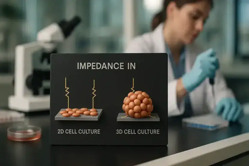
Impedance in 2D vs. 3D Cell Culture
“`html
Impedance in 2D vs. 3D Cell Culture
The advancement of cell culture technologies has revolutionized numerous scientific fields, particularly in pharmaceutical and biotechnology research. As conventional two-dimensional (2D) cell culture methodologies give way to more complex three-dimensional (3D) systems, understanding impedance—the measure of opposition a circuit presents to the passage of alternating current—becomes essential. This article delves into the contrasts between impedance measurements in 2D and 3D cell cultures, exploring their implications for research efficiency and data accuracy. Readers can expect to gain insights into the critical technological advancements shaping this transition.
Common Challenges and Limitations of Traditional Approaches
Impedance Measurement in 2D Cell Cultures
In 2D cell cultures, impedance measurements involve evaluating the electrical resistance across monolayers of cells adhered to flat surfaces. Although this setup provides valuable information on cellular health and proliferation, there are inherent limitations. These include:
- Restricted physiological relevance due to simplified cell attachment and growth patterns.
- Limited mimicry of in vivo environments, reducing predictive validity for drug testing.
- Challenges in modelling complex tissue-specific behaviors.
Despite these limitations, 2D cultures remain a staple in laboratory environments due to their simplicity and cost-effectiveness.
Technological Advances and Automation Trends
Emergence of 3D Cell Culture Systems
The shift towards 3D cell culture systems addresses many of the limitations associated with 2D cultures. In 3D configurations, cells grow in all spatial dimensions, providing a more realistic model of in vivo tissue environments. Impedance measurement in 3D cultures involves capturing data from cells embedded within a matrix or scaffold, often resulting in increased data complexity and a closer approximation of physiological processes. Key advancements include:
- Development of biomimetic materials that better replicate extracellular matrices.
- Integration of advanced imaging systems for enhanced monitoring.
- Automation of culture processes to improve reproducibility and throughput.
These technological strides are crucial for yielding high-fidelity biological insights and enhancing the predictive capabilities of in vitro models.
Practical Examples and Workflows Using Live-Cell Imaging
Role of Incubator-Based Imaging Systems
Live-cell imaging is a transformative tool in both 2D and 3D cell culture paradigms. Systems such as the zenCELL owl, an incubator-compatible live-cell imaging device, facilitate continuous monitoring without disrupting the cell environment. This system enhances traditional workflows by offering automated, high-resolution digital microscopy, thereby increasing data quality and reproducibility.
- Provides non-invasive, real-time tracking of cellular dynamics.
- Enables precise quantitation of cellular impedance in dynamic 3D environments.
- Supports longitudinal studies by maintaining consistent environmental conditions.
Through automation and real-time monitoring, live-cell imaging systems overcome significant analytical challenges posed by traditional culture methods.
Continue reading to explore more advanced insights and strategies.
“`
“`html
Enhancing Data Accuracy in 2D and 3D Cultures
Advanced Analytical Techniques
The accuracy of impedance data in cell cultures is paramount for drawing meaningful conclusions, particularly in pharmacological and toxicological studies. In 2D cultures, impedance measurements can be impacted by cell density and the uniformity of the cell monolayer. In contrast, 3D cultures pose challenges due to the heterogeneity of tissue constructs. However, advancements in analytical techniques have significantly enhanced data accuracy. Techniques such as Fourier Transform Impedance Spectroscopy (FTIS) and Electrochemical Impedance Spectroscopy (EIS) are being increasingly utilized to analyze complex impedance patterns over multiple frequencies, allowing for detailed profiling of cell behavior and interaction.
- Integrate multi-frequency impedance analysis to improve data resolution across different 3D structures.
Optimizing Culture Environments with Biosensors
Integration of Real-time Monitoring Devices
To optimize the culture environments, biosensors have emerged as pivotal tools, providing real-time insights into the physiological conditions of cell models. These sensors measure critical parameters such as pH, dissolved oxygen, and glucose levels. In 3D cultures, the integration of biosensors within scaffolds enables simultaneous monitoring of microenvironmental conditions, ensuring that changes in culture conditions do not adversely affect cell growth or data validity. A biosensor-coupled system in a recent study allowed researchers to maintain cellular homeostasis effectively, thereby achieving consistent cell viability and facilitating long-term experimentation.
- Use biosensor feedback loops to automatically adjust culture conditions and improve cell viability.
Harnessing Machine Learning for Culture Analysis
Application of AI in Impedance Data Interpretation
Machine learning algorithms are revolutionizing the interpretation of impedance data, particularly in complex 3D culture systems. These algorithms can process vast datasets to identify patterns and predict outcomes with a high degree of accuracy. In a clinical research setting, the application of machine learning models reduced the manual analysis time by 70%, leading to faster decision-making in drug development pipelines. By leveraging AI platforms, researchers can enhance the predictive power of their models, focusing on the most promising compounds or interventions.
- Implement machine learning models to detect impedance pattern anomalies, streamlining the validation process.
Synergizing Multi-Omics Approaches
Deepening Biological Insights through Integrated Analysis
The combination of impedance measurement with multi-omics approaches, such as transcriptomics and proteomics, enables a holistic view of cellular dynamics. This integration provides comprehensive insights into the underlying biological responses triggered by different treatments or conditions. For instance, a recent study combined impedance data with RNA sequencing to elucidate the genetic pathways affected by chemotherapeutic agents in 3D tumor spheroids, revealing novel targets for cancer therapy.
- Link impedance data with genomic profiles to create tailored intervention strategies.
Streamlining Workflows through Automation
Leveraging Robotics and AI for Efficient Experimentation
Automation in cell culture experiments not only enhances reproducibility but also significantly decreases the time and resources needed for comprehensive studies. Robotic systems, paired with AI-driven data management tools, automate everything from cell seeding to data acquisition. In a recent pilot study, the deployment of robotic systems in a 3D culture setting increased assay throughput by 80%, allowing scientists to test more variables simultaneously and accelerate research timelines.
- Adopt automated cell culture platforms to minimize human error and increase experimental throughput.
Improving Predictive Validity of Preclinical Models
The Role of 3D Printed Scaffolds
The development of 3D printed scaffolds has opened new avenues for improving the predictive validity of in vitro models. These scaffolds are engineered to mimic the complex architecture of native tissues, enhancing cell differentiation and function. The customizable nature of 3D printing allows for the rapid prototyping of diverse scaffold designs, tailoring them to specific cell types or experimental needs. This capability was demonstrated in a liver toxicity study where 3D printed scaffold models exhibited higher parenchymal cell viability and functionality than traditional 2D cultures.
- Utilize customized 3D printed scaffolds to enhance the physiological relevance of cell models.
Navigating Regulatory Landscapes with Innovative Insights
Aligning Scientific Advances with Compliance Standards
Amid the rapidly evolving landscape of cell culture technologies, aligning with regulatory standards remains crucial. Regulatory agencies globally are beginning to recognize the enhanced predictive capabilities of 3D models. In practice, involving compliance teams in the early stages of 3D model development ensures that innovations align with the latest guidelines, facilitating smoother transitions from research to market. A biopharmaceutical company recently reported reduced approval timelines for their drug candidates by incorporating validated 3D models, underscoring the importance of such alignment.
- Engage with regulatory bodies early in the research and development process to ensure compliance and expedite approvals.
Next, we’ll wrap up with key takeaways, metrics, and a powerful conclusion.
“`
“`html
Advancing toward Personalized Medicine
Customization of Cell Cultures for Individualized Treatments
The integration of personalized medicine into cell culture technologies represents a transformative shift in therapeutic development. Through advances in genomic editing techniques such as CRISPR/Cas9, cell cultures can be tailored to reflect individual genetic variances, thereby accelerating the development of customized treatment regimens. This precision approach enhances the efficacy and safety of new therapeutics by allowing researchers to evaluate drug responses in cultures with patient-specific genetic backgrounds. An emerging trend is the use of organoids derived from patient tissue, offering a powerful platform for disease modeling and personalized drug testing.
- Leverage patient-specific cell lines to increase the relevance and impact of preclinical models.
Exploring the Role of Artificial Organs
The Future of Regenerative Medicine
Artificial organs hold great promise as a frontier in regenerative medicine. These constructs, engineered using advanced 3D bioprinting techniques, offer potential solutions for organ failure by replicating the structure and function of natural organs. The coupling of impedance analysis with artificial organs facilitates the monitoring of tissue development and functionality in real-time, ensuring optimal conditions are maintained for successful integration and performance. A notable breakthrough involved creating a bioprinted heart valve that demonstrated robust endothelialization and mechanical properties, indicating substantial progress toward full-scale organ regeneration.
- Innovate with bioprinting strategies to enhance the viability of artificial organ constructs.
Overcoming Technical Challenges
Continuous Improvement of Methodologies and Technologies
As the complexities of cell culture technologies evolve, overcoming technical challenges remains paramount. Continuous improvement in methodologies, such as enhanced substrate materials and innovative culture ecosystems, is necessary to address issues like cell viability, growth uniformity, and data consistency. Cutting-edge technologies, including real-time imaging and high-throughput screening, are becoming indispensable tools for troubleshooting and optimizing cell culture workflows. A focus on iterative development and feedback mechanisms ensures that these technologies consistently meet the rigorous demands of scientific research.
- Adopt innovative materials and tools to address ongoing technical challenges in cell culture.
Conclusion
The journey through this exploration of impedance in 2D versus 3D cell culture highlights the dynamic intersection of cutting-edge technologies and innovative methodologies. From enhancing data accuracy with advanced analytical techniques to integrating machine learning for efficient data interpretation, the potential to redefine cell culture practices is immense. We have delved into how automation, personalized medicine, and artificial organs symbolize the ongoing transformation in biological research and medical applications.
The significance of these advancements lies not only in overcoming present challenges but also in setting a new standard of precision and reliability in cell culture technologies. As we harness biosensors for real-time monitoring, engage multi-omics approaches for holistic analysis, and align scientific ingenuity with regulatory compliance, the rise of these models underscores a pivotal step toward more predictive, reliable, and impactful scientific inquiry.
This article affirms the remarkable potential within cell culture innovations to fundamentally reshape drug discovery, regenerative medicine, and personalized therapies. As we stride confidently into this new era, let’s embrace the collaborative spirit of scientific exploration, encouraging continuous learning, improvement, and implementation of these technologies.
Engage with the wealth of resources available, and consider how you can incorporate these advancements into your own work, driving your field one step closer to groundbreaking discoveries that stand to benefit humanity in profound ways. Together, let’s pioneer the future of biological research, one cell at a time.
“`
Biological materials as a root cause in failed technology transfer projects

“`html
Biological Materials as a Root Cause in Failed Technology Transfer Projects
Technology transfer within biotechnology and life sciences is a critical process where knowledge and methodologies are transitioned from one laboratory to another or from research institutions to industrial applications. However, not all technology transfer projects achieve their expected outcomes, and one often overlooked factor is the variability and complexity of biological materials. In this article, we will delve into how biological materials can lead to setbacks in technology transfer projects, and strategies researchers can apply to mitigate these challenges.
The Complexity of Biological Materials in Technology Transfer
Understanding Biological Variability
Biological materials such as sera, plasma, and other reagents are inherent components of many cell culture and biotechnology processes. However, their biological nature means they’re subject to variability. Lot-to-lot variability in materials like Fetal Bovine Serum (FBS), for instance, can lead to significant differences in cellular behaviors, impacting the reproducibility of experiments when transferred between labs. This unpredictability poses considerable challenges in achieving consistent results during technology transfer.
- Biological materials can vary significantly between production batches.
- Processes reliant on these materials can produce inconsistent results when transferred.
Continue reading to explore more advanced insights and strategies.
Quality Control and Documentation: Essential Tools for Mitigation
Standardizing Biological Material Use
Implementing rigorous quality control measures and comprehensive documentation is vital in minimizing the impact of biological variability. Batch reservation and testing services can offer stability, by allowing the same batch of biological materials to be used consistently across different sites. Furthermore, detailed documentation aids in tracking deviations and implementing corrective measures. Comprehensive data on the serum’s origin, processing, and quality checks can anchor the transfer process firmly, reducing risk of failure.
- Utilize batch reservation to reduce variability risks in multi-lab setups.
- Leverage documentation for traceability and troubleshooting.
Continue reading to explore more advanced insights and strategies.
Reagents and Their Functional Role in Research Consistency
Ensuring Reproducibility with Precise Reagents
Reagents are fundamental to many biological assays and diagnostics but must be selected with consistency in mind to facilitate successful technology transfer. Ensuring that reagents such as separation solutions maintain known compositions and functions is critical. Advances in incubator-compatible live-cell imaging, such as systems detailed on zencellowl.com, facilitate continuous monitoring of cellular processes, thereby improving reproducibility and documenting subtle differences precipitated by reagent changes.
- Document the specification and source of all reagents used.
- Employ live-cell imaging for real-time process verification.
Continue reading to explore more advanced insights and strategies.
Human-Derived Biologicals: Ethical and Regulatory Dimensions
Addressing Ethical Considerations and Compliance
Incorporation of human-derived biological materials, such as human serum and plasma, necessitates consideration of ethical and regulatory guidelines. Variability in donor samples can affect assay performance, making it imperative to work with certified collections that adhere to ethical standards. Regulatory compliance supports not only ethical research but also ensures that the technology transfer abides by accepted standards, facilitating smoother transitions across geographic and institutional boundaries.
- Ensure compliance with ethical guidelines for donor materials.
- Understand regulatory frameworks affecting material transfer.
Continue reading to explore more advanced insights and strategies.
Scientific Services: Enhancing Stability and Reducing Risk
Custom Solutions for Consistent Outcomes
Partnering with scientific service providers for custom antibody development and biological material sourcing can be instrumental in navigating the challenges posed by biological variability. These services offer tailored solutions that enhance the stability of long-term projects and reduce the risk associated with technology transfers. Expert support in batch testing and documentation fortifies research methodologies, ensuring consistent and reliable outcomes across all project phases.
- Engage in customized sourcing to meet project-specific requirements.
- Apply expert services to validate and stabilize research protocols.
Continue reading to explore more advanced insights and strategies.
“`
“`html
Cross-Disciplinary Collaboration: A Path to Innovation
Leveraging Expertise Across Fields
Successful technology transfer projects often necessitate collaboration across various scientific disciplines. This cross-disciplinary approach leverages the combined expertise of biologists, chemists, engineers, and data scientists to optimize biological materials handling. For example, the Human Genome Project’s success was largely due to the collaborative efforts spanning computational biology to traditional benchwork. Similarly, pooling knowledge and resources in biotechnology can mitigate risks associated with biological variability by integrating innovative analytical techniques and data-driven insights.
- Foster environments that encourage interdisciplinary collaboration to enhance problem-solving.
Data-Driven Decision-Making in Technology Transfer
Utilizing Big Data for Improved Outcomes
Big data analytics has become indispensable in identifying trends and patterns that impact technology transfer outcomes. For instance, examining large datasets of biological material performances across diverse laboratory environments can pinpoint specific factors leading to variability. Machine learning models can further predict the impacts of these variables on project success rates. Initiatives like the European Bioinformatics Institute utilize vast biological databases to enhance reproducibility and standardization across various scientific domains.
- Invest in data analytics tools to improve predictive modeling of biological material performance.
Risk Management in Biological Material Handling
Proactive Strategies for Sustained Success
Risk management is crucial in overseeing biological material logistics. Implementing comprehensive risk assessment methodologies can identify potential failure points early in the technology transfer process. Instituting controls such as contingency planning for batch failures or supplier disruptions ensures adaptation to unforeseen circumstances. Consider the case of pharmaceutical firm Gilead, which effectively mitigated risks through a robust risk management framework during their antiviral drug technology transfers.
- Develop detailed contingency plans to address potential disruptions in biological material supply chains.
Communicating Across International Borders
Effective Communication in Global Transfers
Technology transfer often occurs on a global scale, necessitating the need for clear and effective communication. Differences in language, cultural norms, and scientific standards across countries can introduce errors or misunderstandings. The 2009 technology transfer of a vaccine production process between Indian and European companies underscored the importance of nuanced communication strategies. Engaging professional science communicators and utilizing translation services can bridge these gaps, ensuring clarity and shared understanding.
- Implement standardized communication protocols across all international teams.
Semantics in Scientific Documentation
Enhancing Clarity through Standardized Terminology
Scientific documentation, when inconsistent or ambiguous, can undermine technology transfer efforts. Standardizing terminology used in research documents ensures that all stakeholders have a uniform understanding of protocols and materials. Initiatives like the Open Biological and Biomedical Ontology (OBO) Foundry aim to unify terminologies across biological research, facilitating seamless information transfer and reducing misinterpretation risks.
- Adopt standardized scientific terminologies in training and documentation efforts.
Adaptive Manufacturing Processes
Integrating Flexibility into Production Protocols
Flexible manufacturing processes are essential for accommodating the inevitable variability in biological materials. Implementing adaptive protocols that can adjust to changes in material quality or availability can significantly reduce failure rates in technology transfer projects. The use of modular bioproduction systems, as seen in Genentech’s innovative approaches to drug production, exemplifies how adapting manufacturing frameworks can sustain project momentum and enhance scalability.
- Invest in modular manufacturing systems to enhance adaptability and responsiveness.
Continuous Monitoring and Improvement
Nurturing Sustainable Practices in Research and Production
Continuous monitoring regimens, combined with iterative improvement processes, form the backbone of successful technology transfers. By regularly evaluating outcomes and identifying areas for refinement, organizations can ensure that processes remain optimized and aligned with long-term project goals. The Plan-Do-Check-Act (PDCA) cycle, widely implemented in industrial settings, illustrates how cyclical evaluation fosters enduring project success through continuous enhancement.
- Regularly review and refine processes using cyclic improvement models like PDCA.
Next, we’ll wrap up with key takeaways, metrics, and a powerful conclusion.
“`
“`html
Overcoming Cultural Barriers in Technology Transfers
Building Trust and Understanding Across Divides
In the realm of global technology transfers, cultural barriers present unique challenges that can impact the success of the project. Recognizing and respecting cultural differences while fostering an inclusive atmosphere is essential. Thoughtful engagement strategies, like cultural competency training, can equip teams to bridge diversity gaps. The partnership between American and Japanese firms in biotechnology highlights the effectiveness of cultural sensitivity, which was critical in synchronizing their technology transfer processes.
- Invest in cultural competency training to strengthen global project collaborations.
Harnessing Technology to Facilitate Transfers
Innovative Tools for Enhanced Technology Exchange
Advancements in technology provide an invaluable arsenal of tools that facilitate seamless technology transfers. From advanced simulation software to virtual reality environments, these innovations enhance collaborative efforts by simulating complex biological systems and processes. These technological tools not only decrease the time to transfer but also improve accuracy in translating complex scientific protocols. Companies that deploy such technologies often see improvements in knowledge retention and project outcomes as exemplified by the innovative use of digital twins in biopharma production processes.
- Utilize advanced simulation and digital tools for more efficient transfer processes.
The Role of Leadership in Steering Technology Transfers
Visionary Leadership and Strategic Direction
Effective leadership is a pivotal factor in steering technology transfer projects towards successful outcomes. Strong leaders inspire innovation, encourage diversity of thought, and prioritize strategic resource allocation. Visionary leadership not only guides teams through complex transitions but also anticipates future challenges within the dynamic landscape of biological material handling. The strategic foresight demonstrated by AstraZeneca’s leadership during vital technology transfers showcases how decisive direction can harness collective efforts to deliver groundbreaking advancements.
- Focus on leadership development to cultivate a strategic, innovative project environment.
Conclusion
This article has presented a comprehensive analysis of the myriad factors influencing technology transfer projects involving biological materials. From fostering cross-disciplinary collaboration, leveraging data analytics, and implementing robust risk management strategies to ensuring clear cross-border communication and adaptive manufacturing processes, each element plays a critical role in driving project success.
As organizations navigate the complexities of global transfers, integrating these best practices is paramount. The synthesis of various scientific, technical, and cultural insights provides a cohesive framework to address the inherent challenges in the handling of biological materials. By promoting a culture of continuous monitoring and iterative improvement, companies can maintain agility and respond adeptly to evolving dynamics.
The importance of overcoming cultural barriers cannot be overstated, as it fosters trust and paves the way to sustainable international collaborations. Furthermore, harnessing cutting-edge technologies and visionary leadership ensures that strategic objectives are met with precision and efficiency. Each of these dimensions acts as a cornerstone in rendering technology transfers more predictable and less prone to failure due to the variability of biological materials.
Reflecting upon these insights, stakeholders are encouraged to implement such strategies in their organizational practices. In doing so, they will unlock new potential for innovation and collaboration, driving forward advancements that benefit both science and society at large. Embracing an integrative approach to these transfers, organizations can cultivate environments where groundbreaking discoveries become realities, transcending conventional limitations.
We invite industry leaders, researchers, and practitioners to take these insights to heart and champion efforts in their respective domains. As we continue to push the boundaries of scientific exploration and technological application, let us remain committed to pursuing excellence through cross-disciplinary cooperation, transparent communication, and inclusive practices. Together, we can usher in an era of innovation that not only meets but exceeds the demands of our rapidly evolving world.
Now is the time to act decisively, embrace change openly, and gather momentum in achieving breakthroughs that will define the future of biotechnology and beyond.
“`
Improving Reproducibility in Cell Culture Using Incubator-Integrated Imaging

Improving Reproducibility in Cell Culture Using Incubator-Integrated Imaging
In the rapidly evolving field of cell culture research, ensuring reproducibility and data quality is more critical than ever. As researchers, lab managers, and biotech professionals strive to uncover new biological insights, traditional methods often fall short in consistency and reliability. This article delves into the modern challenges of cell culture, explores technological advances like incubator-integrated imaging, and highlights practical strategies to enhance reproducibility. Whether you’re managing a lab or advancing biotech innovations, uncover how cutting-edge tools are reshaping cell culture research.
Challenges in Traditional Cell Culture Approaches
Inherent Limitations and Varied Outcomes
Traditional cell culture techniques face several challenges that impede reproducibility. Variability in cell growth conditions, human error during handling, and the manual nature of data collection often lead to inconsistent results. Environmental fluctuations, such as changes in temperature, humidity, and CO2 levels, can further disrupt cell viability and data integrity. These variables result in significant gaps between experimental and theoretical reproducibility, undermining research findings.
- Environmental variability affects cell growth consistency.
- Manual data collection increases the risk of human error.
- Inconsistent handling leads to varied experimental outcomes.
Continue reading to explore more advanced insights and strategies.
Advancements in Technology and Automation
Revolutionizing Cell Culture Practices
The integration of innovative technologies and automation into cell culture practices marks a pivotal shift toward enhanced reproducibility. Automated systems minimize human involvement, thus reducing the likelihood of error and improving data reliability. Equipment such as incubator-integrated imaging systems facilitates continuous monitoring under controlled conditions, ensuring a consistent environment for cell growth. These technological advancements not only streamline workflows but also enhance data accuracy and reproducibility.
- Automation reduces human-related inaccuracies in data collection.
- Continuous monitoring ensures stable environmental conditions.
- Advanced imaging techniques offer real-time data insights.
Continue reading to explore more advanced insights and strategies.
Live-Cell Imaging in Practice: Workflow Enhancements
Real-World Applications and Integration
Incorporating live-cell imaging into everyday research workflows offers significant benefits. Researchers can now observe cellular processes in real-time, enabling a deeper understanding of dynamic biological activities. By integrating incubator-based imaging systems, laboratories witness improved workflow efficiency and data fidelity. The zenCELL owl, as an incubator-compatible live-cell imaging solution, provides compact, robust functionality, showcasing an exemplary model of contemporary imaging technology.
- Real-time observation enhances understanding of dynamic processes.
- Incubator-integrated imaging solutions improve workflow efficiency.
- Compact systems like zenCELL owl offer space-saving benefits without sacrificing quality.
Continue reading to explore more advanced insights and strategies.
Incubator-Based Imaging: Elevating Reproducibility
Consistent Monitoring and Enhanced Data Quality
Incubator-integrated imaging is a cornerstone for improving reproducibility in cell culture. By maintaining stable environmental conditions and allowing continuous observation, these systems ensure that cells are not disturbed during critical experiments. This approach not only preserves cell integrity but also provides high-resolution, time-lapse imaging that aids in tracking subtle cellular changes over time. The result is data with higher reproducibility, enabling researchers to draw more accurate and reliable conclusions.
- Maintained stability of culture conditions supports cell integrity.
- Continuous monitoring prevents experimental disturbances.
- High-resolution imaging captures subtle cellular changes.
Continue reading to explore more advanced insights and strategies.
Adopting Scalable Technologies for Enhanced Efficiency
Building Capacity with Scalable Imaging Systems
As research projects grow in complexity and scale, the need for scalable solutions in cytological imaging becomes evident. Scalable technology allows laboratories to quickly adapt to increasing demands without sacrificing quality. Incubator-integrated imaging systems such as the Livecyte and Clarity by Phasefocus exemplify scalable solutions. These systems offer modular configurations, enabling researchers to customize their setup according to specific needs and project requirements. The flexibility provided by scalable systems ensures that research can progress without the friction typically associated with upgrading or expanding traditional equipment.
- Opt for imaging systems that allow for modular expansion to align with laboratory needs.
Leveraging Data Analytics for Improved Decision Making
Turning Complex Data into Usable Insights
Integrating advanced data analytics into cell culture research transforms raw data into actionable insights. High-resolution images generated by incubator-integrated systems contain a wealth of information that, when properly analyzed, can reveal patterns and trends otherwise undetectable. By employing software tools for image analysis and data management, researchers can gain deeper insights into cellular behavior, identify anomalies, and make informed decisions more rapidly. Automation of data interpretation helps to limit human bias, reinforcing the reproducibility of results.
- Implement data analytics software to process and analyze imaging data efficiently.
Implementing Standardized Protocols for Consistency
Ensuring Uniformity Across Research Workflows
One of the significant challenges in cell culture research is maintaining uniformity across various experimental runs. Developing and implementing standardized protocols helps mitigate variability, thus enhancing reproducibility. These protocols should encompass all aspects of cell maintenance, from seeding to imaging and data analysis. Moreover, adherence to standardized procedures ensures that all team members are aligned, facilitating consistency in methodology and technique application. Sharing successful protocols can also contribute to the standardization of practices across research groups globally.
- Create and distribute clear, comprehensive protocols to ensure consistency in workflows.
Utilizing Real-Time Monitoring for Immediate Adjustments
Responsive Adjustments to Enhance Experimental Success
Real-time monitoring offered by incubator-integrated imaging systems empowers researchers to make informed adjustments during the experimental process. This capability allows for immediate modifications in response to environmental shifts or unexpected changes in cell behavior, minimizing potential disruptions. By correcting course in real-time, researchers can enhance the accuracy of their experiments, reduce wastage of precious resources, and improve overall success rates.
- Use real-time monitoring to quickly address issues as they arise, thereby optimizing experimental outcomes.
Supporting Collaborative Research with Cloud-Based Integration
Enhancing Connectivity and Data Sharing
Cloud-based integration in imaging systems promotes collaboration by enabling researchers to access and share data seamlessly across different locations and platforms. This technology facilitates the efficient exchange of information and insights, leading to more cohesive and collaborative research efforts. In multicenter studies, streamlined data sharing via cloud technologies ensures that collaborators contribute to and benefit from collective datasets, driving forward progress in a unified manner. This interconnected approach is essential in today’s globalized research environment.
- Implement cloud-based solutions for efficient data sharing and collaboration across research teams.
Incorporating Training and Skill Development
Building Expertise with Ongoing Education
A critical factor in utilizing advanced imaging technologies effectively lies in the skills and expertise of the personnel operating them. Therefore, investing in regular training and development programs ensures that lab personnel remain proficient with evolving technologies and methodologies. Such initiatives not only improve operational competence but also empower team members to drive innovation within their projects. Regular workshops, certification programs, and peer-based learning sessions can be beneficial in maintaining a high level of expertise across research teams.
- Regularly update and enhance lab staff skills through dedicated training programs on new technologies.
Enhancing Experimental Design Through Integrated Solutions
Strategizing Research with Comprehensive Tools
Integrating incubator-based imaging solutions within a wider suite of experimental design tools provides researchers with a comprehensive approach to study planning and execution. Comprehensive experimental design includes clearly defining objectives, selecting appropriate methodologies, and incorporating technological tools for accurate data collection and analysis. The strategic deployment of integrated solutions allows researchers to identify potential issues in advance, optimize resource allocation, and streamline the entire research process, resulting in a higher likelihood of achieving desired outcomes.
- Approach experimental design holistically by integrating advanced imaging tools from the outset.
Next, we’ll wrap up with key takeaways, metrics, and a powerful conclusion.
“`html
Facilitating Scalability with Flexible Equipment
Preparing for Future Expansion
Scalability is a hallmark of modern research laboratories, adapting them to the ever-evolving demands of science and technology. Flexible equipment solutions, such as adjustable imaging platforms and modular lab furnishings, play a critical role in facilitating this scalability. They allow research spaces to be efficiently reconfigured as project requirements change, ensuring that laboratories remain not only functional but also future-proof. The ability to scale up or down swiftly, with minimal disruption, supports dynamic research agendas and fosters an environment where innovation can flourish.
- Invest in flexible laboratory equipment to accommodate future growth and technological advancements.
Ensuring Data Integrity with Robust Management Systems
Enhancing Reliability Through Structured Frameworks
In research, ensuring the integrity of data is paramount for achieving reproducible and credible outcomes. Utilizing comprehensive data management systems enables researchers to maintain high standards of data quality and security. These systems offer structured frameworks for data entry, storage, and retrieval, along with comprehensive audit trails that document every interaction with the data. As a result, researchers can trust their datasets, streamline the replication of studies, and facilitate seamless information sharing with collaborators, thereby enhancing overall research quality.
- Develop and implement robust data management systems to ensure data integrity and traceability.
Harnessing the Power of Artificial Intelligence in Research
Automating Processes for Enhanced Outcomes
Artificial Intelligence (AI) is revolutionizing how research is conducted, opening doors to unparalleled analytical capabilities. By incorporating AI algorithms into imaging and data analysis processes, researchers can automate tasks previously prone to human error and labor-intensive outcomes. AI-driven tools can dynamically analyze imaging data, predict trends, and suggest potential pathways for exploration. This intelligent automation not only accelerates research timelines but also enhances the depth and accuracy of insights, empowering scientists to make groundbreaking discoveries with confidence and precision.
- Leverage AI technologies to automate and enhance research processes, increasing efficiency and accuracy.
Conclusion
Adopting scalable technologies in cell culture research encompasses integrating advanced imaging systems, embracing cloud-based solutions, and incorporating AI and data analytics. These strategies ensure laboratories are equipped to handle increasing demands and complexity without compromising on quality or efficiency. By fostering environments where adaptability and technological advancement go hand in hand, researchers can push the boundaries of discovery and innovation.
Scalable technologies not only enhance research capabilities but also promote collaboration and reproducibility, which are essential in the modern scientific landscape. Effective data management systems ensure that critical insights are preserved and shared effectively across research teams globally. Meanwhile, standardized protocols and ongoing staff training play a vital role in maintaining consistency, reducing variability, and improving overall research outcomes.
Incorporating these scalable solutions provides laboratories with the ability to adapt, grow, and thrive in an ever-changing field. Researchers are empowered to strategize efficiently, leveraging comprehensive toolsets that optimize their experimental design and execution. Through the integration of real-time monitoring and advanced AI-driven analytics, they can achieve more reliable and insightful results than ever before.
As research continues to evolve, so too must the tools and practices we employ. By adopting these innovations now, laboratories can stay at the forefront of discovery and remain prepared for the challenges of tomorrow’s scientific inquiries. Let us embrace scalability and technology with open minds and a forward-thinking attitude, ensuring our research not only survives but thrives. Take the first step by evaluating current processes and systems, and make strategic decisions to integrate scalable solutions, thus paving the way for a successful and productive future.
“`
Multiwell Plates for Automation Labs

“`html
Multiwell Plates for Automation Labs
The landscape of modern laboratories is rapidly transforming with the integration of automation technologies. Central to this shift are multiwell plates, crucial in facilitating high-throughput screenings and precise data collection. For researchers, lab managers, and biotech professionals, understanding the nuances of developing and utilizing these plates can greatly enhance workflow efficiency and research outcomes. This article delves into the development, production, and application of multiwell plates in automation laboratories, providing an authoritative guide for advanced lab settings.
Challenges with Traditional Multiwell Plate Approaches
The Limitations of Conventional Practices
Historically, traditional multiwell plates have posed several challenges: from issues with reproducibility to limitations in the scalability of experiments. Variability in materials and surface treatments often led to inconsistent results, impacting the reliability of data required for critical research decisions.
- Material Inconsistencies: Impact on data reliability and experiment replication.
- Surface Treatment Challenges: Incapable of supporting diverse assays uniformly.
- Scalability Limitations: Hindrances in expanding high-throughput screening (HTS) capabilities.
Technological Advances and Automation Trends
Integrating Innovation in Lab Environments
The advent of automation in laboratory settings has necessitated the development of advanced multiwell plates tailored for robotic handling and high-speed data collection. Innovations in material science and engineering design have led to enhanced optical clarity, enabling more precise detection and analysis through techniques such as live-cell imaging.
- Advanced Material Selection: Using PS, PP, and COC for enhanced performance.
- Design for Manufacturing (DFM): Optimizing for automation compatibility and robustness.
- Optical Enhancements: Improved clarity for high-sensitivity detections.
Prototyping and Scale-Up for Precision and Reproducibility
From Prototypes to Industrial Production
To effectively transition from conception to production, a structured approach is vital. Initial prototyping involves selecting the right materials and testing surface treatments to meet specific research requirements. Scaling up involves meticulous process validation to ensure batch-to-batch consistency and compliance with regulated production standards.
- Prototyping Strategies: Iterative testing and refinement phases for optimal design.
- Scale-Up Process: Ensuring robust reproducibility and dimensional accuracy.
- Regulation Compliance: Emphasizing cGMP and GMP principles for quality management.
Continue reading to explore more advanced insights and strategies.
“`
“`html
Customization in Multiwell Plate Design
Tailoring Plates for Specific Laboratory Needs
In the ever-evolving landscape of lab automation, customization of multiwell plates is paramount. Custom plate designs allow laboratories to cater to specific assay requirements, from specialized coating needs to unique well geometries. Companies like Greiner Bio-One and Corning have expanded their customization options, offering tailored solutions that enhance the adaptability of multiwell plates to diverse scientific applications. For example, the integration of specialized coatings, such as low-binding or tissue culture treatments, can significantly enhance the quality of cell-based assays.
- Consult with manufacturers to explore customization options.
Automation-Compatible Plate Readers and Handling Equipment
Ensuring Seamless Integration with Robotics
The integration of multiwell plates with automation systems hinges on compatibility with plate readers and handling equipment. Companies must ensure that their equipment can handle variations in plate design without compromising performance. For instance, BMG LABTECH and BioTek Instruments offer advanced plate readers optimized for various plate formats, providing consistent performance metrics essential for high-throughput screenings. These technologies support seamless data collection and synchronization with laboratory information management systems (LIMS).
- Regularly update lab equipment to ensure compatibility with the latest plate formats.
Data Management and Analytics in Automated Systems
Leveraging Software for Enhanced Data Interpretation
Harnessing the power of automated systems extends beyond hardware into sophisticated software solutions. Data management software is essential for collating, analyzing, and securely storing the vast amounts of data generated from high-throughput methods. Platforms such as LabWare and Thermo Fisher’s SampleManager LIMS provide robust analytics capabilities, enabling researchers to conduct detailed analysis and generate actionable insights quickly. This accelerates research timelines and ensures that data integrity is maintained across all stages of experimentation.
- Implement LIMS for streamlined data tracking and reporting.
Environmental Considerations and Sustainable Practices
Innovating with Eco-Friendly Materials and Processes
As laboratories aim to reduce their environmental impact, sustainable practices in the production and disposal of multiwell plates are gaining prominence. Many manufacturers are now focusing on the use of recyclable and biodegradable materials, such as compostable plastics or bio-based polymers. Furthermore, innovations in manufacturing processes, such as using solar energy and reducing water consumption, are aligning production methodologies with global sustainability goals.
- Opt for suppliers committed to sustainability initiatives and certifications.
Quality Control and Assurance in Production
Maintaining Consistency and Compliance
Quality control is a critical component in the production of multiwell plates, ensuring that each plate meets stringent standards. This involves comprehensive testing of material properties, dimensional accuracy, and surface treatment effectiveness. The adherence to ISO standards and the incorporation of automated inspection systems can significantly improve product consistency. For instance, using high-resolution cameras and AI-driven analysis helps in detecting defects or variations at early stages, minimizing wastage and ensuring high-quality outputs.
- Implement regular audits and employ AI for enhanced quality assessments.
Global Market Trends and Future Directions
Navigating Trends in a Evolving Landscape
The global market for multiwell plates is poised for robust growth driven by advancements in drug discovery and personalized medicine. Emerging markets in Asia-Pacific and increased funding for biotech research are further fueling this expansion. Companies investing in R&D for novel plate materials and formats stand to gain a competitive edge. Boston-based MatTek Corporation is an exemplar, having developed innovative multiwell platforms that support advanced tissue models, proving instrumental in toxicology screenings and disease modeling.
- Stay updated on market trends and align R&D efforts with emerging needs.
Case Study: Revolutionizing Cellular Assays
A Real-World Application of Multiwell Plates
In a collaborative project between a leading pharmaceutical company and a biotech startup, advanced multiwell plates played a pivotal role in revolutionizing a high-throughput drug screening process. By utilizing plates with custom coatings and tailored geometries, the teams achieved significant reductions in reagent usage and increased assay sensitivity. This resulted in a 30% improvement in overall throughput, leading to faster identification of potential drug candidates. This case demonstrates the transformative impact of well-chosen multiwell plate designs in critical research endeavors.
- Document and analyze case studies to uncover best practices and insights.
Next, we’ll wrap up with key takeaways, metrics, and a powerful conclusion.
“`
“`html
Multiwell Plate Innovations in Education
Empowering the Next Generation of Scientists
The integration of multiwell plates is not limited to high-level research labs and pharmaceutical companies but is also gaining traction in educational institutions. As educators strive to provide hands-on learning experiences, multiwell plates have become invaluable teaching tools in biology and chemistry curriculums. They offer students the opportunity to engage with real-world laboratory techniques, enhancing their understanding of assay development and data analysis. By introducing students to these tools early on, institutions can foster a generation of scientists who are adept in modern lab practices.
- Incorporate multiwell plates in educational curriculums to enhance practical learning experiences.
Cost-Effectiveness and Economic Impact
Balancing Innovation with Budget Constraints
While multiwell plates present numerous advantages in innovation and customization, their economic impact cannot be ignored. Institutions are constantly balancing the need for advanced technology with budgetary constraints. Cost-effective solutions, such as investing in durable, reusable plates and opting for bulk purchasing, can mitigate expenditure while maintaining quality and functionality. Moreover, companies providing scalable solutions or subscription models offer laboratories economically viable options to access cutting-edge tools without significant upfront costs.
- Evaluate bulk purchasing and subscription models for cost savings.
The Role of Multiwell Plates in Clinical Research
Accelerating Clinical Trials and Diagnostics
Multiwell plates play a crucial role in clinical research by streamlining processes, particularly in drug testing and diagnostic applications. Their capacity for high-throughput screening accelerates the identification of therapeutic candidates and enhances the precision of diagnostic testing. The versatility of multiwell plates allows for extensive testing on a microliter scale, reducing reagent consumption, and ensuring reproducibility—critical factors in fast-paced clinical environments. As personalized medicine continues to grow, multiwell plates will be indispensable in the nuanced requirements of individual patient data and treatment strategies.
- Utilize multiwell plates for efficient, high-throughput diagnostics in clinical settings.
Conclusion
The journey through the evolving landscape of multiwell plates underscores their transformational role across a spectrum of scientific applications. From customization in design to their seamless integration into automated systems, multiwell plates have redefined laboratory processes, empowering both research advancements and educational paradigms. As laboratories pivot towards eco-friendly practices, multiwell plates reinforce their significance through sustainable manufacturing solutions.
The versatility of multiwell plates, as demonstrated through real-world applications and market trends, portrays a promising horizon for scientific innovation. By accommodating unique laboratory needs, ensuring compatibility with robotic handlers, and facilitating superior data management, these plates not only enhance experimental efficiency but also drive down operating costs by minimizing resource usage. Furthermore, the burgeoning markets and ongoing research in plate technology accentuate the dynamic future of multiwell plates, ensuring their relevance and indispensability in modern science.
In closing, embracing the advancements in multiwell plate technology is not merely an option but a strategic imperative for laboratories aiming to remain at the forefront of scientific discovery. By investing in this critical tool, laboratories can achieve unparalleled precision and efficiency, paving the way for accelerated discoveries and breakthroughs. We urge scientists, educators, and industry leaders to continue exploring and implementing the insights and innovations offered by multiwell plates, ensuring we are equipped for the challenges of tomorrow's scientific endeavors.
“`
How serum filtration methods influence protein composition

How Serum Filtration Methods Influence Protein Composition
The Science Behind Serum Filtration
Understanding Filtration Techniques
Serum filtration is primarily implemented to remove unwanted particulates, microbes, and possible contaminants that could interfere with cell culture experiments. Common filtration methodologies include membrane filtration, depth filtration, and tangential flow filtration. Each of these methods employs different mechanisms and pore sizes to achieve varying levels of filtration efficiency, which can, in turn, impact the serum’s protein composition through shear forces, protein binding, and size exclusion.
- Membrane filtration: Utilizes a porous membrane to physically separate materials based on size.
- Depth filtration: Incorporates a thick medium that captures particles throughout its matrix.
- Tangential flow filtration: Allows fluid to smear across the surface, using cross-flow dynamics for more effective separation.
Continue reading to explore more advanced insights and strategies.
Influence on Protein Composition
Impact of Shear Forces
Shear forces exerted during filtration processes can contribute to protein denaturation and degradation, leading to a change in the protein profile of the serum. This is particularly relevant for filters with decreased pore sizes, where increased pressure and turbulence come into play. These mechanical forces can alter sensitive proteins, such as cytokines and growth factors, impacting the biological function of cell cultures reliant on precise protein signaling pathways.
- Proteins critical to cell growth may be selectively filtered out.
- Changes in cytokine levels could influence immune response studies.
Serum Filtration and Quality Control
Ensuring Consistency and Reliability
Filtration processes necessitate stringent quality control measures to maintain consistency across batches. The protein composition of serum products can fluctuate due to different filtration techniques or conditions, including temperature and flow rates. Batch testing and comprehensive documentation are essential for identifying variances and ensuring product consistency, which are crucial for experimental reproducibility in laboratories.
- Perform regular protein assays post-filtration to assess changes.
- Utilize batch records to trace filtration parameters and outcomes.
Continue reading to explore more advanced insights and strategies.
Applications in Cell Culture and Immunology
Tailoring Filtration for Specific Applications
In cell culture and immunological studies, the intended use of serum dictates the specification of filtration methods. For instance, primary cultures might demand a filtration regime that retains critical growth factors, whereas antibody production might prioritize the removal of specific immune mediators. Tailors filtration strategies based on the application ensures that the serum retains the appropriate protein composition for the desired biological outcomes.
- Determine the required protein profile for specific cell types or studies.
- Adapt filtration processes to preserve functional protein integrity.
Continue reading to explore more advanced insights and strategies.
Advancements in Filtration Technology
Continuous Monitoring and Documentation
Advancements in technology, such as the use of incubator-compatible live-cell imaging systems like the zenCELL owl, enable real-time monitoring of cell behavior and the documentation of serum or reagent effects. These systems provide visual documentation of cell culture conditions and help improve the reproducibility of biological workflows through continuous observation, offering significant advantages in the evaluation of serum filtration impacts.
- Improved insight into cell behavior under different filtration conditions.
- Enhanced documentation for traceability and reproducibility.
Continue reading to explore more advanced insights and strategies.
“`html
Innovative Filtration Solutions
Exploring Emerging Technologies
As scientific needs evolve, so too do the technologies supporting them. Innovations in serum filtration are aligning with broader trends in biotechnology, aiming to improve both efficacy and precision. Recent advancements focus on minimizing protein loss while enhancing selectivity through the application of specialized membrane materials and smart filtration designs. Consider, for example, the use of asymmetric membranes that provide a gradient of pore sizes, enhancing the separation of molecules based on size while preserving sensitive proteins typically vulnerable to shear forces or denaturation.
- Leverage specialized membranes to customize protein retention and filtration efficiency.
Impact on Protein Signaling Pathways
Maintaining Integrity in Key Proteins
The consequences of filtration are particularly pronounced in protein signaling pathways, which play pivotal roles in cellular communication. When key proteins such as cytokines or chemokines are inadvertently altered or removed, the effects can ripple across experimental results, leading to misconstrued data. Effective filtration protocols must therefore balance filtration between the elimination of unwanted components and the preservation of essential protein structures critical for accurate signaling.
- Map protein signaling networks to identify and preserve critical proteins during filtration.
Optimizing Filtration through Customization
Tailored Approaches for Diverse Needs
Every research setting may demand unique filtration requirements based on distinct experimental objectives. By employing a customized approach to serum filtration, researchers can align their methods with specific goals. This may involve coalescing different filtration stages or using combinatory processes such as integrating centrifugation with filtration to heighten purity and protein retention. An example might involve modifying flow rates or pressures in tangential flow filtration, ensuring the retention of larger protein molecules that are crucial for a given culture’s proliferation.
- Customize flow parameters to maximize critical protein retention.
Leveraging Data Analytics for Filtration
Enhancing Accuracy through Digital Insights
With the advent of digital tools, data analytics are increasingly employed to optimize and monitor filtration efficacy. By using high-resolution analytics, such as spectroscopy or chromatography, researchers can gain comprehensive insights into protein activity within serum post-filtration. These tools facilitate a better understanding of protein landscape variance, equipping scientists with data that inform more precise adjustments to filtration conditions.
- Incorporate data analytics to refine filtration processes based on empirical protein composition data.
Strategies for Consistent Batch Production
Ensuring Uniformity Across Experimental Batches
A critical challenge in laboratory serum use is maintaining consistency across different batches. Variability can stem from natural differences in source material or fluctuations in filtration processes. To ensure reproducibility, it is vital to implement statistical process control measures, routinely evaluating batches against defined protein content benchmarks. This can be achieved through regular batch sampling and comparison against standardized reference profiles using tools like ELISAs or mass spectrometry.
- Develop stringent benchmarks for protein content based on historical data.
Case Study: Filtration Optimization in Serum Manufacturing
Real-world Applications and Outcomes
Consider a biopharmaceutical company specializing in serum products tailored for stem cell research. By employing a strategic filtration review and applying the insights gathered from data analytics, the company enhanced the retention of essential growth factors by 20%. This was achieved by optimizing the tangential flow filtration stage to minimize protein shear forces. Consequently, product efficacy improved, leading to a 15% increase in product reliability among research clients, illustrating the business and scientific benefits of optimized filtration processes.
- Audit and refine filtration stages to enhance critical protein retention.
Future Directions in Serum Filtration
Pioneering Paths Toward Greater Control
The future of serum filtration sees a convergence with nanotechnology and bioengineering. New methodologies are set to provide even finer control over protein components, offering unprecedented specificity. Expected advances include filtration matrices engineered at the nanoscale for targeting particular molecular signatures, adapting to fluid dynamics in real-time for optimal separation, and integrating AI for predictive adjustments. Such innovations are paving the way for exponential advances in biomedical research fidelity and effectiveness.
- Explore nano-engineered filters for superior selectivity and precision.
Next, we’ll wrap up with key takeaways, metrics, and a powerful conclusion.
“`
“`html
Advancements in Quality Control
Ensuring High Standards in Filtration
Quality control measures in the context of serum filtration are being enhanced with cutting-edge technologies to ensure that the highest standards are upheld. This includes implementing automated monitoring systems that use real-time data to ensure that every stage of the filtration process adheres to predefined quality benchmarks. Innovative quality assurance tools, such as process analytical technology (PAT) frameworks, allow for continuous scrutiny, helping to promptly identify and rectify any deviations from the norm, thus safeguarding the integrity of the filtration process.
- Integrate real-time monitoring systems to ensure adherence to quality standards.
Ethical Considerations in Serum Filtration
Balancing Scientific Advancement with Responsibility
While serum filtration technologies offer immense potential for scientific advancement, they also necessitate careful ethical consideration. Ensuring that the source materials for serum are collected, processed, and utilized responsibly is paramount. Compliance with ethical standards not only addresses humanitarian concerns but also enhances the credibility and acceptance of scientific findings. Transparency in sourcing, along with rigorous adherence to bioethical standards, ensures that these innovative techniques contribute positively and sustainably to scientific progress.
- Prioritize ethical sourcing and processing in filtration protocols.
The Road Ahead: Collaborative Innovation
Synergizing Efforts for Greater Impact
The future of serum filtration will be shaped by collaborative efforts that unite researchers, technology developers, and policymakers. These collaborations are critical for transforming innovative filtration technologies into reliable tools that can meet diverse scientific needs worldwide. By fostering an environment where interdisciplinary exchange is encouraged, stakeholders can collectively overcome current challenges, drive technological breakthroughs, and ensure that the benefits of advanced serum filtration are widely accessible.
- Promote interdisciplinary collaborations for technological advancements.
Conclusion
In exploring the evolution of serum filtration technologies, several key takeaways emerge. The significance of innovative filtration solutions in preserving essential proteins, enhancing experimental reliability, and improving overall research outcomes stands out prominently. Emerging trends in biotechnology, coupled with sophisticated digital tools, are revolutionizing how we approach serum filtration, ensuring greater precision and efficacy in scientific experiments.
As we delve into the impact of serum filtration on protein signaling pathways, the importance of maintaining the integrity of key proteins becomes evident. Customization of filtration processes provides tailored solutions that align with unique experimental requirements, emphasizing that one size does not fit all in biotechnological research. Consistent batch production and ethical sourcing further underscore the commitment to scientific rigor and societal responsibility.
The importance of leveraging data analytics for refined filtration processes cannot be overstated, as it provides empirical insights that drive fine-tuning and optimization of these processes. The case studies and forward-looking advancements highlight the tangible benefits and promising future of serum filtration.
This article sought to illuminate the intricate dynamics and forward-thinking approaches that define serum filtration today. By maintaining high ethical standards, adopting innovative technologies, and fostering collaborative networks, the scientific community is poised to unlock new possibilities in research and application.
For researchers, healthcare professionals, and industry experts, the imperative now is to continue championing these advanced methodologies. Let this be a call to action: embrace the cutting-edge innovations in serum filtration with an eye toward ethical practices and collaborative progress. Together, we can propel the field of biotechnology towards even greater achievements.
“`
Live-Cell Imaging in Cell & Gene Therapy: Managing Critical Culture Phases

“`html
Live-Cell Imaging in Cell & Gene Therapy: Managing Critical Culture Phases
In the rapidly evolving fields of cell and gene therapy, maintaining precise control over cell culture conditions is paramount. Among these, live-cell imaging stands out as an indispensable tool, offering unparalleled insights into cellular dynamics. This article delves into the role of live-cell imaging in optimizing critical culture phases, providing a comprehensive guide for researchers and biotech professionals committed to advancing their work in cell and gene therapies.
Traditional Challenges in Cell Culture and Monitoring
Limitations of Conventional Observation Techniques
Standard methods of cell culture observation have included periodic manual inspections and end-point assays, which are fraught with several limitations. These methods can introduce variability and inconsistency, primarily due to the intermittent nature of data acquisition. Additionally, manual handling increases the risk of contamination, and the discontinuous observations may miss transient cellular events critical to understanding cellular mechanisms.
- High risk of contamination from frequent manual handling
- Potential for missing significant but transient cellular events
- Variability in data due to inconsistent observation intervals
Continue reading to explore more advanced insights and strategies.
Technology and Automation Trends in Live-Cell Imaging
Advancements Facilitating Workflow Efficiency
Recent technological advancements have revolutionized live-cell imaging, making it a cornerstone for effective cell and gene therapy research. Automated systems now allow for the continuous monitoring of cultures, thereby ensuring the consistent acquisition of high-quality data. Automation reduces manual intervention, mitigating contamination risks and improving the reproducibility of experiments. These enhancements are crucial for studies ranging from basic cell biology to complex therapeutic applications.
- Automation ensures consistent and high-quality data capture
- Reduction in manual intervention diminishes contamination risks
- Enhanced reproducibility and reliability of experimental outcomes
Continue reading to explore more advanced insights and strategies.
Integrating Live-Cell Imaging into Culture Workflows
Practical Applications and Benefits
Integrating live-cell imaging into culture workflows offers numerous practical advantages. It allows for real-time monitoring of cell health, morphology, and behavior under various conditions. For instance, in high-throughput screening (HTS), where speed and accuracy are crucial, live-cell imaging provides critical kinetic data that can guide decision-making and facilitate the fast identification of potential therapeutic candidates.
- Real-time monitoring of cellular health and morphology
- Critical for accurate data collection in high-throughput screening
- Enhances decision-making processes and candidate identification
Continue reading to explore more advanced insights and strategies.
Innovations with Incubator-Based Imaging Solutions
Improving Data Quality and Experimental Reproducibility
Incubator-based imaging, such as the zenCELL owl system, represents a significant innovation in live-cell imaging technology. These systems are designed to integrate seamlessly into cell culture environments, providing uninterrupted data collection while maintaining optimal culture conditions. Such platforms support continuous monitoring, minimize disturbances, and ensure high-quality and reproducible data, thus accelerating research outcomes in cell and gene therapy.
- Seamless integration with cell culture environments
- Maintains optimal conditions for continuous data collection
- Enhances reproducibility and accelerates research outcomes
Continue reading to explore more advanced insights and strategies.
“`
“`html
Leveraging Image Analysis Software for Enhanced Data Interpretation
Automating Data Processing for Better Insights
One of the most compelling advancements in live-cell imaging is the integration of sophisticated image analysis software. These tools automate the processing of vast datasets generated by continuous monitoring, transforming complex biological data into actionable insights. Automated image analysis enhances the precision of measurements related to cell count, proliferation rates, and morphological changes, thus reducing human error and subjective interpretation.
- Utilize image analysis software to automate and standardize data processing.
Confocal and Multiphoton Imaging: A Deeper Look
Advanced Imaging Techniques for In-Depth Analysis
Confocal and multiphoton imaging technologies provide an invaluable depth of field, which is particularly beneficial for examining thicker biological samples. These techniques enable researchers to capture high-resolution images of cells in three dimensions, offering an unparalleled perspective on cellular architecture and interactions within their microenvironment. This method has been instrumental in observing live-cell dynamics in intricate gene therapy models.
- Adopt advanced imaging techniques for exploring complex cell structures.
Real-Time Imaging in Stem Cell Research
Monitoring Differentiation and Development
In stem cell research, real-time live-cell imaging offers a decisive edge, allowing scientists to monitor the differentiation and development of stem cells as they occur. This capability is essential for understanding the pathways of cellular differentiation and for optimizing protocols to increase the efficiency and safety of stem cell therapies. For example, researchers can track changes in cell morphology and gene expression over time, providing crucial data for therapeutic advancements.
- Implement real-time monitoring to track stem cell differentiation over time.
High-Content Screening (HCS) in Drug Discovery
Accelerating Drug Development with Live-Cell Imaging
High-content screening combines live-cell imaging with automated analysis, expediting the drug discovery process by facilitating the simultaneous analysis of complex cellular processes across thousands of wells. This approach has been pivotal in identifying new drug candidates for cell and gene therapies, allowing researchers to analyze cell viability, proliferation, and apoptosis in reaction to potential therapeutic compounds in real-time.
- Use high-content screening methods to enhance the efficiency of drug discovery.
Personalized Medicine and Precision Therapies
Customizing Treatments through Cellular Insights
In the realm of personalized medicine, live-cell imaging provides the ability to closely monitor how individual patient-derived cells respond to specific treatments, enabling the tailoring of therapies to maximize efficacy and minimize adverse effects. For instance, oncologists can use live-cell imaging to assess cancer cell response to chemotherapy, adjusting treatments to better suit individual patient needs.
- Leverage live-cell imaging to customize and optimize patient-specific treatments.
Challenges and Solutions in Live-Cell Imaging
Overcoming Technical and Biological Obstacles
While live-cell imaging is a powerful tool, it is not without challenges. Imaging live cells can be complicated by factors such as phototoxicity, photobleaching, and the sheer volume of data produced. However, innovative tools and techniques are continually being developed to address these issues. For example, the use of low-light imaging techniques and advanced data management systems can mitigate these challenges, enabling more sustainable and manageable imaging processes.
- Employ advanced imaging methodologies to reduce phototoxicity and manage data volume effectively.
Case Study: Enhancing CAR-T Cell Therapy
Utilizing Live-Cell Imaging to Transform Immunotherapy
In a groundbreaking case study, researchers leveraged live-cell imaging to optimize Chimeric Antigen Receptor (CAR) T-cell therapy. By observing CAR-T cell interactions in real-time, they could refine genetic modifications to enhance efficacy and reduce side effects. Live-cell imaging allowed for the observation of CAR-T cells’ ability to target and kill cancerous cells effectively, leading to protocol optimizations that significantly improved patient outcomes.
- Apply live-cell imaging to immunotherapy to improve treatment protocols and outcomes.
Next, we’ll wrap up with key takeaways, metrics, and a powerful conclusion.
“`
“`html
Integrating Live-Cell Imaging with Artificial Intelligence
Enhancing Data Interpretation through Machine Learning
The integration of artificial intelligence with live-cell imaging is revolutionizing data interpretation by employing sophisticated algorithms to identify and analyze patterns beyond human perception. Machine learning models can predict cellular behavior, automate image analysis, and streamline data processing, making it possible to manage and interpret the voluminous data generated. By learning from vast datasets, AI can improve the accuracy of predicting cell development pathways, assessing drug efficacy, and even contributing to personalized treatment plans.
- Incorporate machine learning models to enhance data analysis and prediction capabilities.
Ethical Considerations in Live-Cell Imaging
Addressing Privacy and Data Security Challenges
As with any technology that deals with sensitive data, live-cell imaging must be managed with care to ensure privacy and data protection. With the integration of patient-derived cells in the therapeutic process, safeguarding patient information becomes crucial. Developing robust data encryption methods and adhering to strict ethical standards are necessary steps to maintaining trust and ensuring the secure handling of biological and imaging data within live-cell imaging frameworks.
- Develop strict ethical protocols to safeguard data integrity and privacy.
The Future of Live-Cell Imaging in Biotechnology
Pushing Boundaries and Shaping New Horizons
The future of live-cell imaging is poised to unlock unprecedented opportunities in biotechnology and medicine. Emerging technologies such as nanoscopy, super-resolution microscopy, and AI-driven models continue to push the frontiers of what is possible in cellular research. As these innovations evolve, the potential to unravel the complexities of cellular mechanisms further will pave the way for novel therapeutic approaches and a deeper understanding of disease pathology and prevention.
- Explore emerging technologies to drive advancements in cell and gene therapy.
Conclusion
The advancements in live-cell imaging have presented transformative opportunities in cell and gene therapy, offering a granular view into cellular landscapes and their dynamics. Key takeaways from this exploration highlight the importance of leveraging sophisticated image analysis software, real-time monitoring, and high-content screening techniques to enhance research accuracy and efficiency. The integration of advanced methodologies such as confocal and multiphoton imaging, alongside AI and machine learning, further accentuates the way we process and interpret massive datasets for better insights.
In a world increasingly tailored towards personalization, live-cell imaging stands at the forefront of customized medicine, providing invaluable insights that guide patient-specific treatments. Challenges such as phototoxicity and data management are being addressed through innovative techniques, underscoring the field’s dynamic nature and its commitment to overcoming existing barriers.
As the potential of live-cell imaging continues to unfold, its role in pioneering cutting-edge therapies is indisputable. Researchers and clinicians are better equipped than ever to optimize treatment protocols, reduce adverse effects, and push the boundaries of healthcare. The integration of ethical considerations ensures that, while transformative, these advancements are executed responsibly and sustainably.
As we venture further into an era characterized by precision medicine, live-cell imaging will remain pivotal. The relentless pursuit of knowledge and improvement in imaging technologies promises a horizon of breakthroughs in understanding cellular behavior and developing innovative therapies. Let us embrace these advancements, ensuring that we continue to explore and harness the full potential of live-cell imaging, driving progress in biotechnology and medicine.
Stay at the cutting edge, engage with emerging technologies, and make impactful contributions to the fascinating field of live-cell imaging and its vast applications.
“`
