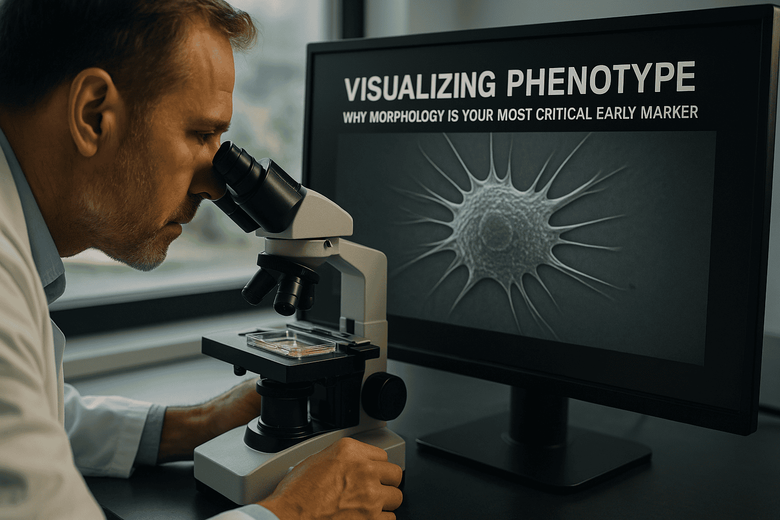
Visualizing Phenotype: Why Morphology is Your Most Critical Early Marker
In the rapidly evolving landscape of cell culture research, the ability to accurately visualize phenotype stands as a pivotal component of experimental success. Morphology, the study of cell form and structure, offers critical insights as an early marker during phenotypic analysis. Researchers, lab managers, and biotech professionals are increasingly recognizing the power of real-time morphological observation in understanding cellular processes, disease progression, and therapeutic responses.
This article delves into the scientific and practical reasons for prioritizing morphology as an essential early indicator, addressing common challenges, exploring technological advancements, and discussing practical applications within modern laboratory workflows.
Häufige Herausforderungen und Beschränkungen traditioneller Ansätze
Limitations of End-Point Assays
Historically, cell morphology has frequently been evaluated using end-point assays. These approaches, while valuable, often provide a limited snapshot of dynamic cellular processes. Static images fail to capture temporal changes and can lead to misinterpretation of data, particularly in heterogeneous populations or when dealing with transient phenotypic states.
- Static images obscure dynamic cellular behavior.
- End-point assays may misrepresent transient or subtle changes.
- Increased risk of data variability due to manual handling.
Challenges in Consistency and Reproducibility
Traditional methods also struggle with issues of reproducibility. Variability can stem from manual sample processing, subjective interpretation, and inconsistent imaging conditions. These factors can obscure meaningful biological insights and hinder the robustness of experimental conclusions.
- Manual processing introduces variability.
- Subjective image interpretation affects analysis consistency.
- Inconsistent imaging conditions reduce data comparability.
Technologische Fortschritte und Automatisierungstrends
Incubator-Based Imaging Systems
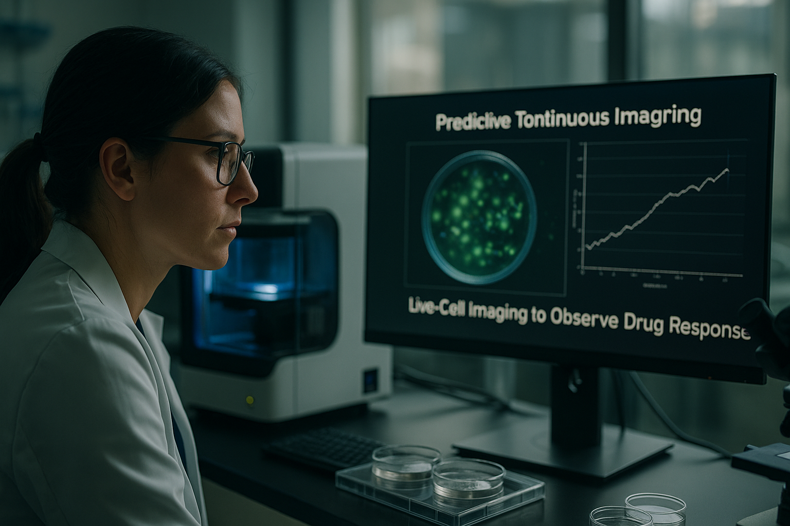
The integration of advanced imaging technologies, such as incubator-based systems, has revolutionized the way researchers approach phenotype visualization. These systems allow for continuous monitoring of cell morphology under tightly controlled environmental conditions, enhancing data consistency and reliability.
- Enables real-time, continuous monitoring within stable environments.
- Reduces user intervention, minimizing data variability.
- Enhances reliability of temporal phenotypic data.
Automation and AI in Morphological Analysis
The advent of automation and artificial intelligence (AI) tools has further facilitated high-throughput morphological analysis. Automated systems minimize human error and enhance reproducibility, while AI algorithms accelerate data analysis by identifying complex patterns and subtle morphological changes that might be undetectable by the human eye.
- Automated imaging increases throughput and reproducibility.
- AI algorithms improve accuracy in detecting subtle changes.
- Facilitates large-scale phenotypic screening and analysis.
Lesen Sie weiter, um tiefere Einblicke und Strategien zu gewinnen.
“```html
Leveraging High-Content Screening for Superior Insights
Integrating Morphology in High-Content Workflows
High-content screening (HCS) systems have emerged as cutting-edge tools in the analysis of cellular morphology. These systems provide a wealth of morphological data, allowing researchers to investigate multiple cellular parameters simultaneously. By incorporating fluorescence imaging and advanced software, HCS enables the extraction of rich phenotypic information that extends beyond basic morphology.
- Integrate diverse data types (e.g., morphological, fluorescence) for comprehensive phenotype profiles.
- Utilize high-content analysis for deeper insights into cellular responses.
- Apply HCS to streamline drug discovery and toxicological assessments.
Harnessing the Power of Computational Algorithms
Data-Driven Morphology Analysis
Computational algorithms now play a pivotal role in morphology analysis. Sophisticated image processing techniques, including machine learning models, allow for the thorough characterization of phenotypic patterns across large datasets. These algorithms help to identify subtle morphological changes and enable predictive modeling of cell behavior.
- Employ machine learning models to predict phenotypic outcomes.
- Enhance data interpretation with algorithm-driven insights.
- Implement automated pipelines for repeatable and scalable analysis.
Real-World Applications in Drug Development
Case Study: Predicting Drug Efficacy
Visualizing cell morphology is crucial in drug development, offering insights that traditional biochemical assays might miss. A notable case involves the use of morphological screening to predict the efficacy of novel cancer therapies. By correlating morphological changes with therapeutic outcomes, researchers can identify promising candidates more efficiently, reducing the time and cost of development.
- Use morphology to correlate drug response with cellular health indicators.
- Enhance therapeutic targeting by understanding cellular dynamics.
- Leverage morphological data to streamline lead optimization processes.
Optimizing Lab Workflows with Integrated Imaging Platforms
Efficiency Gains in Routine Analysis
Integrated imaging platforms have equipped laboratories with tools to optimize workflows by combining high-resolution imaging with automated analysis features. These platforms help reduce manual intervention and cycle times while ensuring data accuracy and reproducibility across experiments.
- Adopt integrated systems to automate routine monitoring tasks.
- Reduce labor-intensive processes through streamlined imaging protocols.
- Ensure data robustness with consistent imaging environments.
Cross-Disciplinary Applications Enhancing Diagnostic Accuracy
Morphology-Driven Diagnostics Innovations
Beyond research, the deployment of morphological analysis has transformed diagnostic methodologies. By integrating quantitative morphological data, it’s possible to improve the precision of disease diagnostics. For instance, morphological assessments are being used to refine cancer grading systems and enhance the sensitivity of early detection techniques for infectious diseases.
- Incorporate morphological data in diagnostic workflows for precision medicine.
- Advance early disease detection with morphology-driven biomarkers.
- Enhance clinical decision-making with data-rich morphological insights.
Maximizing Engagement with AI-Powered Imaging Tools
Increasing Accessibility and Real-Time Decision Making
AI-powered imaging tools have revolutionized accessibility to advanced morphological analysis, enabling real-time decision-making. Researchers can swiftly assess data sets and make informed decisions based on sophisticated AI-processed outputs, propelling advancements in immediate research directives and collaborations.
- Leverage AI-driven tools for real-time data insights.
- Boost collaboration with shared AI-enhanced datasets.
- Implement AI infrastructure to accelerate research findings.
Im Anschluss fassen wir die wichtigsten Erkenntnisse, Kennzahlen und eine wirkungsvolle Schlussfolgerung zusammen.
“`
“```html
Exploring Morphological Deep Learning Techniques
Unveiling Hidden Patterns Through Deep Learning
Deep learning techniques have emerged as transformative tools in morphological analysis, uncovering complex patterns that traditional methods might overlook. By harnessing convolutional neural networks (CNNs), researchers can delve deeper into the myriad layers of cellular structures, discovering unseen correlations between morphology and function. This creates opportunities to redefine our understanding of cellular responses, advancing both academic research and practical applications within clinical settings.
- Utilize CNNs to explore intricate cellular patterns.
- Foster novel insights by integrating deep learning with morphology.
- Advance research outcomes with uncoverable morphological correlations.
Transforming Laboratories with Intelligent Automation
Streamlining Processes Through Smart Automation
The adoption of intelligent automation in morphology analysis is revolutionizing laboratory workflows. By combining automated imaging systems with AI-driven data handling, laboratories can achieve unprecedented efficiency. These innovations reduce human error, enhance accuracy, and allow researchers to focus on analysis and interpretation, catalyzing a shift from reactive to proactive scientific exploration.
- Implement AI-driven automation for increased lab efficiency.
- Decrease errors with precise, automated image analyses.
- Pivot towards proactive, data-driven research methodologies.
Innovative Cross-Sector Applications
Expanding the Impact of Morphology
Morphological analysis is no longer confined to traditional biomedical applications; its impact is being felt across diverse fields, including biotechnology, environmental science, and agritech. For instance, morphology-driven insights are used in crop yield optimization and ecological impact assessments, offering critical data that informs sustainable practices. By continuing to expand these applications, morphology analysis contributes to solving broad, cross-disciplinary challenges facing society today.
- Leverage morphological insights for sustainable agricultural practices.
- Inform environmental sustainability with data-driven morphology.
- Drive innovation across sectors through interdisciplinary morphology applications.
Schlussfolgerung
The intricate world of cellular morphology harbors valuable insights pivotal for scientific advancement and societal progress. Through the course of this article, we have explored how leveraging high-content screening, computational algorithms, AI-powered imaging tools, and deep learning methodologies enhance our ability to decode complex biological processes. These methodologies empower researchers to uncover vital phenotypic markers, offering a rich tapestry of data that sharply informs both research and practical applications.
The integration of morphology within drug discovery, diagnostic innovation, and cross-sector applications underscores its indispensability as a critical analytical marker. Morphology-driven insights facilitate the streamlining of workflows, leading to improved accuracy and efficiency in scientific endeavors. Moreover, intelligent automation and AI have paved the way for real-time decision-making, catalyzing a transformation in how research directives are approached and executed.
In closing, the value of morphological analysis in advancing precision medicine, optimizing laboratory workflows, and enhancing diagnostic capabilities is irrefutable. For scientists and practitioners committed to pushing the boundaries of discovery, morphology stands as a gateway to unprecedented insights and possibilities. Embrace these technologies and methodologies to harness cellular morphology’s full potential. Whether you are driven by the pursuit of cutting-edge research, innovative applications in industry, or transformative healthcare solutions, the future belongs to those who see morphological analysis not just as a tool, but as an essential partner in discovery.
We invite you to continue exploring these advancements, share your experiences, and remain at the forefront of morphological innovation. Unite with a community of like-minded professionals as we redefine the landscape of scientific and clinical inquiry, one morphological insight at a time.
“`