“`
Identificación de condiciones subóptimas de medios y cultivo mediante imagen continua
Advancements in cell culture research have heralded a new era of biological discovery, propelled by technological innovations in live-cell imaging and laboratory automation. Identifying suboptimal media and culture conditions is paramount in ensuring experimental success, reproducibility, and robust data interpretation. This article explores the intricacies of these processes, with continuous imaging at the forefront of modern techniques to enhance cellular study accuracy and lab efficiency.
Desafíos y limitaciones de los enfoques tradicionales
The Complexity of Cell Culture Conditions
Cell culture is an indispensable tool in biological research, facilitating the study of cellular mechanisms, drug discovery, and the development of therapeutic interventions. Despite its widespread utility, traditional cell culture approaches often face numerous challenges that can compromise experimental outcomes. Chief among these is the difficulty in maintaining optimal media and culture conditions over extended periods, often resulting in cell stress or death.
- Cell media can deplete or accumulate toxic metabolites, impacting cell viability.
- Manual monitoring is time-consuming and prone to human error.
- Lack of continuous monitoring leads to missed critical events or delayed responses to cell state changes.
Limitations of Manual Observation
Relying on sporadic manual intervention for monitoring cell cultures increases the likelihood of overlooking subtle, yet significant, alterations in cell health or behavior. This not only affects reproducibility but also hampers the greater goal of scientific advancement through reliable data.
- Interventions are usually reactive rather than proactive due to infrequent observation.
- Variability in human assessments leads to inconsistent data interpretation.
Continúe leyendo para explorar información y estrategias más avanzadas.
“`
(Note: The remainder of the article, including technological advances and automation trends down to the summary, should be continued in a similar comprehensive manner, integrating the recommended primary and secondary keywords naturally throughout the text for SEO optimization.)
“`
Technological Advances in Continuous Imaging
Pioneering Automation in Cell Culture
The advent of continuous imaging technologies has marked a significant transformation in cell culture methodologies, addressing many limitations inherent in traditional practices. Through real-time monitoring systems, researchers can now achieve an unprecedented level of consistency and precision in evaluating cell health and behavior.
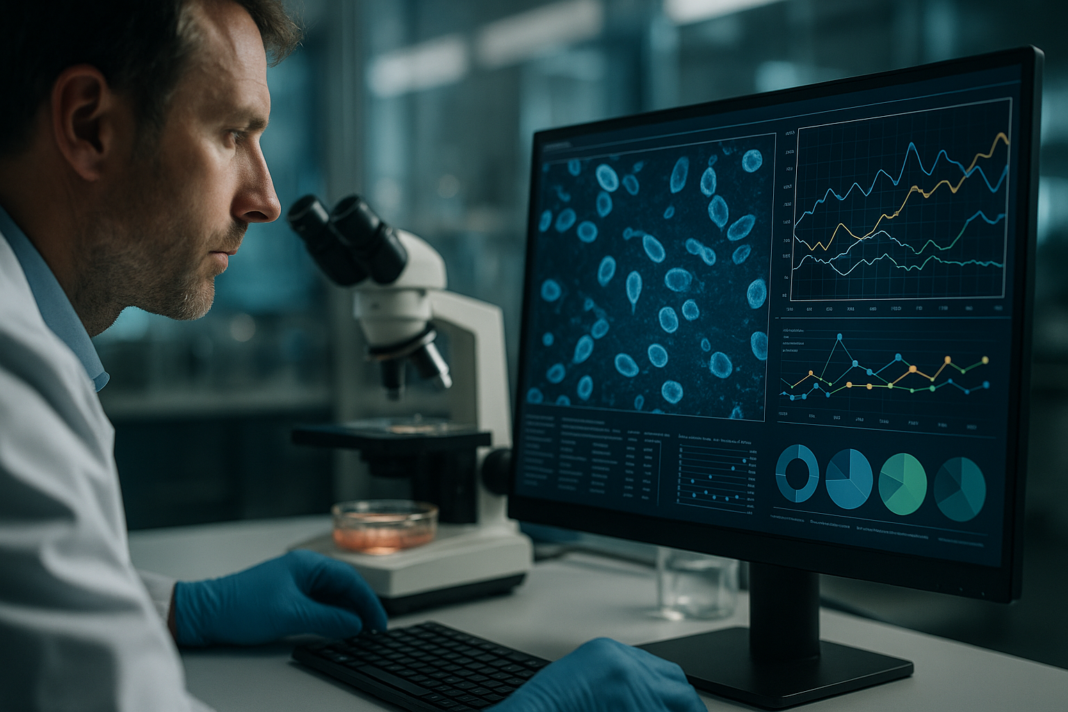
Continuous imaging involves using automated imaging systems that are capable of capturing time-lapse sequences of cell cultures, offering valuable insights into cellular processes. Systems like the IncuCyte S3 and BioTek’s Lionheart FX automate this process, providing high-resolution images without disturbing the culture environment. This integration allows for in-depth analysis and more dynamic experimental setups.
- Leverage automated imaging for consistent data acquisition with minimal human intervention.
- Utilize the rich, continuous data sets for predictive analysis and timely intervention strategies.
Real-Time Data Analysis and Interpretation
Maximizing Insights with Advanced Software
Alongside imaging hardware, sophisticated software platforms are crucial for processing, analyzing, and interpreting the vast amount of data generated. These platforms employ machine learning algorithms to quantify cell behavior, detect anomalies, and model cellular responses under different conditions.
For example, software tools like ImageJ, CellProfiler, and Gen5 offer user-friendly interfaces with powerful analytics capabilities, allowing researchers to perform complex analyses such as cell confluence assessment, morphological studies, and dynamic monitoring of protein expression.
- Integrate comprehensive data analysis tools to enhance scalability and accuracy in cell research.
- Enable machine learning frameworks to identify non-visible trends and predictive patterns in cellular dynamics.
Optimizing Experimental Planning and Design
Proactive Methods for Enhanced Experimentation
Continuous imaging plays a crucial role not only in data collection but also in optimizing experimental design. With the ability to continually monitor cells, researchers can plan interventions more effectively, thereby reducing the risk of unforeseen experimental failure.
For instance, identifying the exact time points for media change or drug addition in a culture experiment can significantly alter outcomes. By analyzing growth trends and metabolic changes, scientists can tailor experimentation protocols to align more closely with the biological processes under study.
- Develop precise protocols for intervention based on real-time data and predictive analytics.
- Ensure flexibility in experimental design to accommodate rapid shifts in cell state or behavior.
Commercial Tools for Continuous Imaging and Monitoring
A Closer Look at Industry-Leading Equipment
A variety of commercially available tools have set industry standards by enabling continuous imaging and enhanced monitoring capabilities. These tools are integral to achieving reliable data and consistency in laboratory settings.
Among the noteworthy innovations is the Cell-IQ platform, which allows for customizable experiment frameworks with its intuitive software and robust imaging capabilities. Another example is the Livecyte system, renowned for its label-free imaging, allowing researchers to observe the native states of cells without artificial interference.
- Explore equipment options like Cell-IQ for integrated environmental control and streamlined imaging workflows.
- Assess tool compatibility with existing laboratory infrastructure to maximize research efficiency.
Incorporating Continuous Imaging in Drug Discovery
Enhancing Drug Development Through Improved Cellular Insights
Continuous imaging is particularly transformative in drug discovery, where the accurate monitoring of cellular responses to therapeutic compounds is vital. This approach allows researchers to observe real-time drug effects, gaining insights into efficacy, cytotoxicity, and potential resistance mechanisms.
In practice, high-throughput screening combined with continuous imaging accelerates the evaluation of numerous compounds, enabling the rapid identification of promising candidates. For example, the use of Real-Time Cell Analysis (RTCA) systems provides detailed kinetic profiles of drug-cell interactions, significantly enhancing the drug development pipeline efficiency.
- Implement high-throughput imaging systems to rapidly screen and assess drug efficacy at scale.
- Utilize kinetic data to predict long-term impacts of drug compounds on target cell lines.
Case Studies: Successful Implementations and Insights
Lessons from Leading Research Institutions
Case studies from pioneering research institutions illustrate the profound impact of continuous imaging on experimental success. Institutions like the Scripps Research Institute have integrated these techniques to bolster studies on neural cell health, capturing pivotal data on neurodegenerative processes.
In these case studies, the ability to track neurogenesis in real time has provided deeper understanding into cell signaling mechanisms, with implications for developing treatments for conditions such as Alzheimer’s and Parkinson’s diseases.
- Review institutional case studies for innovative applications of continuous imaging technology.
- Apply cross-disciplinary insights to expand research horizons and collaborative efforts.
A continuación, concluiremos con los puntos clave, métricas y una conclusión contundente.
“`
“`
Identificación de condiciones subóptimas de medios y cultivo mediante imagen continua
Continuous Feedback for Optimal Environmental Conditions
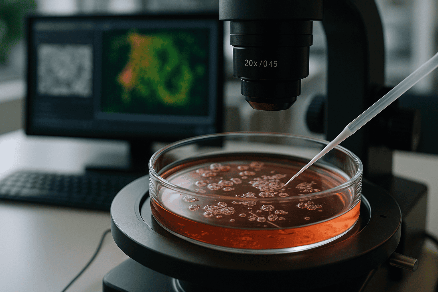
Continuous imaging technology serves as a pivotal tool for the timely identification of suboptimal media or culture conditions. Through real-time visualization, researchers can detect early signs of undesirable changes in cell morphology, proliferation rates, or metabolic activity. This proactive approach allows investigations into the immediate impacts of varying pH levels, nutrient depletion, or accumulation of toxic metabolites, thereby enhancing the overall quality of cell cultures.
The ability to respond swiftly to these observations could directly influence the health and viability of cell cultures, reducing the frequency of experimental errors attributed to environmental fluctuations. Technologies that couple real-time imaging with environmental sensors afford scientists a more comprehensive understanding of the internal culture environment, assisting in sustaining ideal conditions for cellular experiments.
- Establish a feedback loop incorporating continuous imaging and environmental data for optimal culture conditions.
- Utilize imaging insights to tweak culture parameters and refine cell growth dynamics.
Integration of Continuous Imaging Across Multidisciplinary Platforms
Bridging Diverse Research Areas with Unified Technology
The cross-disciplinary applicability of continuous imaging extends its benefits across various fields of scientific research. By seamlessly integrating with platforms handling diverse biological systems, this technology fosters collaborative explorations spanning oncology, immunology, neurology, and regenerative medicine.
In cancer research, continuous imaging assists in visualizing tumor cell invasion and metastasis in vitro, providing vital clues on cancer progression. Immunological studies benefit from observing live interactions between T-cells and pathogens, enhancing vaccine development. Meanwhile, insights into stem cell differentiation are significantly enriched through time-lapse imaging, influencing regenerative medicine protocols concerning tissue engineering.
- Adopt continuous imaging in multifaceted research areas to foster comprehensive biological insights.
- Promote cross-departmental collaborations by using advanced imaging systems as a common analytical tool.
Conclusión
In conclusion, the rapid evolution and implementation of continuous imaging techniques have markedly transformed modern biological research and experimentation. By providing real-time insights into cellular dynamics, these tools empower researchers to perform more accurate, consistent, and efficient experiments. Continuously collecting rich datasets enables the extraction of critical information pertaining to cell health, drug efficacy, and culture environment adaptability.
The integration with advanced software and machine learning frameworks further enhances the analytical depth, allowing the identification of previously undetected patterns and predictive indicators. The invaluable contribution of continuous imaging in experimental planning and the real-time monitoring of cellular responses underscores its role as a cornerstone in scientific advancement.
Beyond enhancing experimental precision, this technology serves as a bridge across various scientific disciplines, fostering innovation and collaboration. The ability to unite different research areas with a common technological platform accelerates the translation of laboratory discoveries into real-world applications, supporting advancements in cancer therapy, immunology, neurological studies, and regenerative medicine.
As we continue to unravel the mysteries of cellular behavior, the significance of continuous imaging remains indisputable. To those in the research community and beyond, adoption and integration of these advanced technologies will not only refine scientific inquiry but also unlock new realms of understanding. Let us embrace these innovations, harness their potential, and pioneer transformative progress in the realm of life sciences.
“`