“`html
From Images to Data: Continuous Live-Cell Imaging for QA and Publications
In the fast-evolving realm of cell biology, the transformation of live-cell images into actionable data has become paramount. From Images to Data: Continuous Live-Cell Imaging for QA and Publications is not merely a trend but a necessity for researchers striving for precision and reproducibility. This article delves into the critical role of continuous live-cell imaging, addressing the challenges of traditional methods, and providing insights into technological advances that are reshaping experimental workflows.
Challenges in Traditional Live-Cell Imaging Approaches
Common Limitations and Their Impact on Research Outcomes
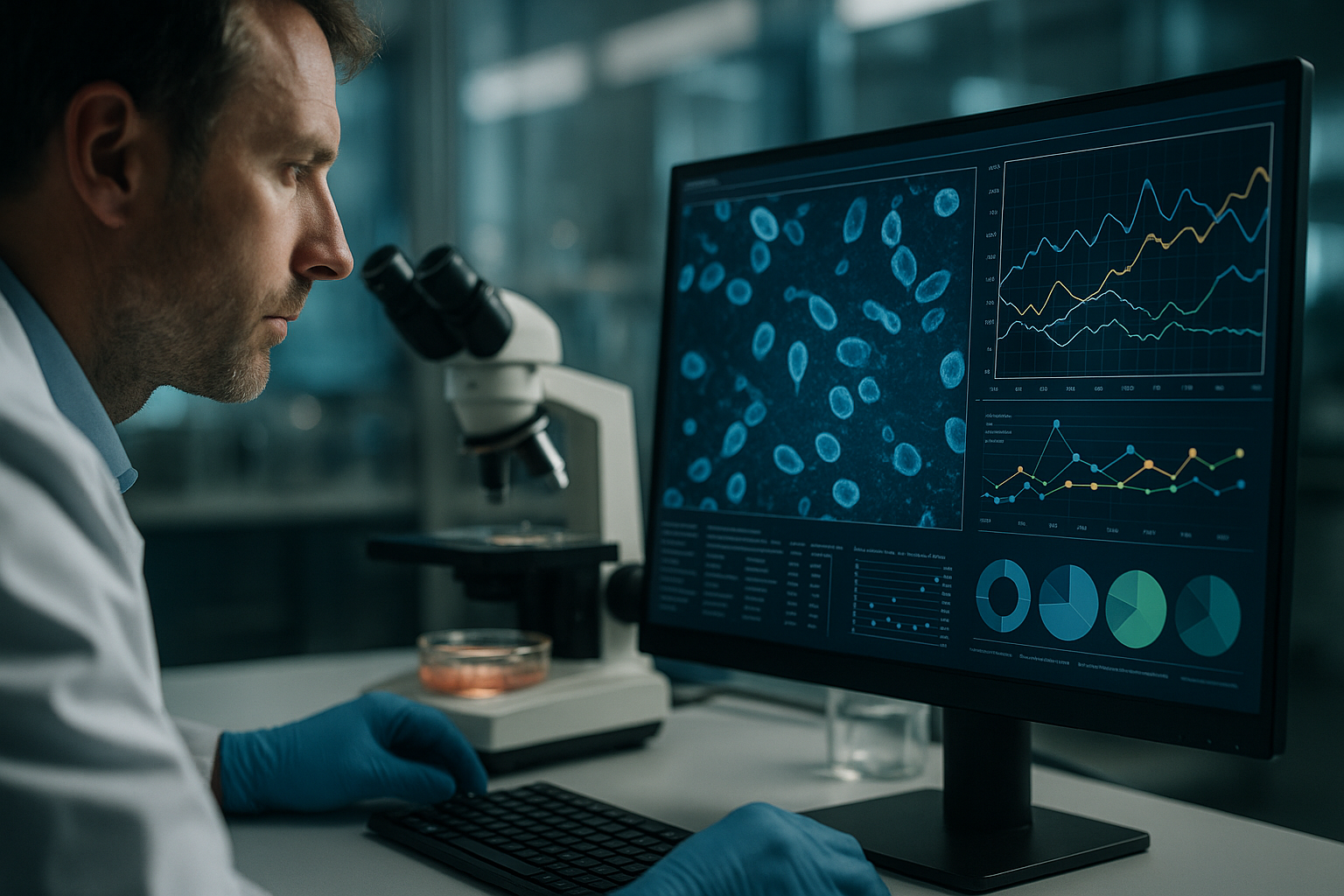
Traditional live-cell imaging methods have presented researchers with numerous challenges. The manual nature of these techniques often leads to inconsistencies, limited resolution, and potential phototoxicity due to repeated exposures. These limitations can result in compromised data quality and a lack of reproducibility, which are pivotal for both quality assurance (QA) and publication standards.
- Lack of continuous monitoring increases the risk of missing critical cell events.
- Manual intervention introduces variability and potential contamination risks.
- Limited data throughput hampers the scope of high-throughput screening (HTS) applications.
Technological Advances and Automation in Live-Cell Imaging
Moving Towards Automation and Enhanced Reproducibility
The advent of automation and advanced imaging technologies is transforming live-cell imaging practices. Tools like the zenCELL owl exemplify compact, incubator-compatible systems that enable uninterrupted monitoring of cell cultures. These advances have made significant strides toward enhancing reproducibility and data quality. Automation reduces human error and allows for seamless integration into laboratory workflows, facilitating data collection that is both continuous and comprehensive.
- Automated systems offer real-time, high-definition monitoring.
- Minimal manual intervention preserves sample integrity.
- Enhanced data analytics tools support sophisticated analyses and robust data interpretation.
Practical Implementations of Continuous Live-Cell Imaging
Streamlining Workflows with Real-World Applications
Incorporating continuous live-cell imaging into laboratory protocols can streamline workflows significantly. For instance, using systems like the zenCELL owl, researchers can observe cell behavior under various conditions in real-time, providing instant feedback for process optimization. This not only enhances quality assurance but also informs decision-making throughout experimental processes.
- Live-cell imaging facilitates the dynamic assessment of drug responses in pharmacological studies.
- Increased data granularity aids in identifying potential errors or anomalies early in the research process.
- Real-time visualization enhances the discovery and development timelines in biotech research.
Continue reading to explore more advanced insights and strategies.
“`
“`html
Building a Robust Data Framework with Live-Cell Imaging
Strategies for Effective Data Management and Integration
As continuous live-cell imaging produces vast amounts of data, developing a robust data framework becomes crucial for effective analysis and integration. Adopting cloud-based storage solutions, coupled with machine learning algorithms, can facilitate the handling of large datasets and enable advanced data mining techniques. Organizations such as pharmaceutical companies and biotech startups are increasingly leveraging these strategies to enhance their research capabilities.
- Implement a centralized data repository for seamless access and collaboration.
Accelerating Research with Machine Learning and AI
Harnessing Artificial Intelligence for Enhanced Insights
Artificial Intelligence (AI) and Machine Learning (ML) are at the forefront of transforming live-cell imaging into a powerhouse of actionable insights. By automating image analysis, AI algorithms can identify patterns and predict cellular behaviors much faster and more accurately than manual methods. A notable example is the use of AI to predict cancer cell metastasis, where AI-driven analysis drastically reduced time consumption and improved prognostic accuracy.
- Integrate AI tools into existing analytical frameworks to amplify research capabilities.
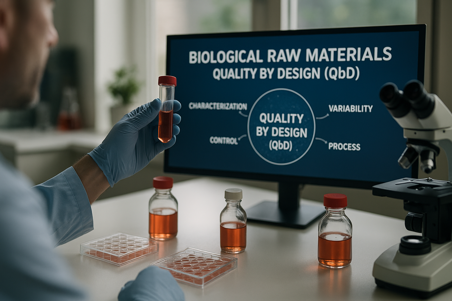
Quality Control and Assurance in Data Collection
Ensuring Precision and Accuracy in Experimental Outputs
Quality assurance in data collection is pivotal for reliable research outcomes. Continuous live-cell imaging systems are equipped with tools that automatically alert researchers to anomalies, allowing swift corrective measures. Pharmaceutical research, for example, benefits from such systems in maintaining the stability of drug screening assays, thus ensuring data accuracy.
- Utilize automated alerts and checks to promptly identify data inconsistencies or errors.
Multidisciplinary Applications of Live-Cell Imaging
Expanding Utility Beyond Traditional Domains
The versatility of live-cell imaging allows its utility to span numerous fields beyond traditional cell biology. In agrigenomics, imaging technologies are applied to study plant cell responses to environmental stressors, providing insights pivotal for developing resilient crop varieties. Similarly, toxicology laboratories utilize these systems for detailed analyses of cellular responses to chemical exposures.
- Explore cross-disciplinary collaborations to maximize the utility of live-cell imaging technology.
Case Study: Revolutionizing Drug Development Processes
Real-Life Applications and Success Stories
One notable case study involves a biotech firm employing continuous live-cell imaging to drastically enhance its drug development pipeline efficiency. By integrating automated imaging systems, the firm reduced lead times for drug efficacy tests by 35%, identified viable drug candidates earlier in the cycle, and improved overall pipeline throughput.
- Invest in automation to realize enhancements in drug discovery timelines and success rates.
Integrating Imaging Tools into High-Throughput Screening
Advanced Techniques for Scalable Research
Integrating live-cell imaging into high-throughput screening (HTS) operations has facilitated more scalable research processes. Technological advancements enable the parallel examination of thousands of samples, enriching data acquisition without the compromise of prolonged exposure times. This approach is increasingly pivotal in large-scale genomic studies where speed and accuracy are of the essence.
- Leverage scalable imaging tools to cater to expansive research studies with efficiency.
Overcoming Implementation Challenges in Live-Cell Imaging
Addressing Practical Barriers with Strategic Solutions
Despite its potential, the implementation of continuous live-cell imaging can present challenges such as high initial costs and technical complexities. Solutions include phased adoption strategies, where systems are integrated incrementally, allowing laboratories to gradually adapt to new workflows and manage costs effectively.
- Consider a phased integration of new technologies to minimize disruption and manage costs.
Next, we’ll wrap up with key takeaways, metrics, and a powerful conclusion.
“`
“`html
Empowering Academic Research Through Live-Cell Imaging
Boosting Publications and Peer Recognition
Academic institutions increasingly recognize the power of continuous live-cell imaging as a pivotal tool for enhancing the quality and depth of research publications. By leveraging the rich data sets generated through these imaging technologies, researchers can uncover novel insights and validate hypotheses with a higher degree of confidence. This method has proven particularly effective in high-impact studies within cellular biology and neurobiology, sharply raising the profile of the institutions involved.
- Focus on integrating live-cell imaging data into academic theses to elevate the standard of research outputs.
The Future of Live-Cell Imaging in Personalized Medicine
Tailoring Treatments with Data-Driven Approaches
In the realm of personalized medicine, live-cell imaging stands poised to play a transformative role by allowing for the individual observation of patient-specific cellular responses. This capability facilitates the customization of therapeutic strategies to maximize efficacy and minimize adverse effects. For chronic diseases, particularly cancer, this precision ensures treatments are finely tuned at a cellular level, offering patients more effective personalized interventions.
- Adopt live-cell imaging to develop bespoke treatment plans that align with individual patient profiles.
Navigating Ethical and Regulatory Considerations
Ensuring Responsible Use of Advanced Imaging Technologies
As live-cell imaging technologies advance, ethical and regulatory considerations become increasingly important. Ensuring transparency in data handling, maintaining patient confidentiality, and adhering to stringent scientific standards are essential for responsible usage. Establishing clear protocols and compliance with international guidelines not only safeguards data integrity but also reinforces trust among stakeholders, from research participants to governmental bodies.
- Implement robust ethical frameworks and regulatory compliance strategies in imaging practices.
Conclusion
The journey through continuous live-cell imaging elucidates a dynamic intersection where science, technology, and data converge to propel research and application to unprecedented heights. By constructing robust data frameworks, integrating machine learning and AI, and expanding the multidimensional applications, researchers can unlock a wealth of insights that drive both innovation and efficacy in diverse fields. From refining drug development processes to enhancing personalized medicine, the potential applications of live-cell imaging are both expansive and profound.
Throughout this exploration, we’ve seen how the strategic implementation of imaging technologies can overcome traditional research barriers, paving the way for more agile, accurate, and insightful scientific inquiries. The blend of high-throughput screening capabilities with AI-enhanced analytical methods epitomizes the remarkable strides being made towards reducing timelines and elevating research standards. Challenges such as initial costs and technical complexities remain, but phased integration strategies and continued technological evolution promise a viable pathway forward for institutions ready to invest in these advancements.
As academic institutions and biotech companies harness these technologies for greater academic impact and commercial benefit, the narrative of live-cell imaging becomes one of unbounded opportunity. The seamless integration of advanced imaging creates fertile grounds for enhancing publication quality, fostering collaborative initiatives, and ultimately, furthering the boundary of what is possible. As we continue to navigate the ethical and regulatory landscapes that accompany this technology, the foundation remains solid for ethical innovation and trust in scientific progress.
In embracing these changes, we invite researchers and institutions alike to reimagine the possibilities within their respective fields. Together, by championing the responsible and innovative application of continuous live-cell imaging, we embark on a transformative journey toward personalized, precise scientific advancements that shape the future of research and medicine. Forward-thinking adoption, coupled with a relentless pursuit of knowledge, will define the next era of scientific inquiry and leave an indelible mark on the world.
“`