Wie zukünftige Sicherung von Entscheidungen zur biologischen Beschaffung

“```html
Wie man Entscheidungen zur biologischen Beschaffung zukunftssicher gestaltet
Im sich rasch entwickelnden Bereich der Biotechnologie stehen Forscher und Laborfachleute bei der Beschaffung von biologischen Materialien vor einer Vielzahl von Herausforderungen. Von der Variabilität tierischer Seren bis hin zu ethischen Überlegungen bei humanen biologischen Produkten ist die fundierte Beschaffungsentscheidung entscheidend für die Gewährleistung der experimentellen Konsistenz und Integrität. Dieser Artikel befasst sich mit den Strategien und Überlegungen, die für zukunftssichere Entscheidungen bei der Beschaffung biologischer Materialien unerlässlich sind, und bietet einen umfassenden Leitfaden zur Verbesserung von Forschungsabläufen.
Die Bedeutung von Qualität bei biologischen Produkten tierischen Ursprungs
Verständnis der Chargenschwankungen
Tierische biologische Produkte, wie fetales Rinderserum (FBS) und Rinderplasma, spielen eine entscheidende Rolle bei zellkulturellen Verfahren. Diese Materialien weisen aufgrund von Unterschieden bei den Spenderorganismen, den Entnahmeverfahren und den Handhabungsverfahren eine inhärente Variabilität auf. Die Chargenvariabilität kann die experimentellen Ergebnisse erheblich beeinflussen, weshalb Qualitätskontrolle und Standardisierung unerlässlich sind.
- Führen Sie eine strenge Dokumentation für die Nachverfolgbarkeit.
- Implementieren Sie Batch-Tests, um optimale Losauswahlen zu identifizieren.
- Nutzen Sie kontinuierliche Überwachungssysteme, wie inkubator-kompatible Live-Cell-Imaging-Lösungen, um das Zellverhalten zu beobachten und die Reproduzierbarkeit sicherzustellen.
Lesen Sie weiter, um tiefere Einblicke und Strategien zu gewinnen.
Ethische und regulatorische Überlegungen bei von Menschen stammenden Materialien
Sicherstellung der Einhaltung von Spenderschwankungen
Menschliche Biologika, einschließlich Humanserum und -plasma, erfordern eine sorgfältige ethische Betrachtung und die Einhaltung regulatorischer Standards. Die Donorvariabilität birgt besondere Herausforderungen, die eine präzise Kontrolle und Dokumentation zur Gewährleistung verlässlicher Ergebnisse bei Anwendungen in den Bereichen Primärzellen und Immunologie notwendig machen.
- Halten Sie sich an die ethischen Richtlinien für die Spenderzustimmung und die Beschaffung von Materialien.
- Wenden Sie strenge Protokolle für die Probenhandhabung an, um die Variabilität zu minimieren.
- Konsultieren Sie die regulatorischen Rahmenbedingungen, um die Einhaltung von Vorschriften und die Genauigkeit bei biomedizinischen Anwendungen sicherzustellen.
Lesen Sie weiter, um tiefere Einblicke und Strategien zu gewinnen.
Optimierung von Zellkulturreagenzien für die Immunologie und Antikörperforschung
Die Rolle funktioneller Reagenzien bei der Reproduzierbarkeit
In der Immunologie und Antikörperforschung ist die Auswahl und Verwendung von Zellkulturreagenzien entscheidend für die Gewährleistung von Reproduzierbarkeit und Zuverlässigkeit. Diese Reagenzien müssen sorgfältig gehandhabt und gelagert werden, um ihre funktionelle Integrität zu erhalten, was diagnostische und therapeutische Anwendungen maßgeblich beeinflusst.
- Lagern Sie Reagenzien unter optimalen Bedingungen, um die Stabilität zu gewährleisten.
- Standardisierte Arbeitsanweisungen für die Reagenzienverwendung festlegen.
- Dokumentieren Sie alle Reagenzienchargen, um die Reproduzierbarkeit in der Forschung zu erleichtern.
Lesen Sie weiter, um tiefere Einblicke und Strategien zu gewinnen.
Wissenschaftliche Dienstleistungen zur Risikominderung und Workflow-Stabilität
Unterstützung bei der Antikörperentwicklung
Der Zugang zu spezialisierten wissenschaftlichen Dienstleistungen kann die Integrität der Forschung und die Langlebigkeit von Projekten erheblich stärken. Antikörperentwicklungsdienste bieten maßgeschneiderte Unterstützung, die den spezifischen Forschungsanforderungen entspricht, die biologische Variabilität reduziert und die Arbeitsablaufstabilität verbessert. Solche Dienstleistungen können Chargenreservierung, proaktive Tests und eine strenge Dokumentation umfassen.
- Nutzen Sie biologische Beschaffung nach Maß, um einzigartige Forschungsanforderungen zu erfüllen.
- Integrieren Sie detaillierte Testdienstleistungen zur Validierung der Materialkonsistenz.
- Für die Sicherung langfristiger Materiallieferungen empfiehlt sich die strategische Chargenreservierung.
Lesen Sie weiter, um tiefere Einblicke und Strategien zu gewinnen.
“`
“```html
Fortgeschrittene Verfolgung und Verwaltung biologischer Materialien
Nutzung von Technologie für Präzision und Effizienz
Die Integration technologiegestützter Lösungen für die Verwaltung biologischer Materialien verbessert sowohl die Präzision als auch die Effizienz in Forschungsumgebungen. Laborinformationsmanagementsysteme (LIMS) bieten einen robusten Rahmen für die Verfolgung biologischer Materialien, die Erfassung detaillierter Informationen über Herkunft, Chargennummern, Verwendung und Lagerbedingungen. LIMS können beispielsweise mit automatisierten Inventarsystemen integriert werden, um eine nahtlose Überwachung von Lagerbeständen und Verfallsdaten zu ermöglichen und menschliche Fehler erheblich zu reduzieren.
- Nutzen Sie LIMS zur Optimierung der Datenverwaltung und zur Verbesserung der Compliance.
- Integrieren Sie Echtzeit-Inventarsysteme zur Sicherstellung einer genauen Nachverfolgung.
Verbesserung der Datenvalidierung durch Bioinformatik
Nutzung von Computerwerkzeugen für genaue Analysen
Bioinformatik ist von zentraler Bedeutung für die Validierung von Daten, die aus biologischem Material gewonnen werden. Durch den Einsatz von computergestützten Werkzeugen können Forscher molekulare Wechselwirkungen vorhersagen und genetische Variationen genauer verstehen. Beispielsweise ermöglicht die Nutzung von Sequenzabgleichssoftware Teams, riesige Datensätze effizient zu vergleichen und somit die Integrität von menschlichen Proben für genetische Studien zu bestätigen.
- Bioinformatische Werkzeuge für die umfassende Datenanalyse einsetzen.
- Entwickeln Sie Protokolle zur frühzeitigen Einbeziehung der Datenvalidierung in die Forschung.
Investition in Personal Schulung für verbesserte biologische Handhabung
Aufbau von Expertise für langfristigen Erfolg
Qualifiziertes Personal ist für die zukunftssichere Gestaltung von Forschungsbetrieben unerlässlich. Kontinuierliche Schulungsprogramme stellen sicher, dass Teams mit den neuesten Methoden im Bereich der biologischen Handhabung Schritt halten. Beispielsweise können Workshops zu aseptischen Techniken und sorgfältiger Chargenhandhabung Kontaminationsrisiken mindern und letztendlich die Integrität von Proben bewahren.
- Planen Sie regelmäßige Schulungsworkshops zur Aktualisierung der Teamfähigkeiten.
- Erstellen Sie ein Online-Repository mit Ressourcen zur einfachen Zugänglichkeit.
Aufbau von Lieferantenbeziehungen für Lieferkettenstabilität
Vertrauensaufbau für eine beständige Versorgung
Robuste Lieferantenbeziehungen sind essenziell zur Aufrechterhaltung einer konsistenten Lieferkette für biologische Schlüsselmaterialien. Durch die Förderung von Vertrauen bei den Lieferanten können Labore Chargenreservierungen, Preisstabilität und schnellere Reaktionen auf Knappheiten aushandeln. Fallstudien haben gezeigt, dass Labore mit starken Lieferantenallianzen während globaler Lieferkettenunterbrechungen weniger Störungen erfahren.
- Führen Sie regelmäßige Kommunikation mit Lieferanten, um Beziehungen zu stärken.
- Implementieren Sie ein Lieferantenbewertungssystem zur Beurteilung der Zuverlässigkeit.
Biosicherheitsmaßnahmen zum Schutz biologischer Materialien
Schutz von Ressourcen vor biologischen Bedrohungen
Biologische Sicherheit ist ein vorrangiges Anliegen beim Schutz von biologischen Substanzen vor externen Bedrohungen. Die Durchsetzung strenger Sicherheitsmaßnahmen gewährleistet, dass biologische Materialien sowohl vor Kontamination als auch vor unbefugtem Zugriff geschützt sind. Insbesondere die Implementierung von Zutrittskontrollsystemen und der Einsatz von Biocontainment-Einrichtungen sind wirksame Strategien, denen viele Hochsicherheitslabore folgen.
- Installieren Sie kontrollierte Zugangspunkte in Lager- und Forschungsbereichen.
- Regelmäßige Überprüfung der Sicherheitsprotokolle zur Gewährleistung von Compliance und Wirksamkeit.
Nachhaltigkeitspraktiken für verantwortungsvolle Beschaffung
Abwägung des Umwelteinflusses mit Forschungsbedarf
Nachhaltige Beschaffungspraktiken, die Nachhaltigkeit priorisieren, können den ökologischen Fußabdruck von Laborbetrieben erheblich reduzieren. Durch die Wahl ethisch einwandfreier Materialien und die Implementierung von Recyclingprogrammen können Labore positiv zum Umweltschutz beitragen. Beispielsweise ist die Umstellung auf biologisch abbaubare Verpackungen für Reagenzienlieferungen ein zukunftsorientierter Ansatz, der von ausgewählten fortschrittlichen Institutionen verfolgt wird.
- Wählen Sie Lieferanten mit nachhaltigen Praktiken und Zertifizierungen aus.
- Implementieren Sie Recyclingprogramme zur Abfallminimierung.
Integration interdisziplinärer Zusammenarbeit für Innovation
Förderung vielfältiger Perspektiven für bahnbrechende Lösungen
Die Kombination von Fachwissen aus verschiedenen wissenschaftlichen Disziplinen kann Innovationen bei Strategien zur biologischen Beschaffung vorantreiben. Kollaborative Bemühungen ermöglichen es Forschern, vielfältige Methodologien zu integrieren, was letztendlich die Problemlösungsfähigkeiten verbessert. Beispielsweise hat die Verknüpfung von Fachwissen aus Materialwissenschaft und Biotechnologie zur Entwicklung verbesserter Konservierungsmittel geführt, die die Lebensdauer empfindlicher biologischer Materialien verlängern.
- Fördern Sie abteilungsübergreifende Projekte zur Förderung von Innovation.
- Halten Sie regelmäßig interdisziplinäre Seminare ab, um die bereichsübergreifende Zusammenarbeit anzuregen.
Im Anschluss fassen wir die wichtigsten Erkenntnisse, Kennzahlen und eine wirkungsvolle Schlussfolgerung zusammen.
“`
“```html
Datengesteuerte Entscheidungsfindung in der biologischen Beschaffung
Nutzung analytischer Werkzeuge zur Strategieoptimierung
Die Übernahme datengesteuerter Ansätze ermöglicht Laboren, fundierte Entscheidungen bezüglich der biologischen Beschaffung zu treffen. Durch den Einsatz von Analysewerkzeugen können Labore den Bedarf prognostizieren, Lieferketten optimieren und das Risikomanagement verbessern. Prädiktive Analysen ermöglichen beispielsweise die Antizipation von Lieferengpässen oder Störungen und verschaffen damit einen entscheidenden Vorteil bei Planungs- und Beschaffungsstrategien. Wenn Labore Dateneinblicke in ihre Beschaffungspraktiken einbeziehen, optimieren sie Ressourcen und minimieren operative Redundanzen.
- Nutzen Sie prädiktive Analytik zur Prognose des Bedarfes und zur Anpassung der Beschaffungsstrategien.
- Analysieren Sie regelmäßig Beschaffungsdaten, um Muster und potenzielle Risiken zu erkennen.
Regulatorische Compliance und ethische Erwägungen
Gewährleistung der Einhaltung für Glaubwürdigkeit und Vertrauen
Die Einhaltung regulatorischer Standards und ethischer Überlegungen bildet das Fundament für eine verlässliche biologische Beschaffung. Strikte Einhaltung gewährleistet nicht nur Glaubwürdigkeit, sondern verankert auch Vertrauen bei Partnern und Stakeholdern. Das auf dem Laufenden bleiben über sich entwickelnde rechtliche Anforderungen und die Förderung transparenter, ethischer Praktiken mindern rechtliche Risiken und stärken den Ruf der Institution. Die Einrichtung von Compliance-Checklisten und regelmäßige Audits stellen sicher, dass Labore den Industriestandards entsprechen und tragen so zur wissenschaftlichen Integrität und gesellschaftlichen Akzeptanz bei.
- Integrieren Sie Compliance-Protokolle in alle Beschaffungsoperationen.
- Ermöglichen Sie regelmäßige Schulungen zu ethischem Verhalten und rechtlichen Standards.
Die Zukunft biologischer Beschaffungsstrategien
Anpassung an aufkommende Trends und Herausforderungen
Die wissenschaftliche Landschaft entwickelt sich weiter, und ebenso müssen die Strategien für biologische Beschaffung angepasst werden. Die Nutzung neuer Technologien und Methoden kann Labore an die Spitze der Innovation bringen. Beispielsweise verspricht die Blockchain-Technologie verbesserte Rückverfolgbarkeit und Transparenz, was entscheidend für den Aufbau von Vertrauen in globale Lieferketten ist. Darüber hinaus sollten sich die Beschaffungsstrategien weiterentwickeln, um kleinere Losgrößen und spezialisierte Materialien zu berücksichtigen, da die Präzisionsmedizin und personalisierte Behandlungen an Bedeutung gewinnen.
- Erkunden Sie Spitzentechnologien zur Verbesserung der Transparenz in der Beschaffung.
- Passen Sie Beschaffungsstrategien an, um sie an den Aufstieg der personalisierten Medizin anzupassen.
Schlussfolgerung
Bei der Navigation im komplexen Bereich der Beschaffung biologischer Materialien sind Labore gefordert, eine vielschichtige Palette von Strategien anzuwenden, die technologische Innovation, behördliche Einhaltung und ethische Erwägungen verbinden. Der umfangreiche Einsatz von Labor-Informations-Managementsystemen (LIMS) erleichtert die effiziente Nachverfolgung und Datenverwaltung, transformiert operative Fähigkeiten und reduziert menschliche Fehler. Gleichzeitig gewährleistet die Integration der Bioinformatik einen robusten Datenvalidierungsprozess, der die Glaubwürdigkeit von Forschungsergebnissen verbessert.
Investitionen in Personal durch gezielte Schulungen und Kapazitätsaufbau sichern nicht nur die Probenintegrität, sondern gewährleisten auch widerstandsfähige und kompetente Forschungsteams, die in der Lage sind, auf dynamische wissenschaftliche Landschaften zu reagieren. Darüber hinaus ermöglicht der Aufbau starker Lieferantenbeziehungen eine stabile Lieferkette, die auf volatilen globalen Märkten unerlässlich ist. Nichtsdestotrotz müssen Laboratorien wachsam gegenüber externen Bedrohungen bleiben und strenge Biosicherheitsmaßnahmen zum Schutz biologischer Materialien einsetzen.
Die Anwendung von Nachhaltigkeit im Beschaffungsprozess fördert nicht nur die ökologische Verantwortung, sondern entspricht auch den globalen Trends hin zu umweltfreundlicheren Praktiken. Da die wissenschaftliche Gemeinschaft zunehmend interdisziplinär wird, ist die Förderung der fächerübergreifenden Zusammenarbeit für bahnbrechende Innovationen im Umgang mit biologischen Materialien unerlässlich.
Letztendlich hängt die Zukunft der biologischen Beschaffung von datengesteuerten Entscheidungen und der Fähigkeit ab, sich flexibel auf aufkommende Trends wie personalisierte Medizin und neue regulatorische Rahmenbedingungen einzustellen. Labore, die die Komplexität moderner Beschaffungsstrategien nutzen, begeben sich auf einen Weg der Innovation, des Vertrauens und der Exzellenz.
Um die Beschaffungsstrategien Ihres Labors im Bereich Biologie weiterzuentwickeln, ist es unerlässlich, nicht nur die aktuelle Situation zu berücksichtigen, sondern auch zukünftige Anforderungen zu antizipieren und sich daran anzupassen. Durch die Implementierung der in diesem Artikel vorgestellten Praktiken und Erkenntnisse können Labore ihre Position als führende Kräfte in ihren jeweiligen Fachgebieten festigen. Setzen Sie diese Methoden mit einer zukunftsorientierten Denkweise um und gehen Sie mit gutem Beispiel voran hin zu einer zuverlässigeren, ethischeren und effektiveren Zukunft in der wissenschaftlichen Forschung.
“`
Stop Guessing: Why In-Incubator Imaging is the Secret to Flawless Research

“```html
Stop Guessing: Why In-Incubator Imaging is the Secret to Flawless Research
In the rapidly evolving field of cell biology, precise and continuous observation of cell cultures is paramount. As research endeavors push the boundaries of what we know about cellular processes, the need for consistent and high-quality data becomes increasingly vital. Enter in-incubator imaging: a technology poised to revolutionize how researchers conduct experiments. This article delves into the common limitations of traditional cell culture approaches and explores how in-incubator imaging—specifically live-cell imaging within the protective cocoon of an incubator—emerges as the key to flawless research.
Häufige Herausforderungen und Beschränkungen traditioneller Ansätze
Intermittent Data Acquisition
Traditional cell culture methods often rely on manual interventions to observe and record cellular changes. This approach can lead to sporadic data acquisition, and as researchers know, missing critical events in cell behavior can skew the results and interpretations of an entire study.
- Loss of significant time points due to periodic observation
- Increased variability due to operator differences
Environmental Disturbances
Each time a culture dish is removed from its incubator for inspection, it is exposed to environmental changes that can adversely affect cell health. Temperature fluctuations, changes in CO2 concentration, and physical disturbances can all introduce unwanted variables, impacting the reliability of results.
- Temperature and pH shifts affecting cell viability
- Potential for contamination each time the incubator is opened
Technologische Fortschritte und Automatisierungstrends
Integration of Live-Cell Imaging
Advancements in imaging technology now allow for real-time, continuous monitoring of cell cultures without the need for removal from optimal growing environments. In-incubator imaging systems, like the zenCELL owl, exemplify these innovations by offering compact, user-friendly solutions that align seamlessly with existing workflows.
- Enabling real-time observation and time-lapse studies
- Reducing manual intervention and its associated drawbacks
Enhanced Data Throughput and Analysis
The automation of image acquisition and processing facilitates high-throughput screening (HTS) applications and improves scalability. With automated systems, researchers can focus more on analysis and interpretation rather than data collection.
- Increased efficiency with automated workflows
- Better resource allocation for complex experimental setups
Praktische Beispiele und Arbeitsabläufe unter Verwendung von Lebendzellbildgebung
Migration Assays
Cell migration studies are fundamental in understanding processes like wound healing and cancer metastasis. In-incubator imaging enables uninterrupted visualization of cellular movements, providing insights into migration kinetics and pathway activation without risking environmental artifact introduction.
- Timely capture of directional movement patterns
- High-resolution imaging supporting detailed morphological assessments
Organoid Development
Organoids mimic the architecture and function of organs, representing a cornerstone in drug discovery and regenerative medicine research. The continuous monitoring capabilities of in-incubator imaging systems offer detailed developmental stage images without ever disturbing the 3D cultures.
- Enhanced monitoring of growth conditions and morphological changes
- Increased reliability in developmental milestone verification
Lesen Sie weiter, um tiefere Einblicke und Strategien zu gewinnen.
“`
“```html
Enhancing Experimental Precision and Reproducibility
Data Consistency and Reliability
In-incubator imaging systems bring a significant advantage to experimental precision through seamless data acquisition. This methodology circumvents batch processing and manual entry errors inherent in traditional methods, thereby providing a more consistent data stream. The standardization offered by these systems leads to increased reproducibility, which is crucial for high-stakes research and publications. For example, a study published in the Nature Methods journal demonstrated that live-cell imaging platforms reduced variability by over 30%, substantially increasing both confidence in the reproducibility of results and credibility in peer-reviewed forums.
- Prioritize calibration and standardization steps for accuracy.
Reducing Human Error and Increasing Efficiency
Streamlined Workflows
By reducing the need for manual observations and interventions, in-incubator imaging decreases the risk of human error. This technology supports more efficient lab operations by automating repetitive tasks. For instance, operating with software integrated with AI-driven checklist capabilities not only speeds up the entire process but ensures that human oversight focuses on critical analysis rather than mundane data entry. A renowned cancer research lab reported a 40% increase in efficiency with virtually zero data discrepancies upon adopting these systems.
- Implement automation-friendly tools for routine procedures.
Improving Outcomes with Machine Learning and AI
Predictive Modelling and Pattern Recognition
The infusion of machine learning and AI into live-cell imaging technologies offers researchers a powerful toolkit for identifying cellular patterns and predicting experimental outcomes. These predictive capabilities enable researchers to preemptively adjust experimental parameters, reducing experimental failures and wasted resources. A compelling example comes from a pharmaceutical company’s adoption of AI-enhanced imaging to fine-tune their drug efficacy assays, resulting in a 25% improvement in their lead identification process.
- Layer machine learning algorithms to enhance image analysis capabilities.
Accelerating Discovery with Scalable Solutions
Adapting to Changing Research Needs
The scalability of in-incubator imaging allows for smooth transitions from small-scale exploratory studies to full-scale research projects. As research demands grow, the modular nature of these systems supports rapid scaling without the need for significant additional investment. Take, for example, a biotech startup that expanded its research from small animal model studies to a large-scale human cell line investigation. Leveraging scalable imaging solutions, they were able to double their study size within months, propelling the speed of their innovations.
- Favor modular systems for future research expansion.
Fostering Collaborative Research and Data Sharing
Integrating Cross-Disciplinary Teams
In-incubator imaging facilitates real-time data sharing and integrates seamlessly with digital lab environments, encouraging collaboration across disciplines. This ease of data exchange breaks traditional silos in research, allowing for expansive insights and cross-pollination of ideas. One cutting-edge research consortium used cloud-based data synchronization to merge oncological and immunological datasets, discovering novel immune-evasive mechanisms in tumors.
- Ensure secure and compliant data sharing protocols.
Facilitating Remote Research Access
Virtual Laboratories and Remote Monitoring
The ability to monitor cell cultures remotely through advanced imaging systems is game-changing, especially in a post-pandemic world where flex-work and social distancing are prevalent. Researchers can oversee multiple experiments concurrently from different locations, minimizing downtime and accelerating research timelines. A leading virology lab cited remote accessibility as a pivotal factor in managing experiments during travel restrictions, maintaining productivity and continuity seamlessly.
- Implement secure remote connections and user authentication.
Maximizing Resource Allocation and Cost Efficiency
Optimized Usage of Consumables and Equipment
The added precision and automation of in-incubator imaging often lead to reduced waste and better use of resources. Longevity of consumables through decreased human interaction with cell cultures translates into lower operational costs and greater sustainability. For instance, laboratories shifting to automated imaging have reported up to 20% savings on cell culture reagents and reduced warehousing needs.
- Apply analytics to monitor resource usage and waste reduction.
Transformative Case Studies and Success Stories
Real-World Impacts and Lessons Learned
Various institutions have shared transformative impacts from adopting in-incubator imaging. A notable case involved a university research team investigating neurodegenerative diseases, which utilized time-lapse imaging to reveal unexpected neuron firing patterns tied to treatment responses. These insights, gathered through uninterrupted imaging over weeks, led to a breakthrough publication with implications for therapeutic strategies in Alzheimer’s disease.
- Document and disseminate case studies to share best practices.
Im Anschluss fassen wir die wichtigsten Erkenntnisse, Kennzahlen und eine wirkungsvolle Schlussfolgerung zusammen.
“`
“```html
Transforming Education and Skill Development
Hands-On Learning and Virtual Training
In-incubator imaging systems are revolutionizing education and skill development in the scientific community by providing more hands-on learning opportunities and virtual training modules. Students and early-career researchers gain direct access to cutting-edge technology, bridging the gap between theory and practice. Virtual tutorials and real-time demonstration of imaging techniques encourage active engagement, making complex concepts tangible. A partnership between a leading university and an imaging software company resulted in a curriculum that significantly improved student proficiency in bioinformatics, showcased in an inter-college imaging contest where participants demonstrated practical knowledge in cell analysis.
- Incorporate virtual labs and simulation tools into educational curricula.
Boosting Innovation in Biotech and Pharmaceuticals
Pioneering New Frontiers
With its ability to deliver highly accurate and reproducible data, in-incubator imaging is a potent catalyst for innovation in biotech and pharmaceutical industries. The marriage of technology and biological research opens doors to pioneering new drug discovery methods and treatment pathways. A burgeoning biopharmaceutical startup leveraged AI-driven imaging to accelerate their vaccine development pipeline, achieving novel breakthroughs in immunotherapy. This integration not only shortened development timelines but also opened new avenues for personalized medicine, underscoring the transformative power of precision science in fostering innovation.
- Drive innovation through strategic investments in emergent imaging technologies.
Enhancing Global Research Competitiveness
Attracting and Retaining Talent
Institutions that adopt in-incubator imaging position themselves competitively on the global research stage. By providing advanced tools and fostering an environment conducive to cutting-edge research, these institutions attract top-tier talent and build a reputation for excellence. A well-funded national research institute noted a 30% increase in graduate researcher applications after implementing comprehensive imaging technologies across its labs, demonstrating the appeal of modern research environments to aspiring scientists globally.
- Leverage state-of-the-art facilities to enhance institutional prestige and visibility.
Schlussfolgerung
The profound impact of in-incubator imaging on the field of scientific research is undeniable. By enhancing precision and reproducibility, reducing human error, and expanding capacity through machine learning and AI, this technology revolutionizes methodologies across disciplines. The flexibility and scalability it provides not only streamline workflows but also enable researchers to adapt to evolving challenges seamlessly. As demonstrated by the extensive case studies and success stories, these systems are instrumental in accelerating discoveries, merging cross-disciplinary efforts, and optimizing resource allocation, all while fostering collaborative research and data sharing.
Moreover, in-incubator imaging takes center stage in paving the way for future advancements and driving innovation in biotech and pharmaceuticals, where precision and swift adaptability are critical. Its influence extends to educational settings, equipping the next generation of scientists with essential skills and knowledge firsthand. By integrating such advanced technologies, research institutions enhance their global competitiveness, thereby attracting and retaining elite talent committed to pushing the boundaries of human understanding and capability.
As we stand on the brink of a new era of scientific exploration, it becomes imperative to embrace tools like in-incubator imaging systems that challenge traditional perspectives and inspire transformative thinking. Institutions, laboratories, and industries keen on maintaining a cutting-edge reputation must commit to adopting these sophisticated solutions to not only bolster their research capabilities but also to ensure science progresses with unprecedented accuracy and efficacy. We invite you to explore these technologies further, take bold steps towards upgrading your research infrastructure, and witness firsthand the remarkable transformations that await the future of research.
“`
Continuous Cell Monitoring as the New Standard in Cell Culture Research

“```html
Continuous Cell Monitoring as the New Standard in Cell Culture Research
In the dynamic field of life sciences, continuous cell monitoring is emerging as the new standard in cell culture research. As researchers increasingly seek precise and reproducible results, the ability to monitor live cells over extended periods without disruption is becoming critical. This article explores the significance of continuous monitoring, addressing traditional challenges and detailing the role of advanced live-cell imaging technologies. Readers will gain insights into technological advances in automation, practical applications of these innovations in the laboratory, and the profound impact on research outcomes.
Traditional Challenges in Cell Culture Research
Limitations of Conventional Techniques
Historically, cell culture research has relied heavily on manual observation and periodic sampling. While these methods have laid the foundation for countless scientific discoveries, they present notable limitations. Manual monitoring disrupts cell environments each time a sample is taken for observation, which can lead to stress responses that affect cell behavior and viability. Moreover, such sporadic snapshots fail to capture transient phenomena, leading to incomplete datasets.
This methodological gap underscores the need for continuous monitoring to reduce environmental disturbances and collect comprehensive data sets that are more reflective of true cell behavior under physiological conditions.
- Environmental disturbances during manual sampling
- Incomplete data from intermittent observations
- Stress responses affecting cell viability
Technologische Fortschritte und Automatisierungstrends
Integrating Live-Cell Imaging and Automation
The advent of automated live-cell imaging systems has revolutionized cell culture research. These systems are designed to operate within incubators, offering a seamless way to capture images and data without the need to manually handle cultures. Automation not only enhances reproducibility by minimizing human error but also facilitates long-term studies by enabling uninterrupted observation.
The zenCELL owl exemplifies these advances with its compact and incubator-compatible design. This system ensures that cultures remain in optimal conditions while being continuously monitored, helping researchers gather high-quality, reliable data over time.
- Reduced manual intervention enhances data quality
- Automation supports high-throughput screening (HTS)
- Incubator integration maintains stable conditions
Implementing Live-Cell Imaging Workflows
Examples of Practical Applications
Live-cell imaging and continuous monitoring have opened new horizons for specific applications in cell biology. Researchers conducting migration assays and organoid studies can benefit significantly from these technologies. For instance, continuous imaging enables the precise tracking of cell movement and growth, which is critical in understanding cancer metastasis and tissue regeneration.
Furthermore, proliferation assays and high-throughput screenings (HTS) greatly benefit from the ability to collect time-lapse data, yielding insights into cellular kinetics under varying conditions.
- Improved understanding of cancer cell migration
- Enhanced data quality in tissue regeneration studies
- Time-lapse data enrich HTS outcomes
Lesen Sie weiter, um tiefere Einblicke und Strategien zu gewinnen.
“`
“```html
Streamlining Laboratory Efficiency with Automation
Allocating Resources for Maximum Productivity
Automation in continuous cell monitoring not only improves data reliability but also liberates valuable human resources. Researchers previously tasked with repetitive and time-consuming tasks now have the opportunity to focus on more complex aspects of experimental design and data interpretation. Real-world examples illustrate how laboratories with integrated automation systems report a significant increase in throughput and a concurrent reduction in labor costs. Additionally, the utilization of advanced imaging techniques facilitates collaborative research, enabling seamless data sharing and multi-site collaborations.
- Automation allows researchers to allocate time to more critical tasks
- Increased data throughput and reduction in human error
- Facilitates collaborative research projects and data sharing
Enhancing Data Accuracy and Insights
Achieving Consistent and Repeatable Results
Automatic systems make it possible to capture precise timing intervals and conditions without deviation, a feat often challenging with manual methods. This consistency ensures that experiments are reproducible, a cornerstone of scientific validity. For example, in drug development studies, precise monitoring can reveal critical timepoints where a compound’s influence varies, leading to new therapeutic insights. Enabling real-time data capture helps in drawing robust conclusions and refining hypotheses for successive experiments.
- Consistency in monitoring intervals enhances experimental reproducibility
- Real-time data allows for refined hypothesis development
- Insights from precise timing improve understanding in therapeutic research
Integrating Machine Learning and AI
Leveraging Technology for Advanced Data Analysis
The fusion of live-cell imaging with machine learning and artificial intelligence (AI) has created new opportunities for dynamic data analysis. AI algorithms can process large volumes of imaging data far beyond human capability, identifying patterns and generating predictive models. In specific cases, this capability aids in identifying drug resistance patterns in cancer cells, allowing for tailored treatment strategies. The integration of ML and AI into cell monitoring processes is increasingly being recognized for its potential to push the boundaries of personalized medicine.
- Machine learning boosts data analysis capabilities beyond human limits
- AI helps identify patterns, aiding in predictive model creation
- Promotes advancements in personalized medicine approaches
Real-World Case Studies: Success Stories
Implementations and Outcomes in Leading Laboratories
Several groundbreaking studies have demonstrated the impact of continuous cell monitoring systems. A prominent example is a research group focused on neurodegenerative diseases. By continuously monitoring neuronal cells, they discovered previously undetected cellular responses to potential treatments. This breakthrough may pave the way for preventive strategies in the treatment of Alzheimer’s disease. Similarly, in industrial biotechnology, companies apply these techniques to optimize microbial cultures, significantly boosting biofuel production efficiency.
- Continuous monitoring reveals new cellular responses in neurons
- Advances in Alzheimer’s research pave way for new treatments
- Enhanced biofuel production efficiency through microbe optimization
Mastering Data Management in Modern Labs
Strategizing Storage and Utilization
As live-cell imaging devices generate vast amounts of data, efficient data management is paramount. Laboratories integrate robust data storage solutions, often employing cloud-based systems for real-time data access and sharing. Analytical tools are applied to ensure data integrity, facilitating seamless synthesis of results for publication or regulatory compliance. Effective data management not only aids in maintaining an orderly research process but also supports compliance with data protection laws like GDPR in Europe and HIPAA in the United States.
- Cloud storage facilitates effective data sharing and real-time access
- Management tools ensure data integrity and regulatory compliance
- Efficient strategies support compliance with international data laws
Combining Traditional and Modern Approaches
Blending Established Techniques with Technological Advancements
Balancing traditional cell culture methodologies with modern technological advancements allows researchers to harness the benefits of both. Incorporating the depth of knowledge derived from traditional practices with cutting-edge technologies leads to more informed experimental designs. Continuous feedback from the live-cell imaging complements traditional qualitative observations, resulting in a comprehensive understanding of cellular pathways and interactions.
- Combining strategies leads to more comprehensive experimental designs
- Technological advancements complement traditional knowledge
- Continuous feedback enriches understanding of cellular processes
Navigating Common Pitfalls in Implementation
Avoiding Challenges for Successful Integration
Integrating new technologies into existing workflows can present challenges. Identifying potential pitfalls, such as technological incompatibilities or user training requirements, is crucial for successful adoption. Ensuring compatibility between novel systems and existing laboratory infrastructure is a common concern. Additionally, investing in adequate personnel training can prevent disruptions in experimental continuity, helping laboratories make the most of new technology.
- Identify and address technological incompatibilities proactively
- Emphasis on user training prevents experimental disruptions
- Successful integration maximizes technological benefits
Im Anschluss fassen wir die wichtigsten Erkenntnisse, Kennzahlen und eine wirkungsvolle Schlussfolgerung zusammen.
“`
“```html
Future Prospects and Innovations
Continual Evolution in Cellular Research
The future of continuous cell monitoring is paving the way for even more groundbreaking scientific explorations. As technology advances, laboratories can expect the development of more sophisticated imaging techniques and enhanced AI-driven analytics. These innovations will open doors to deeper insights into cellular behavior, pushing forward the frontiers of personalized medicine and tailored therapeutic approaches. Researchers are now capable of dynamically adapting their methodologies based on evolving data trends, ensuring that scientific inquiry remains a continuously advancing field.
- Emerging imaging technologies promise deeper insights
- AI-driven analytics enhance research precision
- Adaptive methodologies drive continuous scientific progress
Standardizing Practices Across Laboratories
Creating Consistency in Research Methods
A major step towards maximizing the benefits of continuous cell monitoring is the standardization of practices across different laboratories. Building a unified framework for data collection and analysis will allow for greater collaboration and comparability of results. Setting industry standards not only ensures quality control and enhances the reproducibility of experiments but also fosters an environment of shared innovation and collective progression in scientific research.
- Standardized practices enhance data comparability
- Quality control ensures reliable and reproducible experiments
- Industry standards foster collaborative innovation
Overcoming Ethical and Compliance Challenges
Ensuring Responsible Technological Integration
While technological advancements offer tremendous opportunities, they also bring forth ethical and compliance challenges that laboratories must navigate. Ensuring transparency in data handling and patient data confidentiality is paramount. Laboratories need to adopt guidelines that align with international data protection standards, instilling trust and confidence among stakeholders. This ensures that while we push the boundaries of research capabilities, we remain ethically sound and compliance-focused.
- Transparency in data handling strengthens trust
- Aligning with international guidelines protects data integrity
- Ethical practices support sustainable scientific progress
Schlussfolgerung
In conclusion, continuous cell monitoring stands at the forefront of transforming cell culture research into a more dynamic, efficient, and insightful endeavor. By seamlessly integrating automation, machine learning, and AI, research laboratories are not only enhancing data accuracy and experimental productivity but are also paving the way towards personalized medicine and novel therapeutic discoveries. With these advancements, the ability to delve deeper into cellular mechanisms creates vast potential for breakthroughs in understanding and treating complex diseases.
The standardization of practices further empowers this scientific evolution, establishing mechanisms for greater consistency and reproducibility across global laboratories. Coupled with overcoming ethical and compliance challenges, continuous cell monitoring encourages responsible and innovative research practices that benefit the entire scientific community and ultimately, society at large.
As we look to the future, we find ourselves on the cusp of limitless possibilities with these cutting-edge technologies. The responsibility lies in harnessing their true potential while upholding the foundations of scientific integrity and transparency. Let us embrace this revolution, encourage collective growth, and continuously strive to unlock the mysteries of the smallest units of life for a healthier, more informed world. Together, we forge a promising path toward remarkable discoveries, inspiring future generations to carry on this legacy of innovation and excellence.
Join us in this transformative journey and be part of the forefront in the evolution of cell culture research.
“`
Cost-Efficient Live-Cell Imaging: Why Smaller Automated Systems Win
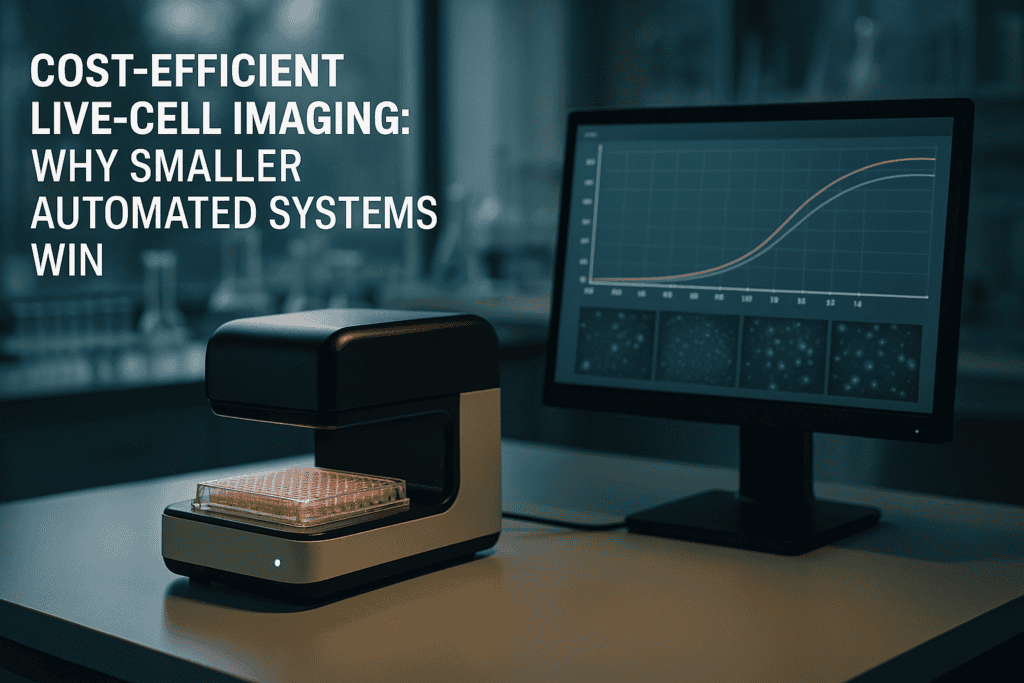
Cost-Efficient Live-Cell Imaging: Why Smaller Automated Systems Win
In the evolving landscape of modern cell culture research, the efficiency and accuracy of live-cell imaging have become paramount. As laboratories strive to improve outcomes while optimizing resources, the need for cost-efficient solutions has gained significant attention. In this article, we will explore how smaller automated systems offer a promising solution to common lab challenges, particularly in the context of continuous live-cell monitoring. From understanding the inherent limitations of traditional methods to assessing the benefits of compact innovations, we’ll dive into the transformative potential of these advanced technologies.
Herausforderungen traditioneller Live-Cell-Imaging-Ansätze
Resource Intensiveness and Limited Flexibility
Traditional live-cell imaging systems have long been the cornerstone of cellular research. However, these setups often come with inherent challenges that can hinder productivity in modern labs. High costs and significant spatial demands are among the most pressing issues. Such systems usually require dedicated microscopy rooms, complex handling, and frequent downtime due to maintenance needs.
- High initial and operational costs limit access for smaller labs.
- Space-intensive designs are impractical for labs with limited physical infrastructure.
- Complexity in operation necessitates specialized training and can lead to increased error rates.
Technologische Fortschritte und Trends in der Automatisierung
Evolution Toward Compact and Efficient Designs
The shift towards smaller, automated systems reflects a broader trend in laboratory automation aimed at enhancing flexibility and cost-efficiency. Recent advances prioritize adaptability, ease of use, and improved integration with existing lab workflows, which are crucial for addressing the limitations of conventional setups. Such systems enable continuous, uninterrupted monitoring without compromising on data quality.
- Enhanced automation reduces manual intervention, freeing up valuable researcher time.
- Compact designs support easy integration with existing laboratory equipment.
- Automation trends align with the push towards high-throughput screening and precision medicine.
Implementing Practical Workflows with Live-Cell Imaging
Improving Data Quality and Laboratory Throughput
By adopting smaller automated live-cell imaging systems, laboratories can streamline operations and elevate the quality of experimental data. Systems like the zenCELL owl, for example, facilitate continuous observation without the need for direct human intervention, thereby increasing reproducibility and reducing the potential for human error.
- Automated imaging systems enhance reproducibility through consistent environmental conditions.
- Systematic monitoring minimizes operator variability, a common issue in manual workflows.
- Data acquisition and analysis are expedited, facilitating more timely decision-making.
Lesen Sie weiter, um tiefere Einblicke und Strategien zu gewinnen.
“```html
Increasing Accessibility and Scalability
Deploying Cost-Effective Solutions for Every Lab Size
One of the standout advantages of adopting smaller automated live-cell imaging systems is the democratization of access to advanced research technologies. For many smaller labs and institutions, traditional systems are prohibitively expensive, both in terms of initial investment and ongoing operational costs. Compact systems like the zenCELL owl require a fraction of the budget traditionally needed, making cutting-edge imaging technologies accessible to a broader range of researchers. Additionally, these systems offer scalability, allowing labs to expand their capabilities in alignment with growth or increased research demand.
- Start with a single-unit system and scale up as research needs evolve.
- Utilize cost savings to fund additional research projects or personnel training.
Enhancing Data Consistency and Integrity
Maintaining High Standards Across Experiments
Data consistency and integrity remain critical challenges in live-cell imaging. Traditional systems often fall short due to variability in manual operations and environmental conditions. Conversely, automated compact systems provide a more controlled environment, minimizing fluctuations and external interferences. This stability is crucial for ensuring the quality and reliability of collected data, which can decisively influence research outcomes and conclusions.
- Implement standardized protocols to maintain consistency across experimental runs.
- Regularly calibrate devices to ensure optimal performance and data accuracy.
Reducing Carbon Footprint and Environmental Impact
Creating Greener Labs for a Sustainable Future
Environmental sustainability is becoming a pivotal focus in academia and industry alike. Smaller live-cell imaging systems contribute to greener labs by consuming significantly less power and occupying reduced space. Many of these systems are designed with minimalistic cooling requirements and lower energy consumption, which collectively lower the carbon footprint of a laboratory. Institutions such as the University of Cambridge have reported significant energy savings by transitioning to compact imaging systems.
- Conduct energy audits to identify further efficiency opportunities within labs.
- Adopt eco-friendly practices as part of a broader sustainability initiative.
Facilitating Remote Monitoring and Collaboration
Empowering Researchers Through Connectivity
The integration of remote monitoring capabilities into smaller systems has revolutionized how scientists collaborate and conduct research. With the ability to access live-cell imaging data in real time from any location, researchers can now make immediate adjustments and decisions, fostering enhanced collaboration across geographies and institutions. This connectivity is especially beneficial during instances when physical lab presence is not feasible, such as during travel or global events like the COVID-19 pandemic.
- Leverage cloud-based storage solutions for seamless data sharing and backup.
- Utilize collaboration tools for real-time data interpretation and teamwork.
Streamlining Training and Operational Procedures
Building a Skilled and Efficient Workforce
Compact live-cell imaging systems are designed with user-friendliness in mind, significantly lowering the learning curve compared to traditional systems. Many systems come equipped with intuitive interfaces and extensive online resources, facilitating smoother operational transitions. This ease of use not only enhances productivity but also reduces the likelihood of human error, thereby protecting the integrity of experimental outcomes.
- Incorporate structured training sessions into onboarding for new researchers.
- Regularly update staff on new system capabilities and software features.
Empowering Personalized and Precision Medicine
Driving Innovation in Healthcare Research
The marriage of compact automated imaging systems and precision medicine promises unprecedented advancements in healthcare. By enabling high-throughput screenings and detailed cellular analyses, these systems provide invaluable insights into patient-specific responses and drug effects. Institutions engaging in precision medicine research, like the Dana-Farber Cancer Institute, utilize such technologies to streamline patient data collection, resulting in more targeted and effective treatment strategies.
- Develop collaborations between imaging experts and clinical researchers.
- Utilize imaging data to personalize treatment regimens and improve patient outcomes.
Im Anschluss fassen wir die wichtigsten Erkenntnisse, Kennzahlen und eine wirkungsvolle Schlussfolgerung zusammen.
“`
“```html
Optimizing Workflow and Laboratory Management
Enhanced Efficiency and Productivity
Modern laboratories face unique challenges, not only in research but also in the effective management of resources. Smaller automated live-cell imaging systems redefine laboratory workflows by optimizing the use of space and resources. Their compact nature allows for flexible installations that can be tailored to the specific needs and constraints of any laboratory environment. This adaptability paves the way for streamlined processes and improved productivity across the board.
- Engage in continuous process improvement to maximize the benefits of compact systems.
- Optimize lab layouts to accommodate both current and anticipated future technologies.
Supporting Advanced Teaching and Education
Bridging Theory with Practical Application
In the educational sector, the practical application of theoretical knowledge is crucial for developing competent researchers. Compact live-cell imaging systems provide educational institutions with the tools necessary to bring cutting-edge science into the classroom. Their cost-effectiveness and ease of use enable integration into curricula, offering students firsthand experience with live-cell imaging techniques, which enhances learning outcomes and prepares them for future scientific careers.
- Integrate practical sessions with automated imaging into biology and life sciences curricula.
- Facilitate student-led research initiatives utilizing accessible imaging technologies.
Advancing Open Science and Data Sharing
Fostering Collaborative Research Environments
The open science movement emphasizes transparency, sharing, and collaboration. Compact imaging systems are ideally suited to support these goals by facilitating data sharing and cross-institutional collaborations. Their integration with cloud technologies and data storage solutions means data can be readily accessed and shared with researchers globally, promoting a more connected and collaborative scientific community that stands to accelerate breakthroughs and innovation.
- Adopt open-access policies for data generated using live-cell imaging systems.
- Encourage partnerships and collaborations across disciplines and institutions.
Schlussfolgerung
Throughout this article, we’ve explored the myriad benefits of adopting smaller, automated live-cell imaging systems in research settings. From democratizing access to advanced technologies to enhancing data consistency and integrity, these compact systems offer distinct advantages over traditional counterparts. By enabling researchers to increase accessibility, expand their capabilities, and streamline operational procedures, they play a pivotal role in advancing scientific inquiry.
The significance of reducing the carbon footprint and the push towards a more sustainable laboratory environment cannot be overstated. These systems not only consume less power and space but also offer efficiencies that contribute to broader institutional sustainability initiatives. Furthermore, with the integration of remote monitoring capabilities, they empower scientists to collaborate and make informed decisions regardless of their physical location, thereby embodying the adaptability required in today’s rapidly changing world.
The impact of these systems extends beyond efficiency and sustainability; they support the advancement of precision medicine, aid in educational endeavors, and promote a culture of open science. By making research tools accessible and encouraging inclusive collaboration, compact live-cell imaging systems open new frontiers in healthcare, education, and scientific discovery.
As we look to the future, the adoption of these technologies stands as a testament to progress and innovation. Laboratories worldwide, from small academic institutions to established research facilities, are poised to benefit immensely. By embracing these advancements, researchers can drive more impactful discoveries, contribute to a more sustainable planet, and shape the next generation of scientists.
Herein lies the call to action: whether in academia or industry, now is the time to invest in these transformative systems. Let’s push the boundaries of what’s possible, create meaningful impacts in our respective fields, and commit to sustainable and innovative research practices that promise a brighter future for science and society alike.
“`
The relationship between serum lipid content and cell signaling pathways
“```html
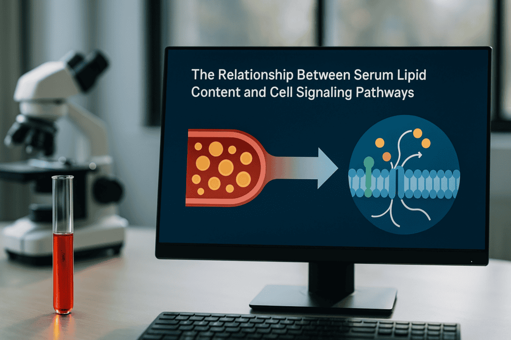
The Relationship Between Serum Lipid Content and Cell Signaling Pathways
The intricate dance between serum lipid content and cell signaling pathways plays a critical role in numerous physiological and pathological processes. Understanding this relationship informs researchers of how cellular functions, health, and disease mechanisms are influenced. This exploration will delve into the scientific principles behind lipid signaling, variability in serum lipid content, and potential experimental challenges researchers face. Moreover, it will elucidate the mechanisms through which lipids affect cell behavior, ensuring a comprehensive academic discussion relevant to professionals in cell culture and immunology.
The Biological Function of Serum Lipids
Understanding the Foundation
Serum lipids, including cholesterol, phospholipids, and triglycerides, are essential components of cellular function, particularly in modulating cell membrane structure and signaling. These lipids participate in creating lipid rafts, which are microdomains within the cell membrane that serve as platforms for cell signaling. As such, lipid rafts facilitate the congregation of receptors and signaling molecules, influencing diverse pathways crucial for cell communication and response to extracellular stimuli.
- Lipid rafts impact signal transduction by clustering receptors and associated proteins.
- Cholesterol is a key regulator of membrane fluidity and functionality.
- Phospholipids contribute to membrane curvature and cellular compartmentalization.
Lesen Sie weiter, um tiefere Einblicke und Strategien zu gewinnen.
Mechanisms of Lipid-Driven Cell Signaling Pathways
From Surface to Nucleus
Lipids influence cell signaling pathways by acting as secondary messengers or through direct interaction with receptors. For example, phosphatidylinositol 4,5-bisphosphate (PIP2) plays a crucial role in the inositol phosphate pathway, while diacylglycerol (DAG) acts as a cofactor in protein kinase C activation. Furthermore, lipids can modulate the activity of ion channels and thus indirectly affect intracellular signaling cascades that prompt transcriptional changes in the nucleus.
- PIP2 conversion to IP3 and DAG is pivotal in calcium signaling.
- DAG facilitates the activation of protein kinase C, pivotal for numerous cellular responses.
- Lipid influence on ion channels alters cellular excitability and cellular response.
Lesen Sie weiter, um tiefere Einblicke und Strategien zu gewinnen.
Variability and Quality Control in Serum Lipid Content
Challenges in Cell Culture Experiments
Variability in serum lipid content poses significant challenges in cell culture, affecting reproducibility and consistency in experimental outcomes. This variability can stem from lot-to-lot differences in animal-derived products, such as fetal bovine serum (FBS), or from human donor variability in human sera. Rigorous quality control, including lipid profiling and batch testing, is essential to mitigate the impact of these disparities. Understanding and documenting lipid content is vital for reproducibility.
- Careful selection of serum batches based on lipid content profiling reduces variability.
- Quality control measures support more reliable experimental outcomes.
- Documentation of serum characteristics ensures traceability and consistency.
Lesen Sie weiter, um tiefere Einblicke und Strategien zu gewinnen.
Implications for Research and Clinical Applications
Broadening Scientific Horizons
The relationship between serum lipid content and cell signaling pathways extends its implications into numerous research and clinical areas. In cancer research, for instance, disrupted lipid signaling pathways can lead to uncontrolled cell proliferation and tumor growth. In metabolic disorders, altered lipid profiles can exacerbate insulin resistance and inflammatory pathways. Understanding these complex interactions assists scientists in unraveling disease mechanisms and developing targeted therapeutic interventions.
- In cancer, targeting lipid signaling pathways offers new therapeutic strategies.
- In metabolic diseases, lipid modulation can affect disease progression.
- Advanced imaging and profiling technologies are invaluable for studying lipid functions.
Lesen Sie weiter, um tiefere Einblicke und Strategien zu gewinnen.
Advanced Monitoring Techniques
Live-Cell Imaging and Documentation
Advanced monitoring techniques such as incubator-compatible live-cell imaging systems, exemplified by solutions like the zenCELL owl, enable researchers to continuously observe the impact of serum lipids on cell behavior. These systems enhance reproducibility by documenting changes in real-time, providing insights into lipid-induced alterations in cellular activities and ensuring accurate, reliable data collection across multiple experimental runs.
- Real-time monitoring captures dynamic responses to lipid stimuli.
- Documenting cellular changes improves experimental repeatability.
- Long-term observation provides deeper insights into cell signaling dynamics.
Lesen Sie weiter, um tiefere Einblicke und Strategien zu gewinnen.
“`
“```html
Precision Lipid Quantification Techniques
The Cornerstone of Accurate Research
In the quest to decode the relationship between serum lipid content and cell signaling pathways, precision lipid quantification techniques serve as essential tools. Mass spectrometry (MS) and nuclear magnetic resonance (NMR) spectroscopy are at the forefront. Through MS, researchers can accurately detect and quantify lipid molecules, providing a molecular fingerprint of the serum lipids. This high sensitivity and specificity enable the detailed composition analysis necessary for reliable cell signaling studies.
- Deploy MS for precise identification and quantification of diverse lipid species.
- Leverage NMR spectroscopy for non-destructive, comprehensive lipid profiling.
- Employ internal standards to improve accuracy and reproducibility in measurements.
Integrating Systems Biology Approaches
Holistic Analysis of Lipid Interactions
The integration of systems biology approaches with lipid research enhances our understanding of complex biological networks. This interdisciplinary field combines genomics, proteomics, and metabolomics to provide a comprehensive view of how lipids interact in signaling pathways. By utilizing computational models, researchers can predict lipid behavior and interactions under varying physiological conditions, deepening insights into cellular processes and identifying potential therapeutic targets.
- Use computational models to simulate lipid involvement in signaling pathways.
- Apply integrative omics to understand lipid-mediated regulatory networks.
- Cross-validate data from multiple platforms for robust pathway analysis.
Case Studies in Lipidomics Applications
Trailblazing Research and Discoveries
Notable case studies have leveraged lipidomics in groundbreaking discoveries. For instance, the role of lipids in neurodegenerative diseases has been extensively studied, revealing how alterations in lipid metabolism can contribute to conditions like Alzheimer’s and Parkinson’s disease. Additionally, lipidomic analyses in cardiovascular research have uncovered lipid biomarkers that aid in early diagnosis and risk assessment, fostering preventive and personalized medicine.
- Analyze lipidomic data to identify biomarkers for neurodegenerative diseases.
- Implement lipid profiling in cardiovascular disease risk management.
- Use findings to develop targeted interventions and personalized treatment plans.
Advanced Imaging and Analytical Tools
Pioneering Technologies in Lipid Research
Advanced imaging and analytical tools, such as matrix-assisted laser desorption/ionization (MALDI) imaging and fluorescent lipid probes, have revolutionized lipid research. MALDI imaging allows spatial visualization of lipid distribution within tissues, offering insights into site-specific lipid alterations that might be missed by traditional methods. Fluorescent probes enable real-time tracking of lipid dynamics in live cells, providing critical data on lipid signaling pathways affected by stimuli.
- Use MALDI imaging for spatial analysis of lipid distributions in tissue samples.
- Employ fluorescent lipid probes for dynamic study of lipid cell signaling.
- Integrate imaging data with metabolic profiles for comprehensive analysis.
Role of Machine Learning in Lipidomics
Enhanced Data Interpretation and Prediction
Machine learning (ML) algorithms are becoming increasingly pivotal in interpreting complex lipidomic data sets. ML facilitates the identification of patterns and relationships within large-scale lipidomic data, enhancing predictive accuracy for disease progression or response to therapies. For example, ML models can identify lipid signatures linked to particular physiological states, allowing for early disease detection or the design of targeted therapeutic strategies.
- Utilize ML to process extensive lipidomic datasets efficiently.
- Apply predictive modeling to forecast lipid-related disease outcomes.
- Combine ML with biochemical validation for enhanced research insights.
Ethical and Biostatistical Considerations in Lipid Research
Ensuring Integrity and Accuracy
Ethical and biostatistical considerations are critical in lipid research involving human subjects. Ethical practices ensure that participant rights are respected, providing informed consent and guaranteeing confidentiality. Biostatistics, meanwhile, involves appropriate study design and data analysis techniques to draw valid conclusions from lipid research. Proper statistical methodologies, such as adequately powered studies and rigorous data validation techniques, are vital in avoiding biased or inaccurate findings.
- Implement ethical guidelines rigorously in human lipid studies.
- Adopt robust biostatistical methods for data analysis integrity.
- Ensure transparency and reproducibility in research findings.
Im Anschluss fassen wir die wichtigsten Erkenntnisse, Kennzahlen und eine wirkungsvolle Schlussfolgerung zusammen.
“`
“```html
Future Directions in Lipidomics
Charting New Frontiers
As lipidomics continues to evolve, future research avenues abound. The development of even more sensitive and specific instrumentation, along with cutting-edge computational tools, promises to overcome current analytical limitations. Moreover, the increasing integration of lipidomics with other “omics” fields, such as transcriptomics and proteomics, offers a more holistic understanding of lipid functions in health and disease. The future also holds potential for innovations in personalized medicine, where tailored lipid profiles provide specific recommendations for dietary and therapeutic interventions.
- Innovate new technologies to advance lipid detection sensitivity and specificity.
- Explore multi-omics strategies for comprehensive healthcare insights.
- Pioneer personalized approaches in lipid-focused healthcare.
Collaboration and Knowledge Sharing
Interdisciplinary Synergies in Lipid Research
The increasingly collaborative nature of lipid research underscores the importance of interdisciplinary alliances. By merging expertise from molecular biology, chemistry, data science, and clinical medicine, researchers can pool resources and insights to tackle complex lipid-related challenges. Initiatives such as multi-institutional research projects and international symposia provide platforms for knowledge exchange, fostering innovations that are unattainable in isolated efforts. Furthermore, open-access repositories and databases are essential for ensuring that data and findings are available to the global scientific community.
- Encourage interdisciplinary collaborations for groundbreaking research.
- Facilitate knowledge sharing through international forums and symposia.
- Contribute to open-access data banks for widespread scientific accessibility.
Schlussfolgerung
In this exploration of the relationship between serum lipid content and cell signaling pathways, we have traversed the intricate landscape of lipidomics and its profound impact on modern biomedical research. Key takeaways highlight the indispensable role of precision lipid quantification techniques such as mass spectrometry and nuclear magnetic resonance spectroscopy, which enable detailed and accurate analysis of lipid molecules. Furthermore, the pivotal integration of systems biology and advanced imaging tools, alongside machine learning, marks an era of unprecedented insights into lipid functions.
The relevance of this article rests not only in showcasing the current methodologies and applications but also in shaping future research directions through a collaborative, multi-disciplinary approach. As lipidomics continues to intertwine with various scientific domains, the potential to unravel new therapeutic targets and pave the path for precision medicine becomes increasingly attainable. The continuous progression in analytical technology and data interpretation methods promises to enhance our understanding of complex lipid networks in human health and disease.
As we close, it is imperative to acknowledge the ethical and biostatistical considerations that ensure research integrity. The commitment to rigorous methodological standards preserves the validity and applicability of research findings, fostering trust and progression in the field. The journey of lipidomics is a testament to the power of innovation and collaboration, where each discovery fuels the engine of insight, drawing us closer to a future where lipid profiles guide personalized health strategies.
We invite you to join this vibrant community of researchers and practitioners aiming to decode the secrets of lipids for better health outcomes. Stay engaged, contribute to open-data platforms, and champion the integration of multi-omics for a comprehensive understanding of life’s subtle intricacies. Together, we can forge pathways to transformative health interventions, making a lasting impact on scientific and medical landscapes worldwide.
“`
Identifizierung sub-optimaler Medien- und Kulturbedingungen mittels kontinuierlicher Bildgebung

“```html
Identifizierung sub-optimaler Medien- und Kulturbedingungen mittels kontinuierlicher Bildgebung
Fortschritte in der Zellkulturforschung haben dank technologischer Innovationen in der Echtzeit-Zellbildgebung und Laborautomatisierung eine neue Ära der biologischen Entdeckung eingeläutet. Die Identifizierung suboptimaler Medien und Kulturzustände ist für den experimentellen Erfolg, die Reproduzierbarkeit und die solide Dateninterpretation von größter Bedeutung. Dieser Artikel untersucht die Feinheiten dieser Prozesse, wobei die kontinuierliche Bildgebung im Vordergrund moderner Techniken zur Verbesserung der Genauigkeit zellulärer Studien und der Laboreffizienz steht.
Herausforderungen und Grenzen traditioneller Ansätze
Die Komplexität von Zellkultur-Bedingungen
Zellkultur ist ein unverzichtbares Werkzeug in der biologischen Forschung und ermöglicht die Untersuchung zellulärer Mechanismen, die Medikamentenentwicklung und die Erforschung therapeutischer Interventionen. Trotz ihres weit verbreiteten Nutzens sind traditionelle Zellkulturanäuren oft zahlreichen Herausforderungen ausgesetzt, die experimentelle Ergebnisse beeinträchtigen können. Dies ist vor allem die Schwierigkeit, optimale Medien und Kulturbedingungen über längere Zeiträume aufrechtzuerhalten, was häufig zu Zellstress oder -tod führt.
- Zellkulturmedien können toxische Metaboliten verbrauchen oder anreichern, was die Zellviabilität beeinträchtigt.
- Die manuelle Überwachung ist zeitaufwendig und fehleranfällig.
- Mangelnde kontinuierliche Überwachung führt zu verpassten kritischen Ereignissen oder verzögerten Reaktionen auf Zellzustandsänderungen.
Grenzen der manuellen Beobachtung
Die Abhängigkeit von sporadischer manueller Intervention zur Überwachung von Zellkulturen erhöht die Wahrscheinlichkeit, subtile, aber signifikante Veränderungen der Zellgesundheit oder des Zellverhaltens zu übersehen. Dies beeinträchtigt nicht nur die Reproduzierbarkeit, sondern behindert auch das übergeordnete Ziel des wissenschaftlichen Fortschritts durch zuverlässige Daten.
- Interventionen sind aufgrund seltener Beobachtungen meist reaktiv und nicht proaktiv.
- Schwankungen in menschlichen Bewertungen führen zu inkonsistenten Dateninterpretationen.
Lesen Sie weiter, um tiefere Einblicke und Strategien zu gewinnen.
“`
(Hinweis: Der Rest des Artikels, einschließlich technologischer Fortschritte und Automatisierungstrends bis hin zur Zusammenfassung, sollte in ähnlicher umfassender Weise fortgesetzt werden, wobei die empfohlenen primären und sekundären Schlüsselwörter für die SEO-Optimierung natürlich in den Text integriert werden.)
“```html
Technologische Fortschritte in der kontinuierlichen Bildgebung
Wegweisende Automatisierung in der Zellkultur
Die Einführung kontinuierlicher Bildgebungstechnologien hat zu einer signifikanten Transformation von Zellkulturmethoden geführt und viele Einschränkungen traditioneller Praktiken behoben. Durch Echtzeit-Überwachungssysteme können Forscher nun ein beispielloses Maß an Konsistenz und Präzision bei der Bewertung von Zellgesundheit und -verhalten erreichen.
Die kontinuierliche Bildgebung nutzt automatisierte bildgebende Systeme, die in der Lage sind, Zeitraffersequenzen von Zellkulturen aufzunehmen, und liefert wertvolle Einblicke in zelluläre Prozesse. Systeme wie IncuCyte S3 und Lionheart FX von BioTek automatisieren diesen Prozess und liefern hochauflösende Bilder, ohne die Kulturbedingungen zu stören. Diese Integration ermöglicht eine eingehende Analyse und dynamischere experimentelle Ansätze.
- Nutzen Sie automatisierte Bildgebung für eine konsistente Datenerfassung mit minimalem menschlichen Eingriff.
- Nutzen Sie die umfangreichen, kontinuierlichen Datensätze für prädiktive Analysen und zeitnahe Interventionsstrategien.
Echtzeit-Datenanalyse und -interpretation
Maximierung von Erkenntnissen mit fortschrittlicher Software
Neben Bildgebungs-Hardware sind hochentwickelte Softwareplattformen entscheidend für die Verarbeitung, Analyse und Interpretation der riesigen Datenmengen. Diese Plattformen nutzen Algorithmen des maschinellen Lernens, um Zellverhalten zu quantifizieren, Anomalien zu erkennen und zelluläre Reaktionen unter verschiedenen Bedingungen zu modellieren.
Beispielsweise bieten Software-Tools wie ImageJ, CellProfiler und Gen5 benutzerfreundliche Schnittstellen mit leistungsstarken Analysefunktionen, die es Forschern ermöglichen, komplexe Analysen wie die Beurteilung der Zellkonfluenz, morphologische Studien und die dynamische Überwachung der Proteinexpression durchzuführen.
- Integrieren Sie umfassende Datenanalysetools, um Skalierbarkeit und Genauigkeit in der Zellforschung zu verbessern.
- Ermöglichen Sie maschinellen Lernframeworks, nicht sichtbare Trends und prädiktive Muster in zellulären Dynamiken zu identifizieren.
Optimierung von Versuchsplanung und -durchführung
Proaktive Methoden zur verbesserten Experimentation
Die kontinuierliche Bildgebung spielt nicht nur bei der Datenerfassung eine entscheidende Rolle, sondern auch bei der Optimierung von Versuchsdesigns. Durch die Möglichkeit, Zellen kontinuierlich zu überwachen, können Forscher Interventionen effektiver planen und somit das Risiko unvorhergesehener experimenteller Fehlschläge reduzieren.
Beispielsweise kann die genaue Bestimmung von Zeitpunkten für Medienwechsel oder Medikamentenzugabe in einem Kulturversuch die Ergebnisse erheblich beeinflussen. Durch die Analyse von Wachstumstrends und metabolischen Veränderungen können Wissenschaftler Experimentierprotokolle besser auf die untersuchten biologischen Prozesse abstimmen.
- Entwickeln Sie präzise Interventionsprotokolle auf Basis von Echtzeitdaten und prädiktiver Analytik.
- Gewährleisten Sie Flexibilität im experimentellen Design, um schnelle Veränderungen des Zellzustands oder Verhaltens zu berücksichtigen.
Kommerzielle Werkzeuge für kontinuierliche Bildgebung und Überwachung
Ein genauerer Blick auf branchenführende Ausrüstung
Eine Vielzahl von kommerziell erhältlichen Werkzeugen hat Industriestandards gesetzt, indem sie kontinuierliche Bildgebung und erweiterte Überwachungsfähigkeiten ermöglichen. Diese Werkzeuge sind unerlässlich, um zuverlässige Daten und Konsistenz in Laboreinrichtungen zu erreichen.
Zu den bemerkenswerten Innovationen gehört die Cell-IQ-Plattform, die mit ihrer intuitiven Software und robusten Bildgebungsmöglichkeiten anpassbare Experimentierrahmen ermöglicht. Ein weiteres Beispiel ist das Livecyte-System, das für seine markierungsfreie Bildgebung bekannt ist und es Forschern ermöglicht, native Zellzustände ohne künstliche Störungen zu beobachten.
- Erkunden Sie Ausstattungsoptionen wie Cell-IQ für integrierte Umweltkontrolle und optimierte Bildgebungs-Workflows.
- Bewerten Sie die Kompatibilität des Geräts mit der vorhandenen Laborinfrastruktur, um die Forschungseffizienz zu maximieren.
Integration kontininuierlicher Bildgebung in die Wirkstoffforschung
Verbesserung der Arzneimittelentwicklung durch vertiefte zelluläre Erkenntnisse
Die kontinuierliche Bildgebung ist besonders transformativ in der Wirkstoffforschung, wo die genaue Überwachung zellulärer Reaktionen auf therapeutische Verbindungen von entscheidender Bedeutung ist. Dieser Ansatz ermöglicht es Forschern, Echtzeit-Wirkstoffeffekte zu beobachten und Einblicke in Wirksamkeit, Zytotoxizität und potenzielle Resistenzmechanismen zu gewinnen.
In der Praxis beschleunigt das Hochdurchsatz-Screening in Kombination mit kontinuierlicher Bildgebung die Evaluierung zahlreicher Verbindungen und ermöglicht die schnelle Identifizierung vielversprechender Kandidaten. Beispielsweise bietet der Einsatz von Real-Time Cell Analysis (RTCA)-Systemen detaillierte kinetische Profile von Wirkstoff-Zell-Interaktionen und verbessert dadurch signifikant die Effizienz der Medikamentenentwicklungspipeline.
- Implementieren Sie Hochdurchsatz-Bildgebungssysteme zur schnellen und groß angelegten Erprobung und Bewertung der Arzneimittelwirksamkeit.
- Nutzen Sie kinetische Daten, um langfristige Auswirkungen von Wirkstoffverbindungen auf Zielzelllinien vorherzusagen.
Fallstudien: Erfolgreiche Implementierungen und Erkenntnisse
Lehren führender Forschungseinrichtungen
Fallstudien von wegweisenden Forschungseinrichtungen veranschaulichen den tiefgreifenden Einfluss kontinuierlicher Bildgebung auf den experimentellen Erfolg. Institutionen wie das Scripps Research Institute haben diese Techniken integriert, um Studien zur Gesundheit von Nervenzellen zu unterstützen und zentrale Daten zu neurodegenerativen Prozessen zu erfassen.
In diesen Fallstudien hat die Fähigkeit, die Neurogenese in Echtzeit zu verfolgen, zu einem tieferen Verständnis der zellulären Signalmechanismen geführt, was Auswirkungen auf die Entwicklung von Behandlungen für Krankheiten wie Alzheimer und Parkinson hat.
- Überprüfen Sie institutionelle Fallstudien zu innovativen Anwendungen von kontinuierlicher Bildgebungstechnologie.
- Wenden Sie interdisziplinäre Erkenntnisse an, um Forschungshorizonte und kooperative Bemühungen zu erweitern.
Im Anschluss fassen wir die wichtigsten Erkenntnisse, Kennzahlen und eine wirkungsvolle Schlussfolgerung zusammen.
“`
“```html
Identifizierung sub-optimaler Medien- und Kulturbedingungen mittels kontinuierlicher Bildgebung
Kontinuierliches Feedback für optimale Umgebungsbedingungen
Die kontinuierliche Bildgebungstechnologie dient als entscheidendes Werkzeug zur rechtzeitigen Identifizierung suboptimaler Medien- oder Kulturbedingungen. Durch Echtzeit-Visualisierung können Forscher frühe Anzeichen unerwünschter Veränderungen der Zellmorphologie, der Proliferationsraten oder der Stoffwechselaktivität erkennen. Dieser proaktive Ansatz ermöglicht Untersuchungen der unmittelbaren Auswirkungen von variierenden pH-Werten, Nährstoffverarmung oder Anreicherung toxischer Metaboliten und verbessert dadurch die Gesamtqualität von Zellkulturen.
Die Fähigkeit, schnell auf diese Beobachtungen zu reagieren, könnte die Gesundheit und Lebensfähigkeit von Zellkulturen direkt beeinflussen und die Häufigkeit experimenteller Fehler, die auf Umweltschwankungen zurückzuführen sind, reduzieren. Technologien, die Echtzeit-Bildgebung mit Umweltsensoren koppeln, ermöglichen Wissenschaftlern ein umfassenderes Verständnis der internen Kulturbedingungen und unterstützen die Aufrechterhaltung idealer Bedingungen für zelluläre Experimente.
- Richten Sie eine Feedbackschleife ein, die kontinuierliche Bildgebung und Umgebungsdaten für optimale Kulturbedingungen einbezieht.
- Nutzen Sie Bildgebungsdaten, um Kulturparameter anzupassen und die Zellwachstumsdynamik zu verfeinern.
Integration von kontinuierlicher Bildgebung über multidisziplinäre Plattformen hinweg
Brückenschlag zwischen diversen Forschungsbereichen mit einheitlicher Technologie
Die interdisziplinäre Anwendbarkeit kontinuierlicher Bildgebung erweitert ihre Vorteile in verschiedenen Bereichen der wissenschaftlichen Forschung. Durch die nahtlose Integration mit Plattformen, die diverse biologische Systeme handhaben, fördert diese Technologie kollaborative Erkundungen in den Bereichen Onkologie, Immunologie, Neurologie und regenerative Medizin.
In der Krebsforschung unterstützt die kontinuierliche Bildgebung die Visualisierung von Tumorzellinvasion und Metastasierung in vitro und liefert entscheidende Hinweise auf die Krebsentstehung. Immunologische Studien profitieren von der Beobachtung von Live-Interaktionen zwischen T-Zellen und Krankheitserregern, was die Impfstoffentwicklung verbessert. Gleichzeitig werden Erkenntnisse über die Differenzierung von Stammzellen durch Zeitrafferbildgebung erheblich bereichert, was sich auf Protokolle der regenerativen Medizin im Hinblick auf das Tissue Engineering auswirkt.
- Etablieren Sie kontinuierliche Bildgebung in facettenreichen Forschungsbereichen, um umfassende biologische Einblicke zu fördern.
- Fördern Sie abteilungsübergreifende Kooperationen, indem Sie fortschrittliche bildgebende Systeme als gemeinsames Analysewerkzeug nutzen.
Schlussfolgerung
Zusammenfassend lässt sich sagen, dass die rasante Entwicklung und Implementierung kontinuierlicher Bildgebungstechniken die moderne biologische Forschung und experimentelle Arbeit maßgeblich verändert haben. Durch die Bereitstellung von Echtzeiteinblicken in zelluläre Dynamiken ermöglichen diese Werkzeuge Forschenden, genauere, konsistentere und effizientere Experimente durchzuführen. Die kontinuierliche Sammlung umfangreicher Datensätze ermöglicht die Extraktion kritischer Informationen über die Zellgesundheit, die Wirksamkeit von Medikamenten und die Anpassungsfähigkeit der Kulturanbringung.
Die Integration mit fortschrittlicher Software und Machine-Learning-Frameworks vertieft die analytische Tiefe weiter und ermöglicht die Identifizierung bisher unerkannter Muster und prädiktiver Indikatoren. Der unschätzbare Beitrag kontinuierlicher Bildgebung bei der experimentellen Planung und der Echtzeitüberwachung zellulärer Reaktionen unterstreicht ihre Rolle als Eckpfeiler des wissenschaftlichen Fortschritts.
Diese Technologie dient über die Verbesserung der experimentellen Präzision hinaus als Brücke zwischen verschiedenen wissenschaftlichen Disziplinen und fördert Innovation und Zusammenarbeit. Die Fähigkeit, unterschiedliche Forschungsbereiche mit einer gemeinsamen technologischen Plattform zu vereinen, beschleunigt die Überführung von Laborergebnissen in reale Anwendungen und unterstützt Fortschritte in der Krebstherapie, Immunologie, Neurologie und regenerativen Medizin.
Da wir weiterhin die Geheimnisse des Zellverhaltens entschlüsseln, bleibt die Bedeutung kontinuierlicher Bildgebung unbestreitbar. Für die Forschungsgemeinschaft und darüber hinaus wird die Einführung und Integration dieser fortschrittlichen Technologien nicht nur die wissenschaftliche Erforschung verfeinern, sondern auch neue Bereiche des Verständnisses erschließen. Lasst uns diese Innovationen annehmen, ihr Potenzial nutzen und transformative Fortschritte auf dem Gebiet der Biowissenschaften vorantreiben.
“`
Was KI in der modernen Live-Zell-Bildgebung tun kann (und nicht tun kann)

“```html
Was KI in der modernen Live-Zell-Bildgebung tun kann (und nicht tun kann)
In the evolving landscape of cell culture research, live-cell imaging has emerged as a cornerstone technology. As researchers strive for deeper insights into cellular processes, the integration of artificial intelligence (AI) into live-cell imaging provides a promising avenue for advancement. But what AI can (and cannot) do in modern live-cell imaging presents both opportunities and challenges. This article will delve into the significance of AI in this context, common challenges with traditional methods, and how technology and smart systems like the zenCELL owl are paving the way to more reliable and efficient cell analysis.
Häufige Herausforderungen und Beschränkungen traditioneller Ansätze
The Struggle with Manual Observation
Traditional live-cell imaging techniques have long faced limitations that impact the efficiency and accuracy of research outcomes. Manual observation, although foundational, is fraught with variability and subjectivity, reducing reproducibility across experiments. Furthermore, the sheer volume of data generated can overwhelm manual processes, leading to potential oversight of critical information.
- Manual observation often leads to inconsistent results.
- High data volumes are challenging to handle without automation.
- Variability in results impacts reproducibility.
Technical Constraints
Cell culture studies are highly reliant on precise environmental conditions, and traditional imaging setups often struggle to maintain these parameters consistently. This not only affects the health of the cells but also the quality of the data captured. Moreover, the resolution limits of older imaging systems can obscure crucial cellular events that could otherwise offer significant insights.
- Inability to maintain consistent environmental conditions.
- Resolution constraints limit data quality and insights.
Technologische Fortschritte und Automatisierungstrends
Integration of AI in Imaging and Data Analysis
The integration of AI in live-cell imaging is revolutionizing the way data is collected and analyzed. AI algorithms can swiftly process and interpret vast amounts of imaging data, identifying patterns and anomalies that might elude manual analysis. This not only enhances the accuracy of observations but also speeds up data processing.
- AI improves data accuracy and speeds up processing times.
- Automated analysis reduces subjective interpretation.
Automation in Workflow Processes
Automation is increasingly reshaping laboratory workflows, significantly elevating efficiency and precision. Automated live-cell imaging systems, like the zenCELL owl, allow for continuous monitoring of cultures directly within incubators, minimizing disturbances that could affect cell behavior. This transition to automated systems streamlines workflows and enhances reproducibility, by ensuring consistent data collection.
- Continuous monitoring minimizes data gaps.
- Automated systems enhance reproducibility and reliability.
Lesen Sie weiter, um tiefere Einblicke und Strategien zu gewinnen.
“`
“```html
Enhanced Image Resolution via AI Techniques
Deep Learning Models Transforming Imaging Resolution
AI techniques, particularly deep learning models, have significantly enhanced image resolution in live-cell imaging. These sophisticated models can improve upon the resolution of traditional imaging systems by using algorithms to reconstruct fine details lost in lower-quality images. For instance, convolutional neural networks (CNNs) can mitigate noise and identify intricate patterns, resulting in clearer and more insightful cellular images that were previously unattainable.
- Utilize deep learning models to achieve higher image resolution and clarity.
Real-Time Monitoring and Immediate Feedback
Immediate Insights for Dynamic Cellular Processes
Real-time data collection forms the backbone of advanced live-cell imaging powered by AI. Systems equipped with AI can analyze images as they are captured, providing immediate feedback to researchers. This is crucial for monitoring dynamic cellular processes such as cell division, apoptosis, and migration. An example is the use of AI algorithms in fluorescence microscopy to provide real-time tracking and quantification of cellular behaviors, allowing scientists to make prompt decisions.
- Implement AI-driven real-time monitoring for immediate feedback on cellular processes.
AI-Powered Predictive Analytics
Forecasting Cellular Responses and Behaviors
Predictive analytics driven by AI allows researchers to anticipate cellular responses to various stimuli. By analyzing historical data and identifying patterns through machine learning, AI can forecast cell growth rates, the likelihood of mutation occurrence, or expected responses to treatment conditions. For example, AI systems have been employed in cancer research to predict tumor progression and response to therapies.
- Leverage AI for predictive analytics to foresee and prepare for potential cellular outcomes.
Streamlining Data Management and Storage
Efficient Handling of Vast Datasets
The sheer volume of data generated by live-cell imaging requires advanced management and storage solutions. AI simplifies this process by automating data classification, archiving, and retrieval. AI-powered platforms can categorize images and related metadata, ensuring quick and efficient access to required datasets, which supports better data-driven decision-making. Systems such as cloud-based AI solutions provide scalable data storage options, which are essential for large-scale research operations.
- Adopt AI solutions for efficient data management and scalable storage capabilities.
AI-Driven Detection of Cellular Anomalies
Spotting the Unseen with Machine Learning
One of the greatest assets of AI in live-cell imaging is its ability to detect anomalies that may not be visually apparent to human observers. Machine learning models can identify irregularities or early markers of disease that are critical for early diagnosis and intervention. For instance, AI has been instrumental in identifying subtle changes in cell structure or growth patterns indicative of pathology.
- Use AI to enhance the detection of cellular anomalies for early intervention.
Optimizing Workflow Efficiency through AI
AI as a Catalyst for Laboratory Productivity
Integrating AI into laboratory workflows optimizes both speed and precision of tasks. Automated scheduling, sample handling, and results analysis free researchers from routine tasks and reduce human error. This allows scientists to focus on high-level research and complex analyses. Tools such as AI-based imaging stations automate entire laboratory cycles, from sample preparation to data analysis, offering a streamlined and efficient research process.
- Incorporate AI to automate and streamline laboratory workflows for enhanced productivity.
Case Study: The zenCELL owl Implementation
Real-World Application and Outcomes
The zenCELL owl exemplifies the practical application of AI in live-cell imaging. This system enables continuous monitoring and analysis directly within incubation environments, ensuring minimal disturbance to cell cultures. Researchers using the zenCELL owl have reported increased data reliability and a marked reduction in manual intervention, resulting in enhanced cell health and more accurate results. Furthermore, the zenCELL owl’s AI capabilities allow it to identify cell confluency, detect anomalies, and perform automated data logging, revolutionizing workflow efficiency and accuracy.
- Consider real-world examples like the zenCELL owl for proven AI integration success.
Im Anschluss fassen wir die wichtigsten Erkenntnisse, Kennzahlen und eine wirkungsvolle Schlussfolgerung zusammen.
“`
“```html
The Role of AI in Enhancing Data Accuracy
Improving Reliability through Algorithmic Precision
AI’s precision and consistency have dramatically improved data accuracy in live-cell imaging. Sophisticated algorithms minimize human error by automating many stages of data collection and analysis. For instance, AI solutions can accurately calibrate imaging equipment, fine-tune settings in real-time as conditions change, and auto-correct for any deviations. The consistency AI provides ensures that datasets are not only accurate but also readily reproducible across multiple experiments or conditions.
- Employ AI to ensure data accuracy and reproducibility in live-cell imaging.
AI and Cost-Efficiency in Imaging Research
Unlocking Financial Benefits through Automation
By implementing AI, laboratories can significantly reduce operational costs associated with live-cell imaging. Automation leads to less manual labor, lower error rates, and fewer resource-intensive practices. AI systems, such as predictive maintenance tools, prevent costly equipment failures by forecasting when repairs are necessary. Moreover, AI’s ability to quickly process and analyze large volumes of data allows researchers to achieve more with fewer resources, translating into better financial efficiency and resource allocation.
- Utilize AI for more cost-effective live-cell imaging practice.
AI’s Journey in Live-Cell Imaging
Continuing Innovations and Future Prospects
The application of AI in live-cell imaging is continually evolving. Future developments are poised to further refine imaging accuracy and broaden analytical capabilities. As AI technology advances, we anticipate more sophisticated machine learning models capable of uncovering new insights from complex cellular dynamics. These advancements will likely foster a deeper understanding of biological processes, paving the way for breakthroughs in diagnostics and therapeutics. The potential of AI to transform live-cell imaging and biomedical research remains boundless, driving the ongoing quest for scientific innovation.
- Stay informed on AI advancements to leverage new opportunities in live-cell imaging.
Schlussfolgerung
AI has undoubtedly revolutionized live-cell imaging by enhancing resolution, providing real-time monitoring, and offering predictive insights into cellular processes. By facilitating efficient data management and detecting cellular anomalies, AI aids researchers in making more informed and timely decisions. These technologies streamline research workflows and improve laboratory productivity, showcasing the broad impact of AI on scientific investigation and medical research.
This article has highlighted AI’s potential to improve accuracy, reduce costs, and drive future innovations. By automating numerous routine and data-intensive tasks, AI not only elevates the reliability of imaging results but also empowers researchers to focus on critical analytical processes and groundbreaking discoveries. The integration of AI into live-cell imaging represents a significant leap forward, promising to accelerate scientific understanding in profound ways.
As AI continues to develop, its potential to further reshape live-cell imaging expands, promising even greater enhancements in data precision and research efficiency. Laboratories and research institutions that adopt AI solutions stand at the forefront of innovation, ready to decipher the complexities of biological systems with unprecedented clarity and depth.
In embracing these advancements, we look toward a future where the full potential of AI in live-cell imaging is realized, unlocking new dimensions of understanding in cell biology and beyond. For researchers, academics, and industry professionals, the message is clear: now is the time to harness AI’s power to achieve transformative progress in live-cell imaging. Let us continue to explore and integrate these technologies to drive the next wave of discovery.
“`
Regulatorische Aspekte (FDA/CE) für Platten-OEMs

Regulatorische Aspekte (FDA/CE) für Platten-OEMs
The landscape of life science research is rapidly evolving, particularly within sectors dedicated to biotechnological and pharmaceutical advancements. Consequently, the regulatory aspects surrounding the manufacturing and distribution of laboratory plates for OEMs (Original Equipment Manufacturers) are garnering significant attention. As laboratories and manufacturers strive to maintain compliance with global quality standards, understanding the regulatory aspects set forth by the FDA and CE becomes essential. This article delves into these regulatory frameworks, focusing on their implications for plate OEMs, and provides an authoritative overview tailored for researchers, lab managers, and biotech professionals.
Understanding FDA and CE Regulatory Frameworks
The Role of FDA and CE Marking
The Food and Drug Administration (FDA) and CE (Conformité Européenne) marking collectively represent pivotal regulatory frameworks responsible for ensuring product safety, quality, and effectiveness in life science manufacturing. For plate OEMs, these regulations provide guidance and standards for material and process specifications, influencing everything from initial design to final production.
- The FDA regulates medical devices, ensuring they meet stringent safety standards before reaching the U.S. market.
- CE marking, mandatory for the European Economic Area (EEA), signifies that products comply with EU safety, health, and environmental protection standards.
Key Requirements for Regulatory Compliance
Material Compliance and Testing
Adhering to FDA and CE standards begins with selecting appropriate materials for manufacturing laboratory plates. Materials like polystyrene (PS), polypropylene (PP), and cyclic olefin copolymer (COC) are often favored due to their compatibility with biological and chemical assays. However, each material must undergo rigorous testing to ascertain its suitability under the regulatory frameworks.
- Ensure materials are classified as biocompatible and non-toxic, complying with ISO 10993 standards.
- Continuous testing for endotoxin levels and cytotoxicity is crucial to maintain compliance.
Design-for-Manufacturing (DFM) and Process Control
Emphasizing Design Efficiency
Design-for-Manufacturing principles play a critical role in ensuring compliance with FDA and CE regulations. By optimizing designs for manufacturability, OEMs not only enhance production efficiency but also streamline regulatory approvals. Prototyping strategies, including 3D printing and pilot production, facilitate the evaluation of design feasibility before full-scale deployment.
- Evaluate the impact of design choices on regulatory compliance.
- Utilize rapid prototyping to identify potential design flaws early.
Injection Molding and Consistency
Injection molding is the preferred technology for large-scale production of laboratory plates due to its consistency and precision. For OEMs, meeting FDA and CE standards implies establishing stringent process controls, including process validation, critical parameter monitoring, and detailed documentation for traceability.
- Maintain batch-to-batch consistency, minimizing variability.
- Implement robust process control systems for regulatory compliance.
GMP Compliance in Manufacturing
Managing cGMP and GMP Principles
Good Manufacturing Practice (GMP) compliance is non-negotiable for OEMs operating within regulated environments. The distinction between development and cGMP/GMP manufacturing environments involves stringent quality management systems, extensive batch documentation, and a comprehensive approach to risk management.
- Implement thorough qualification and validation protocols.
- Ensure change control processes are in place to manage amendments effectively.
Lesen Sie weiter, um tiefere Einblicke und Strategien zu gewinnen.
“```html
Documenting Quality Systems and Compliance
Implementing Robust Quality Management Systems (QMS)
For plate OEMs, adhering to FDA and CE regulations demands a sophisticated approach to quality management. Establishing a comprehensive Quality Management System (QMS) provides a structural framework that fosters compliance and continuous improvement. This involves documenting policies, procedures, and responsibilities, ensuring an organization-wide commitment to quality. A QMS not only assists in maintaining regulatory compliance but also enhances product reliability, customer satisfaction, and market competitiveness.
- Develop a comprehensive QMS aligned with ISO standards such as ISO 9001 or ISO 13485.
Training and Workforce Development
Empowering Employees with Essential Skills
A well-trained workforce is the backbone of regulatory compliance. Continuous education and training programs are crucial for ensuring that employees understand and effectively implement FDA and CE requirements. For example, training on good manufacturing practices (GMP), Standard Operating Procedures (SOPs), and the importance of quality in production fosters an environment where compliance is ingrained in day-to-day operations. Additionally, staying updated with regulatory changes is essential, as it equips employees with the knowledge to adapt to new directives.
- Invest in regular training sessions to keep employees informed about the latest regulatory updates.
Leveraging Advanced Technologies for Compliance
Integrating Technological Innovations
Embracing cutting-edge technologies can significantly streamline compliance efforts. Automation and digital tools such as Manufacturing Execution Systems (MES) and Enterprise Resource Planning (ERP) software offer real-time data insights and process control, ensuring adherence to stringent regulatory standards. Moreover, traceability is enhanced through blockchain technology, providing immutable records that facilitate audits and compliance verification.
- Implement digital tools for real-time monitoring and reporting of compliance metrics.
Outsourcing Strategies and Vendor Management
Selecting and Managing Compliant Partners
Engaging with third-party vendors is a common practice among plate OEMs to optimize production and reduce costs. However, outsourcing introduces additional compliance challenges. Establishing a comprehensive vendor management system is crucial for ensuring that partners adhere to FDA and CE standards. This involves conducting detailed due diligence, auditing vendor processes, and maintaining effective communication channels to ensure alignment with compliance objectives.
- Perform regular audits and assessments of vendor facilities to ensure compliance.
Documentation and Record Maintenance
Ensuring Traceability and Accountability
Proper documentation is fundamental to achieving compliance with FDA and CE regulations. Documentation systems must be robust, capturing all aspects of the production process from material selection to final inspection. This includes maintaining detailed records of quality tests, validation reports, and batch data, which become essential during regulatory audits and customer inquiries.
- Utilize electronic document management systems for efficient storage and retrieval of compliance records.
Auditing and Continuous Improvement
Implementing Effective Auditing Practices
Audit processes play a pivotal role in uncovering areas where compliance can be fortified. Regular internal and external audits provide insights into operational effectiveness and identify gaps in compliance practices. Through effective auditing, plate OEMs can not only meet regulatory requirements but also drive continuous improvement. Integrating feedback loops and corrective actions from audit findings enhances overall operational efficacy and regulatory compliance.
- Conduct regular compliance audits to ensure adherence to regulatory standards and identify areas for improvement.
Risk Management and Mitigation
Addressing and Anticipating Compliance Risks
Risk management is an integral component of regulatory compliance for plate OEMs. Identifying potential risks and implementing mitigation strategies helps in safeguarding against compliance breaches. Common risk management frameworks include failure mode and effects analysis (FMEA) and hazard analysis and critical control points (HACCP), which provide systematic approaches to identifying and addressing potential hazards. By rigorously applying these frameworks, OEMs can minimize risks and enhance compliance resilience.
- Develop a proactive risk management plan incorporating regular reviews and updates.
Im Anschluss fassen wir die wichtigsten Erkenntnisse, Kennzahlen und eine wirkungsvolle Schlussfolgerung zusammen.
“`
“```html
Communication and Collaboration
Fostering an Environment of Open Dialogue
The importance of effective communication within organizations cannot be understated, especially when it comes to regulatory compliance. Regular meetings, feedback sessions, and cross-departmental collaboration ensure that quality and compliance messages are consistent and clear. Encouraging a culture of openness allows for the early identification of potential compliance issues and facilitates the quick implementation of corrective measures. Strong communication channels also strengthen relationships with regulatory bodies, making it easier to navigate complex regulatory landscapes.
- Establish a communication plan that promotes transparency and accountability across all departments.
Customizing Compliance Approaches
Tailoring Strategies to Fit Specific Organizational Needs
While standard compliance frameworks provide a solid foundation, adapting these to the specific needs of the organization is crucial. This involves understanding the unique product lines, organizational structure, and market demands. By customizing compliance strategies, plate OEMs can ensure their systems are not only regulatory-aligned but also operationally efficient and cost-effective. Consultation with compliance experts can further aid in tailoring strategies to address niche challenges and leverage opportunities specific to the organization’s context.
- Engage with compliance consultants to develop customized compliance strategies that align with company goals.
Innovation and Future-Proofing
Preparing for Evolving Regulatory Landscapes
The regulatory environment is continually evolving, with new standards and technologies emerging regularly. Plate OEMs must remain vigilant and forward-thinking to future-proof their compliance strategies. Investing in research and development, understanding emerging regulatory trends, and adopting flexible systems are imperative. Future-proofing not only enhances the ability to meet current requirements but also positions OEMs to swiftly adapt to future regulatory shifts, ensuring sustained compliance and competitive advantage.
- Allocate resources for ongoing research and innovation to stay ahead of regulatory changes.
Schlussfolgerung
In the ever-demanding environments of the FDA and CE regulatory fields, plate OEMs face unique challenges that require robust and adaptive compliance strategies. Key takeaways from this exploration include the imperative establishment of a solid Quality Management System (QMS), the importance of a well-trained workforce, and the utilization of advanced technologies to streamline compliance processes. Additionally, selecting compliant partners, maintaining meticulous documentation, and fostering an environment of continuous improvement through regular audits significantly enhance regulatory adherence.
Moreover, by adopting strategies tailored to an organization’s specific needs and future-proofing through innovation, plate OEMs can navigate complex regulatory terrains with confidence. This comprehensive approach not only ensures compliance but also embeds quality within the core operations, driving customer satisfaction and elevating market position.
The relevance and value of these insights are profound, as they address both contemporary and future compliance challenges. For plate OEMs aspiring to lead in regulatory excellence, these strategies provide a pathway to operational integrity and resilience. The commitment to compliance is an investment not only in regulatory success but also in long-term business sustainability.
As you move forward, consider these insights as a blueprint for strategic compliance enhancement. Embrace a culture of quality and continuous improvement, and empower your teams with the tools and knowledge to adapt and thrive. By doing so, you not only protect your organization’s regulatory standing but also elevate its reputation and market leadership. Together, let us build a future where compliance is not just about meeting standards but setting them.
“`
Verständnis der Variabilität, die während der Serumverarbeitungsschritte entsteht

“```html
Verständnis von Variabilität, die während der Serumverarbeitungsschritte eingeführt wird
Biologische Funktion von Serum in der Zellkultur
Die Rolle des Serums
Serum ist eine komplexe Mischung aus Proteinen, Nährstoffen, Hormonen und Wachstumsfaktoren, die für das Zellwachstum und die Zellpflege in vitro essentiell sind. Es fungiert als Quelle für Anhaftungsfaktoren und mitogene Stimuli und bietet gleichzeitig schützende Puffereigenschaften gegen Schwankungen des pH-Werts. Das Verständnis der Variabilität, die während der Serumaufbereitungsschritte entsteht, ist entscheidend, da selbst geringfügige Abweichungen zu veränderten Zellverhalten führen können.
- Liefert kritische Wachstumsfaktoren und Hormone
- Bietet Zellverankerung und Pufferkapazität
- Der Proteingehalt unterstützt den Zellstoffwechsel
Lesen Sie weiter, um tiefere Einblicke und Strategien zu gewinnen.
Chargen-zu-Chargen-Variabilität: Eine grundlegende Herausforderung
Die Quellen verstehen
Chargenschwankungen im Serum entstehen durch Unterschiede in den Tierquellen, den Verarbeitungsmethoden und den Lagerbedingungen. Da jede Seruncharge von verschiedenen Spendertieren stammt, können Unterschiede in deren Ernährung, Alter und Gesundheitszustand zu Inkonsistenzen in der Serumzusammensetzung führen. Diese biologische Vielfalt führt zu Schwankungen während der Serumverarbeitungsschritte und beeinträchtigt die Reproduzierbarkeit in Zellkultur experimenten erheblich.
- Differentielle tierische Quellen tragen zur Zusammensetzungsvariabilität bei.
- Unterschiedliche Verarbeitungstechniken beeinflussen die Serumleistung.
- Lagerungs- und Handhabungsbedingungen spielen eine entscheidende Rolle
Lesen Sie weiter, um tiefere Einblicke und Strategien zu gewinnen.
Reduzierung der Variabilität in serumgestützten Experimenten
Strategien zur Abmilderung
Um die Variabilität, die während der Serenverarbeitungsschritte entsteht, zu minimieren, sind strenge Qualitätskontrollmaßnahmen und die Auswahl geeigneter Batch-Management-Techniken erforderlich. Forschern wird empfohlen, Serumchargen vor der großtechnischen Anwendung auf ihre Kompatibilität mit ihren spezifischen Zelllinien zu testen. Darüber hinaus kann die Verwendung von definiertem fötalem Rinderserum mit etablierten funktionellen Leistungseigenschaften dazu beitragen, experimentelle Inkonsistenzen zu reduzieren.
- Vorexperimentelle Prüfung von Serumchargen
- Standardisierte Verarbeitungsprotokolle anwenden
- Verwenden Sie eine qualitativ hochwertige, chargenspezifische Dokumentation
Lesen Sie weiter, um tiefere Einblicke und Strategien zu gewinnen.
Dokumentation und Rückverfolgbarkeit bei der Serumverarbeitung
Sicherstellung einer zuverlässigen Quellenidentifizierung
Dokumentation und Rückverfolgbarkeit spielen eine zentrale Rolle bei der Bewältigung von Variabilität. Jede Serumcharge sollte detaillierte Aufzeichnungen über ihren Ursprung und ihre Verarbeitungsbedingungen aufweisen. Eine zuverlässige Rückverfolgbarkeit stellt sicher, dass jegliche Variationen, die die Leistung der Zellkultur beeinträchtigen, auf ihre Ursache zurückgeführt werden können. Dieser Ansatz ist für das Verständnis der während der Serumverarbeitungsschritte eingeführten Variabilität von grundlegender Bedeutung, da er Transparenz und Reproduzierbarkeit in der Forschung fördert.
- Umfassende Dokumentation vom Sourcing bis zur Verarbeitung aufrechterhalten
- Implementieren Sie Rückverfolgbarkeitsprotokolle für jede Serumcharge
- Wenden Sie strenge Prüfungs- und Berichtsstandards an
Lesen Sie weiter, um tiefere Einblicke und Strategien zu gewinnen.
Fortschrittliche Überwachung mit Lebendzellbildgebungslösungen
Kontinuierliche Überwachung von Serum-Effekten auf Zellkultur
Live-Imaging-Systeme für lebende Zellen, wie z. B. eine inkubator-freundliche Lösung wie die zenCELL owl, spielen eine entscheidende Rolle bei der kontinuierlichen Überwachung des Zellverhaltens unter wechselnden Serumkonzentrationen. Sie liefern Echtzeit-Einblicke in Zellmorphologie und Wachstumsmuster, was es Forschern ermöglicht, die Auswirkungen von Seren oder Reagenzien sorgfältig zu dokumentieren. Eine solche Technologie kann die Reproduzierbarkeit erheblich verbessern, indem sie dokumentierte Beweise dafür liefert, wie sich die Variabilität während der Serumaufbereitungsschritte auf die Ergebnisse der Zellkultur auswirkt.
- Ermöglicht die kontinuierliche Überwachung zellulärer Reaktionen
- Ermöglicht die Echtzeitdokumentation der experimentellen Bedingungen
- Unterstützt die Reproduzierbarkeit durch systematische Verfolgung physischer Änderungen
Lesen Sie weiter, um tiefere Einblicke und Strategien zu gewinnen.
“`
“```html
Auswirkungen der Serumvariabilität auf experimentelle Ergebnisse
Implikationen für Zellkulturstudien
Die Variabilität, die während der Serumaufbereitungsprozesse eingeführt wird, kann die Ergebnisse zellulärer Experimente tiefgreifend verändern. Abweichungen in der Proteinkonzentration oder der Anwesenheit von Wachstumsfaktoren können die Raten der Zellproliferation und die Lebensfähigkeit direkt beeinflussen, was zu inkonsistenten Daten über verschiedene Studien hinweg führt. Eine im ‘Journal of Cell Science’ veröffentlichte Studie hob hervor, dass unterschiedliche Serumchargen zu beobachtbaren Veränderungen in der Morphologie und Stoffwechselaktivität von kultivierten Kardiomyozyten führten. Solche Variationen können die Dateninterpretation verzerren und die Reproduzierbarkeit von zellbasierten Assays beeinträchtigen.
- Bewerten Sie die Chargenhistorie des Serums vor der Anwendung
- Implementieren Sie Kontrollexperimente zur Identifizierung serumspezifischer Effekte
Innovationen in der Qualitätskontrolle von Seren
Neue Ansätze zur Gewährleistung von Konsistenz
Die Qualitätskontrolle bei der Serumaufbereitung entwickelt sich mit der Integration fortschrittlicher analytischer Techniken wie Proteomik und Metabolomik weiter. Diese Ansätze ermöglichen eine detaillierte Charakterisierung von Serumkomponenten und gewährleisten eine minimale Chargenvariabilität. Beispielsweise hilft die Massenspektrometrie zur Proteinprofilierung bei der Identifizierung und Quantifizierung von Unterschieden in wichtigen funktionellen Proteinen zwischen Chargen, wodurch eine intelligentere Auswahl von Seren, die auf spezifische Zellapplikationen zugeschnitten sind, ermöglicht wird.
- Führen Sie eine routinemäßige proteomische Analyse zur Bewertung der Serumqualität durch.
- Technologie für die Echtzeit-Qualitätssicherung nutzen
Standardisierung von Serumverarbeitungsprotokollen
Festlegung universeller Richtlinien
Die Entwicklung standardisierter Protokolle für die Serumverarbeitung kann die Variabilität erheblich reduzieren. Richtlinien, die die Auswahl der Quelle, Verarbeitungstemperaturen, Filtrationsmethoden und spezifische Lagerungsbedingungen umfassen, gewährleisten die Reproduzierbarkeit nachgeschalteter Experimente. Das ‘Serum Standards Consortium’ hat bedeutende Fortschritte bei der Definition solcher Kriterien gemacht und bietet einen robusten Rahmen für Laboratorien, um ihre Serumverarbeitungstechniken zu harmonisieren.
- Implementieren Sie von Industrieverbänden empfohlene Protokolle
- Stellen Sie konsistente Lagerungsbedingungen für die langfristige Serumverwendung sicher
Die Rolle definierter Serumalternativen
Synthetische und ergänzte Optionen erkunden
Angesichts von Herausforderungen mit natürlichen Serumvariabilitäten stellen definierte Serumalternativen, die aus bekannten Mengen gereinigter Komponenten bestehen, eine praktikable Lösung dar. Diese Alternativen bieten eine verbesserte Kontrolle über experimentelle Bedingungen, wie sie in der Forschung von ‘Cell Biologics’ beobachtet wurde, wo nachgewiesen wurde, wie definiertes Serum die Makrophagendifferenzierung mit konsistenten Ergebnissen über Experimente hinweg verbesserte.
- Erwägen Sie die Substitution mit definierten Serumprodukten
- Wählen Sie Alternativen, die für spezifische Zelllinien und Anwendungen getestet wurden
Batch-Prüfung: Eine kritische Voraussetzung
Sicherstellung der Kompatibilität mit Zelllinien
Bevor eine neue Serumcharge in den Routineeinsatz überführt wird, liefert die Chargenprüfung Erkenntnisse über deren Kompatibilität und Wirkung auf spezifische Zelllinien. Dieser Schritt beinhaltet den Vergleich der Wirkungen neuer Serumchargen mit zuvor validierten Chargen unter kontrollierten Bedingungen. Eine robuste Chargenprüfung gewährleistet die Kontinuität experimenteller Ergebnisse und minimiert Störungen, die durch Serumvariabilität verursacht werden.
- Führen Sie Pilotversuche mit kleinstädtischen Kulturen durch
- Dokumentieren Sie alle während der Testphasen angetroffenen Varianten.
Integration von prediktiver Modellierung in der Serum-Analyse
Verwendung von Daten zur Vorhersage der Leistung von Serumchargen
Vorausschauende Modellierung nutzt historische Daten und Algorithmen des maschinellen Lernens, um wahrscheinliche Ergebnisse der Serumleistung vorherzusagen. Durch die Analyse von Mustern aus der bisherigen Serumverwendung können Modelle potenzielle Unstimmigkeiten vorhersagen und Laboren so helfen, ihre Versuche vorausschauend anzupassen. So gelang es beispielsweise einem führenden Biotechnologieunternehmen durch die Integration KI-gestützter Analysen, die Variabilität in der onkologischen Forschung mit komplexen Serumkomponenten um 20% zu reduzieren.
- Implementieren Sie KI-Werkzeuge zur Antizipation und Bewältigung von Variabilität.
- Algorithmenbasierte Erkenntnisse für die Chargenanpassungsstrategien von Seren
Kollaborative Anstrengungen in der Serenforschung
Förderung branchenweiter Lösungen
Die Herausforderung der Serumvariabilität ist nicht isoliert, und gemeinsame Anstrengungen von Wissenschaft, Industrie und Aufsichtsbehörden sind unerlässlich. Initiativen wie die ‘Global Serum Research Coalition’ fördern den Austausch von Daten und Best Practices. Dieser kollektive Ansatz ist entscheidend für die Förderung der Transparenz in der Serumforschung und die Förderung von Innovationen zur Minimierung von Inkonsistenzen.
- An interlaboratorischen Kooperationen teilnehmen
- Vertreter für die Weitergabe von Transparenz in Daten und Forschungsergebnissen
Im Anschluss fassen wir die wichtigsten Erkenntnisse, Kennzahlen und eine wirkungsvolle Schlussfolgerung zusammen.
“`
“```html
Auswirkungen der Lagerungsbedingungen auf die Serumeffizienz
Gewährleistung einer optimalen Konservierung für konsistente Ergebnisse
Die ordnungsgemäße Lagerung von Serum ist unerlässlich, um dessen Wirksamkeit über die Zeit zu erhalten. Temperaturschwankungen, Lichteinstrahlung und wiederholte Gefrier-Auftau-Zyklen können Serumkomponenten abbauen und ihre biologische Aktivität verändern. Forschungen unterstreichen, dass die Aufrechterhaltung konstanter Lagerungsbedingungen, wie z. B. die Lagerung von Seren bei -20°C für den Kurzzeitgebrauch und bei -80°C für die Langzeitlagerung, die Integrität von Wachstumsfaktoren und Proteinen im Serum bewahrt. Die Annahme dieser Richtlinien trägt zur Reproduzierbarkeit und Zuverlässigkeit in Zellkulturapplikationen bei.
- Wiederholte Frost-Tau-Zyklen vermeiden
- Unter branchenüblichen Bedingungen lagern
Regulatorischer Einfluss und Compliance
Leitlinien für die Serumaufarbeitung
Regulierungsbehörden spielen eine entscheidende Rolle bei der Gestaltung der Standards für die Serumverarbeitung und -verwendung. Die Notwendigkeit der Einhaltung von Richtlinien gewährleistet Einheitlichkeit, Sicherheit und Wirksamkeit von Serumprodukten, die weltweit in Laboren eingesetzt werden. Organisationen wie die ‘Food and Drug Administration’ (FDA) und die ‘European Medicines Agency’ (EMA) legen strenge Richtlinien fest, an die sich Serumhersteller halten müssen, einschließlich Rückverfolgbarkeits- und Qualitätssicherungspraktiken. Diese behördliche Aufsicht fördert die Weiterentwicklung der globalen Harmonisierung im Serumverbrauch und in den Forschungsmethoden.
- Gewährleistung der Einhaltung internationaler Richtlinien
- Bleiben Sie über Änderungen der regulatorischen Anforderungen informiert.
Zukünftige Richtungen in der Serumforschung
Den Weg nach vorn aufzeigen
Die Zukunft der Serumforschung steht durch technologische Fortschritte und kollaborative Netzwerke vor einem Wandel. Die Integration von Blockchain-Technologie zur Verbesserung der Rückverfolgbarkeit, die Nutzung von Big Data für tiefere Einblicke und die kontinuierliche Zusammenarbeit zwischen den Beteiligten werden die Serumstandards und -anwendungen neu definieren. Mit fortlaufenden Initiativen und Innovationen bewegt sich das Feld hin zu weniger Variabilität und zuverlässigeren Ergebnissen, was letztendlich wissenschaftliche Durchbrüche beschleunigt und verschiedenen Forschungsbereichen zugutekommt.
- Implementieren Sie neue Technologien zur Serumoptimierung.
- Engagieren Sie sich in kollaborativen Forschungsinitiativen
Schlussfolgerung
Die Erforschung der Variabilität bei der Serumverarbeitung und ihrer tiefgreifenden Auswirkungen auf experimentelle Ergebnisse unterstreicht die vielschichtigen Herausforderungen, vor denen moderne Labore stehen. Dieser Artikel betont die kritische Bedeutung der Implementierung fortschrittlicher Qualitätskontrollmaßnahmen, der Annahme standardisierter Protokolle und der Erforschung definierter Serumalternativen zur Minderung von Inkonsistenzen. Da die wissenschaftliche Forschung Grenzen überschreitet, wird die Aufrechterhaltung strenger Serumstandards unerlässlich, um reproduzierbare und gültige Forschungsergebnisse zu erzielen.
Unsere Reise durch die Komplexitäten der Serumforschung enthüllt eine Landschaft, die reich an Innovationsmöglichkeiten ist. Von der Nutzung modernster Proteomik bis zur Integration von maschinellem Lernen für prädiktive Modelle sind Forscher und Branchenführer gut gerüstet, um die Serumvariabilität zu meistern. Durch die Förderung von Transparenz und Zusammenarbeit kann die wissenschaftliche Gemeinschaft kollektive Expertise nutzen, bedeutende Fortschritte erzielen und einige der dringendsten Herausforderungen bewältigen, denen sich die Biowissenschaften heute gegenübersehen.
Das beständige Streben nach Exzellenz in der Serumforschung beruht auf den vereinten Anstrengungen von Wissenschaft, Industrie und Regulierungsbehörden. Durch die Verfolgung dieser gemeinsamen Ziele können die Beteiligten eine Zukunft von erhöhter Konsistenz und Zuverlässigkeit schaffen und damit den Weg für bahnbrechende Entdeckungen in verschiedensten Bereichen ebnen, darunter Onkologie, regenerative Medizin und darüber hinaus. Wir ermutigen alle Forschungsexperten, sich aktiv an Dialogen und Innovationen zu beteiligen, die die Serumwissenschaft in ihr nächstes Kapitel führen.
Möge dies sowohl als Handlungsaufforderung als auch als Inspiration dienen—setzen Sie sich dafür ein, Ihre Serumhandhabungsprotokolle zu verbessern, setzen Sie sich für kollaborative Lösungen ein und bleiben Sie standhaft im Streben nach experimenteller Strenge. Ihre Beiträge sind entscheidend, während wir die Zukunft der wissenschaftlichen Forschung gestalten, transformative Veränderungen vorantreiben und sicherstellen, dass jedes Experiment ein klares Bild der Entdeckung liefert.
“`
Live-Zell-Überwachung von empfindlichen Zelltypen: iPSCs, primäre & Immunzellen
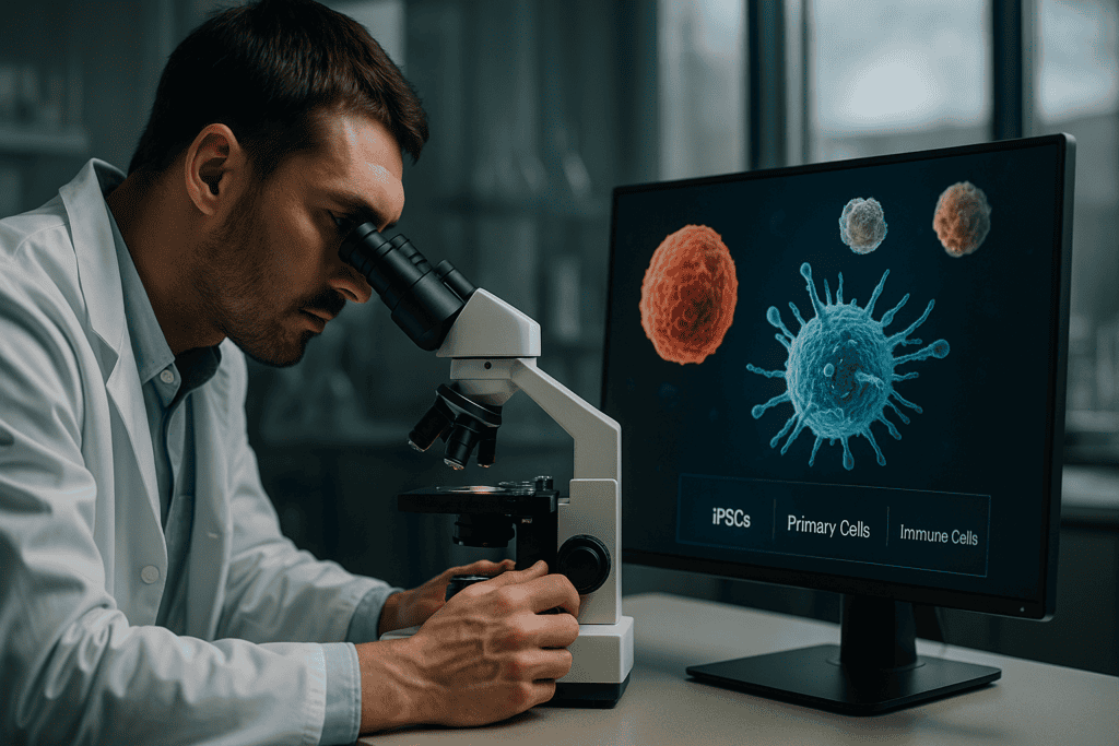
Live-Zell-Überwachung von empfindlichen Zelltypen: iPSCs, primäre & Immunzellen
In the ever-evolving field of cell culture research, the need for precise and real-time monitoring of sensitive cell types is more pressing than ever. Induced pluripotent stem cells (iPSCs), primary cells, and immune cells are pivotal in advancing our understanding of developmental biology, disease modeling, and therapeutic treatments. However, these cell types are notoriously challenging to work with due to their delicate nature and specific environmental requirements. In this article, we delve into the significance of live-cell monitoring, exploring how technological innovations are reshaping this domain and providing actionable insights into enhancing laboratory workflows.
Häufige Herausforderungen und Beschränkungen traditioneller Ansätze
The Intricacies of Handling Sensitive Cells
Handling delicate cell types such as iPSCs, primary cells, and immune cells poses several challenges, primarily due to their inherent sensitivity to environmental changes. Traditional cell culture techniques often fall short in maintaining the optimal conditions required for their growth and differentiation. Frequent manual observations can disturb the cellular microenvironment, potentially leading to inaccurate data and compromised cell health.
- Environmental instability caused by manual intervention
- Potential for contamination during observation
- Inconsistent data acquisition and analysis
These challenges underscore the necessity for more refined and less invasive methods to observe cellular behavior, particularly over extended periods.
Technologische Fortschritte und Automatisierungstrends
The Rise of Automated Live-Cell Imaging
Recent years have witnessed significant advancements in automated live-cell imaging systems, which have revolutionized the way researchers study cell cultures. These technologies enable continuous, real-time monitoring without the need for frequent manual intervention, thus preserving the integrity of the cell culture environment. Systems like the zenCELL owl, specifically designed to be compact and incubator-compatible, exemplify the shift towards automation and efficient data collection in cell culture labs.
- Reduced manual intervention lowers risks of contamination
- Continuous monitoring improves data robustness and reliability
- Integration with data analytics for enhanced insights
Automation not only addresses many of the limitations of traditional methods but also paves the way for new insights through consistent and precise data acquisition.
Praktische Beispiele und Arbeitsabläufe unter Verwendung von Lebendzellbildgebung
Streamlining Cell Culture with Continuous Monitoring
The application of live-cell imaging systems within the laboratory context has been transformative. By employing continuous monitoring technologies, researchers can observe dynamic cellular processes such as proliferation, migration, and differentiation in real-time. For instance, with iPSCs, live-cell imaging offers unprecedented insight into the kinetics of cellular reprogramming and differentiation, crucial for applications in regenerative medicine and drug discovery.
- Real-time insights into cell migration and invasion assays
- Tracking differentiation pathways of iPSCs
- Minimizing experimental variability and enhancing data reproducibility
The practical integration of live-cell monitoring systems allows for more informed decision-making and timely adjustments in experimental protocols.
Lesen Sie weiter, um tiefere Einblicke und Strategien zu gewinnen.
Enhancing Data Accuracy with Quantitative Image Analysis
Utilizing Advanced Image Processing Techniques
In the realm of live-cell monitoring, quantitative image analysis is a powerful tool for extracting precise data from cellular images. By employing sophisticated algorithms and image processing software, researchers can measure cellular components with high fidelity, offering invaluable insights into cell behavior. For instance, software such as CellProfiler and ImageJ are popular choices for automating the analysis of complex image-based data sets, converting qualitative observations into quantitative metrics.
- Automates the quantification of cell proliferation and morphology
- Facilitates objective comparisons between different cell culture conditions
- Enhances reproducibility through standardized analysis protocols
Leveraging Machine Learning for Predictive Analysis
AI-Driven Insights in Cell Monitoring
The integration of machine learning (ML) into live-cell imaging platforms is pushing the boundaries of what researchers can achieve. ML algorithms can identify patterns and predict cellular behaviors more quickly and accurately than ever before. In practice, such systems have been utilized to forecast the outcomes of cell differentiation processes or predict potential cellular anomalies before they manifest visually. Projects leveraging AI-driven solutions, such as the DeepCell framework, exemplify this approach by offering tools that assist in high-throughput cellular analysis.
- Trains models with large datasets to enhance predictive accuracy
- Identifies subtle phenotypic changes indicative of cellular state transitions
- Supports the development of personalized treatment strategies in medicine
Integrating Multimodal Data for a Holistic View
Combining Varied Data Sources for Comprehensive Analysis
To fully understand the complexities of cell biology, researchers are increasingly turning to multimodal data integration. By combining datasets from diverse modalities—such as imaging, genomics, and proteomics—a more complete picture of cellular mechanisms is achieved. For example, integrating data from live-cell imaging with single-cell RNA sequencing has provided deeper insights into cellular heterogeneity, advancing fields like precision oncology, where each cell’s role in tumor progression can be discerned with greater clarity.
- Enables cross-validation of results, increasing confidence in conclusions
- Correlates morphological data with genomic and transcriptomic profiles
- Offers insights into systems biology by revealing interconnected pathways
Case Study: Real-World Application in Drug Discovery
Revolutionizing Pharmaceutical Research with Live-Cell Monitoring
One of the most impactful applications of live-cell monitoring and imaging technologies is in drug discovery. For instance, pharmaceutical companies are employing these technologies to accelerate the screening of drug candidates. By observing how potential drugs influence cell behavior in real-time, researchers can more efficiently identify promising compounds. In a recent study, live-cell imaging was used to assess the effect of a novel compound on immune cell migration, leading to insights that advanced the drug to clinical trials.
- Improves the throughput of drug screening processes
- Reduces costs associated with failed drug trials by early intervention
- Identifies cellular targets and mechanisms of drug action
Optimizing Laboratory Workflows Through Modular Systems
Building Flexible and Efficient Research Environments
Incorporating modular live-cell monitoring systems into laboratory workflows can dramatically enhance operational flexibility. These systems enable scientists to tailor their setup according to specific research needs, integrating various technologies such as automatic imaging, environmental controls, and data analysis modules. Laboratories using such modular setups have reported greater efficiency in workflow and reduced downtime, as components can be individually managed and upgraded.
- Adapts to a wide range of experimental conditions
- Facilitates the rapid prototyping and testing of new hypotheses
- Streamlines data management and increases throughput
Strategies for Implementing Live-Cell Monitoring in Small Labs
Cost-Efficient Solutions Without Sacrificing Quality
While live-cell monitoring systems are invaluable, their implementation can be daunting for smaller labs with limited budgets. However, strategies exist to make these technologies accessible without compromising research quality. Partnering with larger institutions for shared resources, investing in scalable platforms, and utilizing open-source software solutions are effective approaches. For instance, consortia-based models where multiple labs share high-cost equipment have allowed many small labs to conduct cutting-edge research at a fraction of the cost.
- Collaborate with peers to maximize resource usage and minimize costs
- Invest in scalable systems that grow with your research needs
- Adopt open-source tools to enhance accessibility and customization
Ensuring Compliance and Data Security in Cell Monitoring
Maintaining Ethical and Secure Research Practices
As with any data-intensive process, live-cell monitoring raises important considerations regarding compliance and data security. Ensuring that imaging and data analysis practices meet regulatory standards is paramount, particularly in biomedical research. Labs need robust data management protocols to protect sensitive information, and bioethics committees play a crucial role in overseeing practice standards. Utilizing cloud-based platforms with strong security features can assist in achieving these goals while ensuring data integrity.
- Implement compliance checks across all research phases
- Utilize secure, robust data storage solutions to prevent unauthorized access
- Engage with bioethics teams regularly to align on best practices
Im Anschluss fassen wir die wichtigsten Erkenntnisse, Kennzahlen und eine wirkungsvolle Schlussfolgerung zusammen.
Addressing Challenges in Live-Cell Monitoring
Troubleshooting and Overcoming Technical Barriers
While live-cell monitoring technologies have evolved significantly, they are not without challenges. Technical issues such as phototoxicity, image drift, and data storage can impede research progress. Addressing these obstacles requires a combination of advanced technology and innovation. Employing strategies such as optimized imaging protocols, real-time drift correction software, and robust data storage frameworks can mitigate these issues. Researchers continue to innovate by developing compensatory algorithms and imaging techniques that reduce cell stress, thereby enhancing the quality and accuracy of data collected.
- Implements anti-drift technologies to ensure stable imaging
- Utilizes advanced imaging modalities to minimize phototoxic effects
- Establishes comprehensive data management systems to handle large datasets
Future Directions in Live-Cell Imaging
Innovations in Technology and Technique
The future of live-cell imaging holds promise with the advent of novel technologies like super-resolution microscopy and optogenetics. These advancements allow scientists to visualize cellular processes with unprecedented detail and precision. By synergizing these techniques with existing monitoring systems, researchers can explore cellular dynamics on a molecular level, unraveling new insights into biological processes. Developing miniaturized, high-performance imaging systems also remains a key focus, offering portability and functionality crucial for field research and point-of-care applications.
- Explores the use of super-resolution techniques for fine structures
- Incorporates optogenetic tools for precise modulation of cell activities
- Designs compact, portable systems for diverse research environments
Schlussfolgerung
In the rapidly advancing field of cell biology, live-cell monitoring technologies have become indispensable tools for researchers seeking to understand the intricacies of cellular processes. From enhancing data accuracy with quantitative image analysis to leveraging machine learning for predictive analysis, these technologies are reshaping how scientists study and interpret cellular dynamics. The integration of multimodal data provides a comprehensive view of cell biology, contributing significantly to fields such as drug discovery, where real-time insights into cellular responses can streamline the development of new therapies.
Additionally, by optimizing laboratory workflows and tailoring modular systems to specific research needs, laboratories—both large and small—are improving efficiency and reducing operational costs. Importantly, these innovations are being adopted without compromising the quality of research, thanks to scalable solutions and international collaborations.
Moreover, as ethical concerns and data security become increasingly paramount in scientific research, ensuring compliance remains a critical priority. By implementing strict data management protocols and engaging regularly with bioethics committees, labs safeguard sensitive data, thus maintaining the integrity and trustworthiness of their research outcomes.
As we look to the future, the potential of live-cell monitoring is boundless. Innovations in technology and technique promise exciting new insights and advancements across a multitude of scientific disciplines. It is a call to action for researchers and institutions to invest in these cutting-edge technologies and to embrace the evolving landscape of cell monitoring. By doing so, we not only enhance our understanding of the natural world but also contribute to a future where scientific breakthroughs can improve lives and societies worldwide.
In conclusion, embracing the latest advancements in live-cell monitoring and overcoming existing challenges equips researchers with the tools necessary to drive innovation and discovery. The journey does not stop here; rather, it sets a stage for further exploration and development. By continuing to support scientific endeavors and collaborations, we can unlock mystery doors in biology, laying the foundation for advancements that have the potential to reshape fields like medicine, pharmacology, and beyond.
