
“```html
Automation for Everyone: Why Complex Imaging is No Longer Just for Specialists
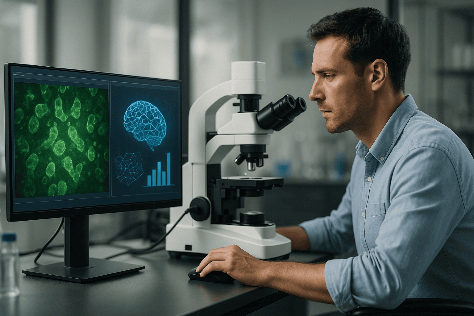
The landscape of life sciences and biotechnology is rapidly evolving, driven by technological innovations that are reshaping how researchers approach cellular studies. One such revolution is occurring in the realm of complex imaging. Traditionally the domain of specialists, advanced imaging technologies are now accessible to a broader range of users thanks to automation. This shift is particularly significant in modern cell culture research, where the ability to continually monitor live cells with precision and efficiency can be transformative. This article examines why complex imaging is now within reach for everyone, outlines the traditional challenges researchers face, and explores the latest advancements that democratize these technologies.
Herausforderungen und Grenzen traditioneller Ansätze
Complexity and Skill Dependency
Historically, complex imaging technologies required skilled personnel with significant expertise, making them inaccessible to many labs. These traditional systems often involved intricate setups and demanding operational protocols that discouraged widespread use. Researchers faced several challenges, including:
- High costs associated with specialized training and maintenance
- Time-consuming manual processes that reduced throughput
- Potential for human error affecting data quality and reproducibility
Such challenges have limited the ability of many researchers to exploit the full potential of live-cell imaging in their work.
Technologische Fortschritte und Automatisierungstrends
Opening Doors with Automation
Recent technological advances have paved the way for automation to revolutionize lab workflows, making complex imaging more accessible. Automation minimizes human intervention, reducing the possibility of error and improving consistency in data acquisition. Key trends driving this change include:
- Development of user-friendly software with intuitive interfaces
- Integration of AI and machine learning for enhanced data analysis
- Design of compact and scalable imaging systems for widespread adoption
These innovations not only democratize technology but also empower researchers to focus on data interpretation rather than technical execution.
Praktische Beispiele und Arbeitsabläufe unter Verwendung von Lebendzellbildgebung
Streamlined Processes and Enhanced Data Collection
With automation, live-cell imaging no longer requires the hands-on expertise once deemed necessary. For instance, systems like the zenCELL owl provide continuous monitoring capabilities that adapt to a variety of research scenarios. In practical terms, live-cell imaging enables workflows such as:
- Longitudinal studies of cell behavior over extended periods without manual interference
- Real-time observation of cellular responses to external stimuli or drug treatments
- High-throughput screening (HTS) applications that demand rapid and repetitive imaging
Such workflows highlight the potential of automated imaging systems to streamline operations and enhance scientific outputs.
Wie Inkubator-basierte Bildgebung die Reproduzierbarkeit und Datenqualität verbessert
Eliminating Environmental Variability
The incorporation of imaging systems directly into incubators plays a pivotal role in improving data quality and reproducibility. By maintaining a consistent environment, these systems eliminate the variability that can arise from transferring cultures between different conditions. Advantages include:
- Stable temperature, humidity, and CO2 levels throughout the imaging process
- Minimal disruption to cell culture conditions ensuring natural cell behavior
- Reduced risk of contamination with contained setups
Incubator-based imaging therefore provides researchers with the confidence that observations reflect true biological processes.
Lesen Sie weiter, um tiefere Einblicke und Strategien zu gewinnen.
“`
“```html
Advanced Imaging Techniques in Automation
Pioneering the Future of Research
The development of advanced imaging techniques integrated with automation has paved the way for cutting-edge research methodologies. Systems are now equipped with enhanced functionalities that further empower researchers to conduct more sophisticated experiments without the need for extensive manual adjustments. These techniques include fluorescence imaging, confocal microscopy, and multi-photon imaging, each offering precise insights into cellular mechanisms from a new perspective while remaining accessible to non-specialists.
- Fluorescence imaging enables visualization of specific cellular components through targeted dyes or fluorescent proteins.
- Confocal microscopy provides enhanced resolution by eliminating out-of-focus light, ideal for 3D structures.
- Multi-photon imaging allows for deeper tissue penetration with less photodamage, beneficial for live specimens.
Combining these techniques with automation pushes the boundaries of what researchers can achieve, making it feasible for more laboratories worldwide to utilize these sophisticated tools.
High-Content Screening and its Role in Drug Discovery
Accelerating Pharmaceutical Development
High-content screening (HCS) is an automated process that allows researchers to analyze the effects of thousands of compounds on live cells simultaneously. This approach has proven invaluable in the pharmaceutical industry, notably in drug discovery and toxicology studies. By incorporating imaging systems that automate data collection and analysis, HCS offers a comprehensive view of cellular responses, helping to identify promising drug candidates early in the research process.
- HCS enables the identification of active compounds, reporter genes, and pathway analysis with high efficiency.
- Automated image analysis reduces subjective bias and enhances reproducibility across experiments.
- Integration with machine learning can further refine the screening process by predicting outcomes based on previous data.
This technology not only accelerates the drug development pipeline but also reduces costs associated with traditional methods.
Adopting Machine Learning for Enhanced Imaging Analysis
Elevating Insights with Data-Driven Approaches
Machine learning (ML) is increasingly becoming a cornerstone of automated imaging analysis, enhancing the interpretive power of collected data. ML algorithms can process vast amounts of information at speeds unachievable by humans, recognizing patterns and anomalies with precision. This ability makes it possible to refine datasets to highlight significant changes that might otherwise go unnoticed, thereby improving the accuracy of research outcomes.
- ML techniques can classify cell types, detect rare events, and quantify material properties more effectively.
- Deep learning models, such as convolutional neural networks, enhance image recognition capabilities.
- ML facilitates adaptive imaging, where systems optimize imaging parameters in real-time based on preliminary results.
The integration of machine learning not only provides a deeper understanding of complex biological systems but also allows for the development of predictive models that can forecast cell behavior under varying conditions.
Cost-Effective Solutions for Smaller Labs
Bringing Innovation to Every Researcher
While state-of-the-art imaging systems sound financially daunting, innovations in design and development have introduced cost-effective solutions tailored for small labs and early-career researchers. These solutions maintain high performance without compromising on budget constraints, giving a broader demographic access to advanced capabilities.
- Modular imaging components allow labs to customize their systems according to specific research needs, minimizing upfront expenditures.
- Leasing models and shared equipment infrastructures help distribute costs more evenly over time.
- Open-source software paired with affordable hardware solutions offers viable alternatives to expensive proprietary systems.
This democratization of imaging technology ensures that breakthroughs can emerge from any lab, regardless of size or funding.
Real-World Application: Success Stories from the Field
Transforming Research Outcomes with Automated Imaging
Numerous research institutions have adopted automated imaging technologies with significant success, illustrating the real-world impact of these advancements. For example, a leading university used integrated imaging systems to expedite their research on neurodegenerative diseases, resulting in faster identification of potential therapeutic targets. Another biotech startup streamlined its cancer research by automating cellular imaging processes, yielding insights that shortened their drug development timeline by months.
- Institutions report a 30% increase in data throughput and a 20% reduction in experimental time by adopting automation.
- Automated workflows have enabled multi-disciplinary collaborations by providing standardized data outputs across global teams.
- Case studies show marked improvements in results, often achieving publication-quality data with greater efficiency.
The empirical evidence underscores the transformative power of automation and its ability to improve research quality and speed.
Im Anschluss fassen wir die wichtigsten Erkenntnisse, Kennzahlen und eine wirkungsvolle Schlussfolgerung zusammen.
“`
“```html
Integrating Robotics for Seamless Operations
Precision and Efficiency in Modern Laboratories
Integrating robotics with automated imaging not only enhances operational efficiency but also ensures precision, thus transforming experiment workflows. Robotics facilitate the handling of delicate sample manipulation that would be challenging without automation, allowing labs to operate with higher speed and accuracy.
- Robotic systems automate repetitive tasks, freeing researchers to focus on data analysis and interpretation.
- Coordinated robotics and imaging systems provide scalable solutions for varying research demands, optimizing lab resources.
- Enhanced precision through robotics eliminates human error, ensuring replicable and reliable results.
The synergy between robotics and imaging empowers labs to scale up their operations, aligning with the rapid pace of scientific discovery.
Addressing Barriers to Adoption
Solutions for Universal Accessibility
While the advantages of automated imaging systems are clear, certain barriers such as cost, training, and resistance to change must be addressed for universal adoption.
- Developing robust training programs eases the transition, equipping researchers with the necessary skills to leverage new technologies effectively.
- Collaborative partnerships with tech companies can provide affordable equipment and software access for academic institutions.
- Promoting success stories within the scientific community encourages adoption by showcasing tangible benefits and innovative breakthroughs.
By overcoming these obstacles, the adoption of automated imaging can become a universally embraced standard in research endeavors.
Future Prospects of Automated Imaging
Charting the Course for Innovation
The future of automated imaging lies in its continuous evolution, promising to revolutionize scientific research further. With advances in artificial intelligence, quantum computing, and nanotechnology, the horizon gleams with new possibilities.
- AI-driven imaging systems will enable even deeper analyses of biological phenomena at an unprecedented scale.
- Miniaturized, portable imaging devices can expand accessibility, allowing in-field research across diverse environments.
- Interdisciplinary collaborations will accelerate advancements, blending insights from computer science, engineering, and life sciences.
Embracing these emerging technologies, researchers are poised to unlock further layers of complexity within biological systems, providing insights that could redefine our understanding of life itself.
Schlussfolgerung
As we wrap up our exploration of automated imaging technologies, it becomes clear that these advancements are not merely incremental improvements but transformative forces in the research landscape. The amalgamation of automation, machine learning, and advanced imaging techniques spearheads a new era of discoveries that bridge past constraints and usher in a future where research is faster, more efficient, and more accessible than ever before.
Key takeaways from this article highlight the dynamic potential of automated imaging systems, from streamlining drug discovery processes to enhancing our understanding of complex biological systems with unparalleled precision. We have seen how cost-effective solutions and flexible infrastructures have democratized access, making high-level research capabilities available to smaller labs and novice researchers. The utilization of robotics and machine learning has not only augmented operational precision but also shed light on hidden patterns that could lead to monumental breakthroughs.
The relevance and value of these technologies cannot be overstated. Across disciplines, the integration of automated imaging is playing a pivotal role in accelerating scientific progress, bridging gaps within the global research community, and fostering collaborations that lead to innovative insights. This transformation is providing the scientific community with tools that were once the domain of a privileged few, ensuring that expertise and creativity, rather than budget and size, dictate the future of discovery.
Moving forward, let this discussion serve as a catalyst for change and inspiration. We stand on the cusp of a technological renaissance that challenges us to think bigger and reach farther. Equip yourselves with these advances, and do not shy away from adopting the changes that these automated systems represent. Whether you are a seasoned researcher or just beginning your scientific journey, embrace the tools of today to forge the innovations of tomorrow.
In conclusion, automation is not the end but rather the beginning of an era defined by unprecedented possibilities in research, inviting us all to contribute to a collective vision that transcends conventional limitations and explores boundless scientific frontiers.
“`





