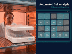
zenCELL owl enables continuous, non-invasive monitoring of oocytes and embryos inside the IVF incubator. Label-free brightfield imaging — from fertilisation to blastocyst stage — without removing embryos from their optimal environment.
Traditional IVF monitoring requires removing embryos from the incubator for observation — exposing them to temperature changes, CO₂ fluctuations and mechanical stress. zenCELL owl eliminates this entirely. The device sits inside your IVF incubator and images continuously, 24/7, without any disturbance to the embryo culture environment.
Embryos never leave the incubator. Temperature, CO₂ and humidity remain stable throughout the entire culture period — from fertilisation to transfer.
No fluorescent labels, no phototoxicity. Embryos are imaged in their natural state using transmitted light brightfield microscopy — safe for downstream use.
Every developmental stage is captured and documented automatically. Furthermore, timelapse videos are generated for each well — providing a complete developmental record.
The dedicated IVF software version includes real-time measurement tools — draw a circle to measure oocyte diameter, or a line to measure zona pellucida thickness directly in the software.

From wildlife conservation to clinical IVF research — zenCELL owl enables continuous embryo monitoring across a broad range of species and research contexts.
Monitor oocyte maturation and early embryo development in endangered or rare species — koala, kangaroo, and other marsupials. Non-invasive continuous imaging reduces handling stress and maximises developmental data.
Bovine, equine, porcine and ovine embryo monitoring. Automated timelapse documentation of cleavage stages and blastocyst development — for research, breeding programmes and embryo quality assessment.
Evaluate culture media, incubator conditions and IVF protocols with continuous objective data. Compare developmental kinetics across treatment groups simultaneously — 24 wells monitored in parallel.
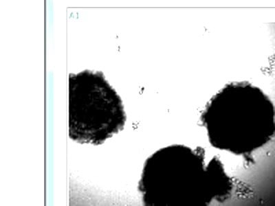
Observe cumulus cell expansion, compaction and detachment during in vitro maturation. Additionally, monitor cytoplasmic activity and germinal vesicle breakdown in real time.
Test the effect of different culture media formulations, oxygen concentrations or compounds on embryo development — with continuous objective brightfield data across all wells simultaneously.
Document timing of first cleavage, compaction, cavitation and hatching. Furthermore generate timelapse videos and export measurement data as CSV for statistical analysis.
The dedicated IVF software includes real-time measurement tools (diameter, distance) and an optimised interface for embryo monitoring. Installs in minutes on any Windows PC.
The device fits on any standard incubator shelf — 18×18×10 cm, 1.5 kg. One USB-C cable to your PC. Allow 2 hours for temperature equilibration before the first experiment.
Place embryos or oocytes in your chosen vessel format. Set your imaging interval (10 minutes recommended for IVF). Use live view to adjust focus manually for optimal image quality.
Monitor embryo development from any device, anywhere. The software runs continuously without incubator opening. Timelapse videos and images are automatically saved at every time point.
Export timelapse videos as AVI, individual images as PNG or JPG, and measurement data as CSV. All data is stored locally — no cloud subscription required.

Key specifications for embryo and oocyte monitoring applications.
Monitor 24 positions in parallel — ideal for comparative IVF research across multiple treatment groups.
1.2 × 0.9 mm field of view. Sufficient for oocyte and early embryo imaging in standard IVF culture vessels.
10-minute imaging interval captures all key developmental events without generating excessive data volumes.
IVF version includes real-time measurement tools — diameter, distance — not available in the standard cell culture version.
Automated timelapse video generation per well. Additionally, individual images exported as PNG or JPG.
Uninterrupted imaging for days to weeks. PC must remain active — disable sleep mode for long experiments.
Purchase from €14,000 or rent from €290/month. IVF software version included at no extra cost.
One cable connection. Windows 10/11. No internet required. Data stays local in your lab.
"Although the oocyte didn't mature completely to do ICSI, it's fascinating to see the cumulus cells moving and detaching over time, the cytoplasmic activity, and the germinal vesicle. It's a really great video! Thank you for the device!"
Embryo monitoring in IVF refers to the continuous observation of oocyte maturation and early embryo development from fertilisation to the blastocyst stage. Traditional IVF monitoring requires periodically removing embryos from the incubator for observation under a standard microscope — each removal introduces temperature shock, CO₂ fluctuation and mechanical stress that can negatively impact developmental outcomes.
Time-lapse embryo monitoring uses continuous imaging to capture every stage of embryo development without incubator disruption. Furthermore, it provides objective, quantitative kinetic data on cleavage timing, compaction and cavitation that correlates with embryo viability. zenCELL owl enables time-lapse monitoring in any standard IVF incubator — without requiring a dedicated time-lapse incubator system.
Wildlife IVF presents unique challenges — rare species, limited oocyte availability and the critical importance of minimising handling. zenCELL owl is particularly well-suited for wildlife IVF research: it monitors up to 24 wells simultaneously, generates automatic timelapse videos, and eliminates incubator openings during observation. Additionally, the dedicated IVF software version includes measurement tools for zona pellucida thickness and oocyte diameter assessment.
zenCELL owl is compatible with standard 24-well plates (Ø 16mm) and petri dishes (17–35mm diameter) — the most commonly used vessel formats in IVF research. For small oocyte volumes, placing drops in the centre of the well maximises imaging coverage. The live view function allows manual focus adjustment for optimal embryo positioning before starting the recording.
Download our application note on live cell imaging for IVF and reproductive science. Includes protocol, software settings and real imaging data from oocyte monitoring experiments.
30 minutes via MS Teams. We show you zenCELL owl imaging real samples live — including the IVF software version with measurement tools. No commitment required.