🔪 Application — Scratch Assay & Wound Healing

Scratch Assay —
Record Every Movement. Calculate Speed Accurately.

Automated wound healing and migration assay monitoring — 24 wells in parallel, continuous kinetic data, real-time gap closure analysis. No manual imaging. No missed time points.

24
assays in parallel
5 min
imaging interval
gap closure time
0
manual imaging steps
zenCELL owl scratch assay — automated wound healing monitoring incubator microscope
Background

What Is a Scratch Assay?

The wound healing, migration assay or scratch assay is a standard method to analyze cell migration in vitro. The dynamics of cell migration into a cell-free area are monitored and quantified to analyze migration characteristics and calculate gap closure time.

📹
Dynamic Data for Dynamic Processes

With conventional manual microscopy it is not possible to capture every dynamic change at the cell level. zenCELL owl records images every 5 minutes — capturing every cell movement in detail, even during long-term analyses over days.

Wound Healing Speed — Accurately Calculated

Observe and control every time point of the wound healing process retrospectively. Calculate gap closure speed by analysing the increase of confluence in the gap area. Determine t½ gap closure time automatically.

🔄
24 Assays Under Identical Conditions

Simultaneous analysis of cell morphology and confluence in up to 24 wells under identical incubator conditions. Optimises comparability of results and reproducibility of data across conditions, drugs and cell lines.

Practical example

L929 Fibroblasts — 24-Hour Gap Closure Timelapse

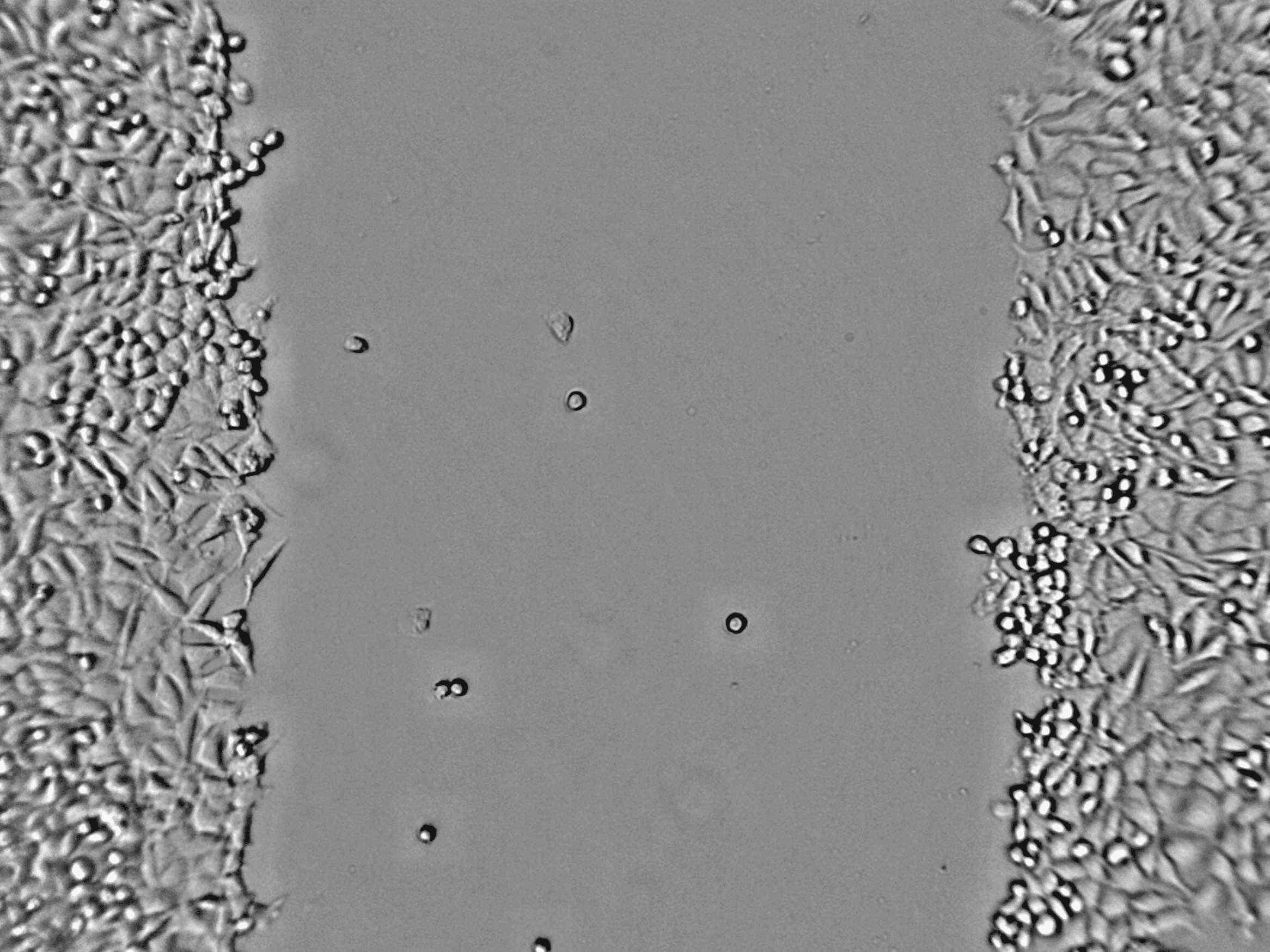
A migration assay of L929 fibroblasts was performed over 24 hours. The gap was inserted in a confluent monolayer at time zero. Within 24 hours, continuous cell migration results in complete closure of the gap — captured automatically at every time point.

L929 scratch assay 0 hours — gap created in confluent monolayer
0 hours
L929 scratch assay 1 hour — cells start migrating at gap edge
1 hour
L929 scratch assay 3 hours — visible gap closure beginning
3 hours
L929 scratch assay 12 hours — halfway gap closure
12 hours
L929 scratch assay 18 hours — advanced gap closure
18 hours
L929 scratch assay 24 hours — complete gap closure
24 hours

L929 mouse fibroblasts — scratch assay timelapse over 24 hours. Gap inserted at 0h by pipette tip. Complete closure visible at 24h. Digital phase-contrast imaging. Scalebar: 200 µm.

zenCELL owl gap closure speed calculation — t1/2 relative gap area plot

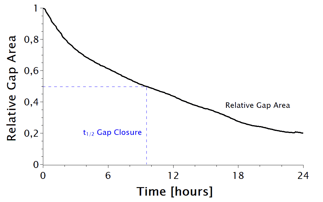
Relative gap area as a function of time — t½ gap closure determined automatically from confluence data

Calculate Gap Closure Speed Automatically

Cell coverage data from zenCELL owl is used to calculate the Relative Gap Area at every time point. Plotted as a function of time, the half gap closure time (t½ Gap Closure) is determined directly from the confluency curve.

  • Relative gap area calculated per image automatically
  • t½ gap closure time determined from growth curve
  • Compare gap closure speeds across 24 conditions
  • Export data to CSV for statistical analysis
  • Full retrospective analysis of any time point
📄

Application Note — Scratch Assay

Full protocol, gap closure analysis method and data for scratch assays with zenCELL owl.

Download PDF →
Step-by-step protocol

Running a Scratch Assay — Complete Protocol

From cell seeding to final data export — the complete automated scratch assay workflow with zenCELL owl. Every step documented, every time point captured.

Step 01
Seed Confluent Monolayer

Seed cells in 24-well plate and grow to full confluency. Use coated plates for optimal adhesion and consistent monolayer formation.

Step 02
Create the Scratch

Use a pipette tip for standard assays — or the zenCELL Scratch Maker (light-based, ECM-preserved) for physiological migration studies. Full 24-well plate in 60 seconds.

Step 03
Wash & Add Treatment

Wash away detached cells. Add drug treatment, inhibitor, or growth factor at defined concentrations across 24 wells.

Step 04
Place in zenCELL owl

Transfer plate to zenCELL owl inside the incubator. Define imaging interval (5–30 min). Continuous monitoring starts automatically — no further manual steps needed.

Step 05
Automated Gap Closure Analysis

Software calculates Relative Gap Area at every time point automatically. Growth curves generated per well. t½ gap closure determined from confluence data — no manual measurement.

Step 06
Export & Compare Results

Export CSV data and timelapse videos. Compare gap closure speeds across all 24 conditions. Full retrospective analysis of any time point — publication-ready data.

Method comparison

Scratch Methods — Side by Side

Not all scratches are equal. The method used to create the gap fundamentally affects the biology of the migration assay.

MethodCell DamageECMReproducibilityThroughputCost
Pipette TipHigh — cells destroyedDestroyedLow — operator-dependentManual · slowVery low
Ibidi InsertNoneNo coating possibleHighManual · moderateHigh per plate
✓ Scratch MakerNone — light-basedFully intactVery high — identical every time24 wells in 60 secOne-time device

Full method comparison: Migration Assay Methods compared →

Take it further

Upgrade to Physiological Scratching — Scratch Maker System

The example above uses a manual pipette tip scratch. For publication-quality, physiological migration data, the zenCELL owl Scratch Maker uses light-based technology — preserving cells and ECM completely.

Light-Based vs. Pipette Tip — What Changes

A manual pipette tip scratch destroys the cell layer and ECM. The Scratch Maker uses photonic technology to create a precise, reproducible gap without any mechanical contact. 24 identical scratches in 60 seconds.

  • No cell damage — cell layer beneath preserved
  • ECM fully intact — physiologically relevant surface
  • No cell debris in the gap
  • 24 identical scratches in 60 seconds
  • Mask-guided — same position every time
Learn more about the Scratch Maker →
🧪
Pipette Tip
Cell damage · ECM destroyed · debris
💡
Scratch Maker
No damage · ECM intact · clean gap
Also compare all methods on our Migration Assay Methods comparison page — pipette tip, Ibidi insert, robotic system and Scratch Maker side by side.
50+
peer-reviewed publications cite zenCELL owl — including scratch assay and wound healing studies
View all publications →
FAQ

Scratch Assay — Common Questions

What is the difference between a scratch assay and a wound healing assay?+
The terms are used interchangeably. Both describe an in vitro method where a gap is created in a confluent cell monolayer and its closure is monitored over time to study cell migration. The wound healing assay term emphasises physiological relevance, particularly for skin biology and regenerative medicine research.
How does zenCELL owl calculate gap closure speed?+
zenCELL owl measures cell confluence in the gap area at every imaging interval (minimum 5 minutes). This data is used to calculate the Relative Gap Area as a function of time. From this curve, the half gap closure time (t½ Gap Closure) is determined — giving a precise, quantitative measure of migration speed that can be compared across 24 conditions simultaneously.
Can I run 24 scratch assays simultaneously?+
Yes. zenCELL owl monitors up to 24 wells simultaneously under identical incubator conditions. This allows comparison of different cell lines, drug concentrations, or treatment conditions in a single experiment — eliminating the variability of sequential testing.
What is the Scratch Maker and how does it improve scratch assay results?+
The zenCELL owl Scratch Maker uses light-based photonic technology to create scratches without mechanical contact. Unlike a pipette tip, it preserves the cell layer beneath the gap and leaves the ECM intact — providing physiologically relevant migration conditions. 24 identical scratches in 60 seconds. See the Scratch Maker product page for full details.
Is there an application note for scratch assays with zenCELL owl?+
Yes — download the full application note above. It covers the complete protocol, gap closure analysis method, t½ calculation, and practical data from L929 fibroblast scratch assays.

See Scratch Assay Monitoring Live —
Free Remote Demo

Watch zenCELL owl track gap closure in real cells inside a real incubator. On request via MS Teams. No obligation.

On request via MS Teams No commitment 50+ publications Application Note available
Vue en direct

See zenCELL owl
live, 30 min, free online

Real cells. Real data. Real comparison. Watch AI cell counting, XYZ stage and full software live inside a real incubator — twice a week via MS Teams.

Full course of live cell imaging

Get your free live online demonstration of the zenCELL owl directly inside an incubator, available twice per week.

Remote-Demo

or