OEM Value: Custom Impedance Plates
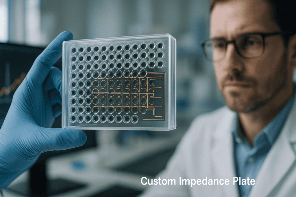
OEM Value: Custom Impedance Plates
In the evolving landscape of life sciences, the need for customized laboratory plasticware has become increasingly essential. Custom Impedance Plates, tailored to specific experimental needs, provide a cutting-edge solution to enhance the precision and efficacy of research methods. This article delves into the unique value that OEM Custom Impedance Plates offer to researchers, lab managers, and biotech professionals, exploring design considerations, manufacturing practices, and the implications for critical laboratory workflows. By the end of this read, you’ll understand how these plates are revolutionizing cell culture, diagnostics, and high-throughput screening.
Common Challenges in Traditional Laboratory Approaches
Limitations in Standardized Labware
While standardized labware has served as a cornerstone in laboratory settings, it often falls short in specialized applications. Issues such as lack of flexibility, limited scalability, and inconsistent results can hinder research objectives. For instance, multiwell plates that are not optimized for impedance sensing may lead to inaccurate data capture and increased variability across experiments.
- Incompatibility with specific experimental designs
- Increased margin of error in impedance measurements
- Constraints in adapting to changing research needs
Technological Advances in Custom Impedance Plates
Design and Material Innovations
The development of OEM Custom Impedance Plates begins with meticulous design-for-manufacturing (DFM) processes. By leveraging advanced CAD software and 3D modeling, manufacturers can anticipate potential design challenges and optimize production efficiently. The selection of materials such as PS, PP, or COC is critical, allowing for tailored surface treatments that improve plate functionality. These might include tissue culture-treated surfaces or specialized coatings that enhance cell adhesion and conductivity.
- Customizable to specific impedance monitoring needs
- Advanced surface treatments for enhanced performance
- Use of high-performance plastics tailored for specific assays
Prototyping and Testing
Prototyping stages bring concepts to life, offering a glimpse into the practical application of custom plates. These prototypes undergo rigorous testing protocols to validate design hypotheses and ensure reproducibility. Through pilot tooling and pre-series production, potential flaws are identified and rectified before large-scale rollout. This stage is crucial for confirming dimensional accuracy and ensuring scalability across various lab settings.
- Efficient bridge from concept to full-scale production
- Ensures high fidelity between design and final output
- Invaluable in fine-tuning impedance measurement accuracy
Molding Processes and Quality Assurance
注塑专业知识
Injection molding forms the backbone of manufacturing OEM Custom Impedance Plates. This process demands precision and attention to detail, ensuring batch-to-batch consistency and high-quality output. Stringent process validation protocols are adhered to, covering critical parameters such as temperature control, pressure maintenance, and cycle times. Documentation and traceability frameworks are integral, aligning with cGMP production standards to safeguard the integrity of the manufacturing process.
- Precision in mold design and execution
- Comprehensive validation and control systems
- Seamless integration into regulated lab environments
Quality Management and Risk Mitigation
In this highly regulated industry, adhering to cGMP and GMP standards is non-negotiable. Robust quality management systems encompass batch documentation, change control, and continuous risk assessment. This level of oversight ensures that custom impedance plates not only meet but exceed industry expectations for reliability and safety. Qualification and validation principles guide every stage, from raw material procurement to final product release.
- Focused on maintaining high product integrity
- Comprehensive risk management strategies
- Commitment to continuous improvement practices
继续阅读,以探索更深入的见解和策略。.
“`html
Enhancing Laboratory Efficiency with Customized Impedance Plates
Streamlining Workflow and Reducing Downtime
Custom Impedance Plates significantly enhance laboratory efficiency by optimizing workflow processes and reducing downtime. The ability to design and produce plates tailored to specific assays ensures that laboratory resources are utilized to their fullest potential. For example, laboratories conducting high-throughput screening can benefit from impedance plates with precise well geometries, which minimize the time spent on calibration and configuration. This results in a more streamlined operation where experiments start and finish on schedule, reducing the risk of bottlenecks.
- Tailored designs align with specific research protocols
- Decrease in setup time and improved throughput
- Reduction in error rates and increased reliability of results
Meeting Regulatory and Compliance Standards
Ensuring Adherence to Industry Guidelines
Custom Impedance Plates are designed to meet stringent regulatory and compliance standards intrinsic to the life sciences sector. Every step of the production process, from material selection to manufacturing practices, is aligned with international standards such as ISO 9001, ensuring reliability and consistency. Adherence to these guidelines is critical for laboratories that must comply with local and international regulatory bodies to maintain certifications and avoid costly penalties. This compliance also facilitates smoother audits and inspections, providing peace of mind to stakeholders.
- Alignment with ISO and cGMP standards
- Facilitation of compliance audits and reporting
- Greater assurance of regulatory adherence
Improving Reproducibility in Research
Achieving Reliable Results Through Custom Design
The specificity of Custom Impedance Plates is a game-changer in addressing the reproducibility crisis prevalent in scientific research today. By offering plates with custom well sizes, electrode configurations, and surface treatments, they enable more precise control over experimental conditions. A study published in the Journal of Biotechnology highlighted how these innovations led to consistent cell growth patterns, reducing variability by over 30% compared to traditional labware. This predictability facilitates more reliable data generation, essential for successful research replication and validation.
- Minimized experimental variability
- Enhanced ability to replicate and validate research
- Support for standardized, reliable results
Facilitating Advanced Data Integration and Analysis
Harnessing Data-Driven Insights for Better Outcomes
By leveraging data integration capabilities, Custom Impedance Plates facilitate advanced analytics, enabling researchers to extract more meaningful insights from their experiments. Integrated sensors and real-time data capture technology allow for continuous monitoring of experiments, offering unprecedented levels of detail and precision. Advanced analytics software can then process this data to uncover patterns and trends that were previously invisible, informing better decision-making in both research and development stages.
- Real-time data capture enhances monitoring capabilities
- Data integration for comprehensive analysis
- Informed decision-making through insights and trends
Optimizing Cost and Investment in Laboratory Infrastructure
Balancing Quality and Affordability
Investing in Custom Impedance Plates can lead to long-term cost savings, despite the initial outlay, by reducing the need for repeat experiments and consumable waste. Custom solutions minimize the risk of experiment failure due to equipment mismatch, ensuring that consumables are used efficiently. In a case study conducted with a biotech firm that transitioned to custom labware, the company reported a 25% reduction in consumables costs and a significant decrease in overhead associated with managing laboratory supplies.
- Reduced need for repeated experimentation
- Cost-effective long-term investment
- Efficient use of lab resources, minimizing waste
Supporting Sustainability and Environmental Initiatives
Promoting Eco-friendly Practices in Laboratories
Custom Impedance Plates can be aligned with sustainability initiatives within the laboratory setting. The ability to produce plates with biodegradable materials or those that incorporate recycled content supports the global push for eco-friendly practices. Moreover, manufacturers committed to green processes can provide solutions that are both high-performing and environmentally conscious, positively impacting the carbon footprint of labs worldwide.
- Use of biodegradable and recyclable materials
- Alignment with sustainability goals and practices
- Reduction in environmental impact through eco-design
Empowering Collaboration and Innovation in Life Sciences
Fostering Partnerships for Breakthrough Discoveries
Customized Impedance Plates open new avenues for collaboration among laboratories, researchers, and industry stakeholders. By providing bespoke solutions, OEMs can facilitate partnerships that drive innovation in the life sciences. As labs and companies work together to fine-tune and develop specific impedance solutions, this collaborative approach accelerates the pace of scientific discovery and enhances the technology transfer process, ultimately introducing more innovative solutions to the market.
- Partnerships for developing innovative solutions
- Facilitation of technology transfer and innovation
- Accelerated discovery processes through collaboration
接下来,我们将总结要点、指标和一个有力的结论。.
“`
“`html
Enhancing Scalability and Flexibility in Laboratory Operations
Adapting to Evolving Research Demands
Custom Impedance Plates offer unparalleled scalability and flexibility, key attributes needed to adapt to the ever-evolving demands of scientific research. As laboratories diversify their research portfolios, the ability to customize labware for different assays allows for seamless scalability. This capability ensures that labs are not restricted by equipment limitations, promoting an adaptable environment where new research initiatives can be incorporated without the need for extensive infrastructure overhauls. Furthermore, this flexibility extends to accommodating various experimental designs, supporting expansive research possibilities.
- Adaptation to changing research needs
- Facilitation of diversified research initiatives
- Streamlined infrastructure expansion for scalability
Ensuring Ergonomic Design and User-Friendliness
Enhancing User Experience and Operational Safety
Optimizing the ergonomic design of Custom Impedance Plates can significantly enhance user experience and operational safety within laboratory environments. By tailoring the plate designs to the ergonomic needs of users, laboratories can reduce the occurrence of user-related errors and fatigue-related mishaps, enhancing overall safety and efficiency. Ergonomically designed equipment ensures a more comfortable and intuitive operation, leading to improved handling precision and satisfaction for laboratory personnel. This focus on user-friendliness helps cultivate a more productive and harmonious workplace.
- Ergonomic designs for improved user handling
- Reduction in user error rates
- Promotion of safety standards and user satisfaction
Boosting Laboratory Innovation through Training and Resources
Equipping Laboratories for Future Challenges
Training and resource availability are crucial in maximizing the potential of Customized Impedance Plates. By offering specialized training programs, laboratories can ensure that staff is well-versed in the operation and maintenance of custom labware, thereby leveraging its full capabilities. Access to comprehensive resources and expert support during equipment integration fosters an environment of continuous learning and innovation. This proactive approach not only prepares laboratories to face future challenges but also instills a culture of excellence and adaptability.
- Training programs for effective equipment use
- Access to comprehensive support resources
- Fostering a culture of continuous improvement
结论
Custom Impedance Plates represent a transformative step in laboratory operations, offering a multitude of benefits that enhance productivity, compliance, and sustainability. As demonstrated through our extensive exploration, these bespoke solutions ensure alignment with research protocols, reduce operational costs, and contribute to environmental initiatives, all while supporting reproducible and innovative scientific explorations. The adaptability, regulatory compliance, and user-centric design of custom plates underscore their long-lasting impact on laboratory efficiency.
The ability to develop plates tailored to specific research needs ensures that laboratories remain agile and responsive to the dynamic landscape of the life sciences sector. By minimizing bottlenecks, reducing error rates, and supporting sustainable practices, such customized solutions bolster both operational excellence and environmental responsibility. This article has highlighted the key facets of customized labware that empower laboratories to attain not merely efficiency but a higher echelon of scientific rigor and responsibility.
As the life sciences industry continues to evolve, embracing innovations like Custom Impedance Plates will remain crucial for laboratories wishing to stay at the forefront of research. We encourage laboratories to invest in customized solutions that propel them toward groundbreaking discoveries and operational superiority. By fostering a collaborative and adaptive workspace, these investments will yield dividends in scientific advancements and productivity, making a significant impact on global health and environmental stewardship.
With a commitment to quality, compliance, and sustainability, Customized Impedance Plates emerge as indispensable allies in the pursuit of scientific excellence. Laboratories poised to leverage these innovations will find themselves equipped to lead the charge in the ever-evolving realm of scientific discovery. Engage with us on your journey toward enhancing laboratory capabilities and exceeding industry standards.
“`
冻融循环对血清性能的影响
冻融循环对血清性能的影响
生物血清是哺乳动物细胞培养系统中宝贵的组成部分,它们富含生长因子、激素和营养物质,支持细胞增殖和功能。然而,处理和储存方法——尤其是冻融循环——会显著影响动物源性和人源性血清的性能。对于使用胎牛血清 (FBS)、人血清或血浆衍生试剂的研究人员来说,理解反复冻融对血液的生物学和理化影响对于确保可重复性、最小化变异性和维持细胞培养的功能完整性至关重要。本文探讨了冻融循环改变血清性质的机制,回顾了已记录实验的证据,并概述了细胞培养工作流程中血清储存和处理的最佳实践。.
易受冻融降解的血清成分
蛋白质、脂质和生物活性分子
生物血清包含蛋白质、脂蛋白、生长因子、激素和小分子的复杂混合物。这些成分对与冷冻和解冻相关的物理应力敏感。当血清冷冻时,冰晶会破坏蛋白质的三级结构,使生长因子和酶变性。含脂质的分子,如低密度脂蛋白 (LDL),可能会聚集或氧化,影响其生物功能。重复的冷冻-解冻循环会加剧这些影响,并可能导致:
- 血清蛋白(包括白蛋白和免疫球蛋白)的沉淀或聚集
- 脂质过氧化和脂蛋白颗粒的不稳定
- 酶活性的丧失(例如,碱性磷酸酶、酯酶)
- 敏感细胞系生长促进活性的降低
即使血清成分的细微变化也会对细胞活力、形态和基因表达产生下游影响。例如,原代免疫细胞和干细胞尤其容易受到批次间差异和营养物质不稳定的影响。.
继续阅读,以探索更深入的见解和策略。.
冻融作为实验变异性的一个贡献因素
细胞培养工作流程中的不一致来源
细胞培养中最严峻的挑战之一是维持实验的可重复性。血清(如 FBS 或人血清)等生物材料引入的变异性已有详细记载。然而,一个更微妙且常被忽视的误差来源在于,由于分装不当或操作不一致而导致的反复冻融循环。这些问题导致:
- 重复或测定之间的细胞响应差异
- 细胞因子或抗体产生方面意料之外的差异
- 批次不稳定性在纵向研究中
在学术和工业实验室中,实验需要可追溯的工作流程。如果血清在不同天数或不同人员进行了多次解冻,可能会发生粘度、浊度或营养完整性方面的非有意变化。这些变化可能会影响下游的敏感检测,例如流式细胞术、免疫测定或活细胞成像方案。.
连续成像系统,例如 zenCELL 貓頭鷹 支持对细胞健康和形态的实时、兼容孵化器监测,并提供了一种可视化差异性能的宝贵方法,这些差异性能可能与冻融引起的血清降解有关。.
继续阅读,以探索更深入的见解和策略。.
人类和动物来源血清的注意事项
血清类型的差异性冻融敏感性
冻融循环的影响因血清的生物来源和处理方法而异。胎牛血清是最常用的补充物之一,在分发前经过无菌过滤和严格的质量控制。然而,它仍然含有易于降解的不稳定成分。同样,人类来源的生物制品——如凝固后的人血清或汇集人血浆——根据捐赠者变异性、冷冻前储存时间以及凝固方法,可能表现出不同的稳定性特征。.
- FBS 富含对成纤维细胞、上皮细胞和杂交瘤至关重要的生长因子。多次冻融会降低其丝裂原性。.
- 人体血清,常用于培养淋巴细胞或单核细胞,在反复冻融后可能会显示出细胞因子含量和补体活性的改变。.
- 含有纤维蛋白原或凝血蛋白的血浆衍生试剂在凝血特性上可能会发生不可逆的变化。.
对于研究人员来说,在采购动物血清或人类血浆等材料时,审查供应商提供的文件和质量控制措施至关重要,例如可从以下渠道获取的: shop.seamlessbio.de, 以评估不同血清类型的推荐储存和处理方案。.
继续阅读,以探索更深入的见解和策略。.
血清处理和储存的最佳实践
通过小心分装最大限度地减少降解
避免冻融损伤最有效的方法是在收到血清后立即将其分装成小份、单次使用的体积。此做法能够随着时间的推移保持材料的生物活性,同时为实验设计提供灵活性。.
- 使用适用于低温储存的专用冻存管
- 血清应储存在 -20 °C 或 -80 °C,具体取决于所需的保质期
- 在 2–8 °C 的冰箱中或在室温下缓慢解冻分装物,避免高温。
- 避免再次冷冻;使用后丢弃剩余部分
预热血清过快或反复加热-冷却循环可能会增加蛋白质变性。此外,使用耐高温的实验室耗材——例如来自 shop.innome.de—有助于确保解冻过程的一致性并降低污染风险。.
将血清批号、储存历史和冻融循环的记录整合到标准操作规程中,可提高可追溯性并支持受监管工作流程的可重复性。.
继续阅读,以探索更深入的见解和策略。.
质量控制和风险缓解策略
确保血清随时间的性能
为了减轻冻融循环对血清性能的影响,机构实验室和生物生产设施通常会实施质量保证策略,其中包括:
- 关键批次的批量预留策略,确保长期可用性
- 使用靶向细胞系或测定法对血清批次进行预鉴定
- 冻融后细胞生长、形态和活力的功能测试
- 保留分析证书、可追溯性文件和内毒素报告
科学服务提供商可以通过提供定制的测试方案、血清池化解决方案以降低变异性以及对关键材料进行长期冷冻储存来支持此类工作流程。这些措施在抗体开发项目和免疫学检测方面尤为重要,因为在临床前阶段保持一致性至关重要。.
在依赖细胞因子反应的免疫学检测中,冻融伪影会改变血清中生长因子的基线水平,从而影响结果的解读,这突显了严格的操作流程的重要性。.
通过采取全面的血清管理实践并了解冻融降解的细胞影响,研究团队可以最大程度地减少实验伪影,并支持强有力的生物开发工作。.
为新批次实施血清鉴定规程
通过一致的批次测试来降低性能变异
在将新批次的血清整合到实验流程之前,通过标准化的功能测试对每个批次进行预先鉴定至关重要。该策略涉及使用已定义的细胞系——例如 CHO、HEK293 或间充质干细胞——来评估血清的功能活性。标准可能包括增殖率、形态、代谢活性(例如 MTT 或 alamarBlue 测定)以及细胞特异性标志物的表达。通过将新批次的结果与合格的参考标准进行比较,研究人员可以检测批次间的差异并减轻冻融相关的损伤。.
- 设计和实施一个批次比较试验,使用相关的细胞模型和基线对照。.
利用自动化和温度跟踪优化存储工作流程
利用自动化控制工具增强一致性
现代实验室自动化系统可以帮助消除人为错误并保持血清材料的完整性。温度监测工具(包括数字数据记录器和智能冰箱系统)可以精确跟踪储存条件。集成解决方案,如低温库存平台或冰箱管理软件(例如,Zebrabase 或 Quartzy),可以实现实时警报、库存可追溯性和特定批次的温度曲线,从而降低意外解冻的风险(例如,在取用过程中或设备故障时)。.
- 使用无线温度探头和自动记录来维护存储历史记录和合规性。.
在实验室和团队之间实现解冻方案标准化
通过控制解冻动力学来防止不一致
解冻方案在人员、部门或研究地点之间存在差异,是血清降解的一个隐藏来源。例如,一些技术人员可能在温水中快速解冻血清,而另一些技术人员可能使用冷藏方法。这些不一致的做法可能由于不同的热应激对敏感生长因子而产生不同的生物学结果。标准操作程序(SOP)应明确定义解冻温度范围、时间窗口和混合技术,以及冻融后检查标准,如浊度或蛋白质沉淀。.
- 创建实验室范围内的标准操作规程,并辅以视觉指南或视频,以确保协议的统一性。.
集成数字化可追溯性和统计跟踪
使用元数据监控血清相关趋势随时间的变化
实施数字化文档系统,无论是集成在实验室信息管理系统(LIMS)中,还是使用基于云的电子表格,都可以实现对血清批号、使用日期、冻融历史和实验关联的可靠追踪。随着时间的推移,这些数据可用于统计分析血清状况与测定变异性之间的相关性。例如,一家生物制药实验室可能会发现,特定的解冻周期可以预测在杂交瘤培养中转染效率降低或抗体滴度下降。.
- 记录关键血清细节(批号、体积、分装日期、冻融次数)以及实验结果。.
应用血清池化以减少生物学变异
通过混合多种批次来实现一致性
混合同一供应商的多个血清批次可以消除由捐赠者差异或冻融应力引起的生物学波动。这种做法尤其有利于需要大量一致培养基的转化研究。通过创建一个混合母批次(例如,混合五个认证的 FBS 批次),实验室可以稳定细胞因子水平、离子浓度和批次行为。这种方法在生物测定开发、造血干细胞培养和体外毒理学测试中特别有用。.
- 与提供预汇集血清的供应商合作,或支持质检合格批次的定制汇集。.
使用无血清适应来降低风险
将高灵敏度细胞系转为无血清培养基
对于受血清变异不利影响的细胞类型——例如CAR-T细胞、iPSC衍生的神经元或原代肝细胞——逐渐适应无血清或化学成分明确的培养基可能是一种解决方案。成分明确的培养基消除了由血清成分降解引起的代谢不确定性。然而,这种转变需要逐步降低血清浓度,并补充重组生长因子和预先优化的添加剂。成功的适应可以显著减少敏感工作流程中由冻融引起的性能漂移效应。.
- 进行一个为期 2-3 周的血清逐步撤除过程,监测形态和倍增时间。.
用活细胞成像可视化降解效应
捕捉到解冻血清的实时性能变化
冻融相关血清效应的量化并不局限于终点测定。连续细胞监测平台——例如 zenCELL 貓頭鷹 成像系统——允许用户实时观察不同血清批次或解冻次数如何影响细胞铺展、贴附和形态。在一个案例研究中,研究人员评估了同一批次的两个血清分样:一个新鲜解冻,另一个经历了三次冻融循环。延时成像显示,多次解冻的样本的细胞铺展速度降低,细胞质颗粒度发生改变,这与下游活力指标和细胞因子分泌率的降低相关。.
- 结合活细胞成像技术,直接观察血清完整性如何影响早期细胞行为。.
培训实验室人员实施血清管理
在实验台层面建立质量控制文化
无论存储系统或标准操作规程多么完善,人为因素常常导致血清意外损坏。侧重于血清管理的培训项目有助于实验室人员识别冰冻-解冻降解的细微迹象,如粘度增加或浑浊,并强化最佳实践,包括解冻后的正确混合、避免污染以及实时记录。实践研讨会、动手操作血清示范以及新技师的入职标准,都有助于保持结果的一致性和材料的长期完整性。.
- 开展强化培训课程和内部审计,以确保血清处理程序持续合规。.
接下来,我们将总结要点、指标和一个有力的结论。.
以定量指标对冻融影响进行基准测试
使用可复现的端点评估血清功能
为了有效评估冻融循环对血清性能的影响,实验室应在所有评估中实施标准化的定量指标。常见的性能基准包括倍增时间、细胞传代数(PDLs)以及通过MTT、刃天青或葡萄糖消耗测定评估的代谢活性。此外,实验室可以利用实验特异性结果,例如报告系中的荧光素酶活性或杂交瘤培养中的抗体产量,将血清质量直接与方案成功率联系起来。这些指标不仅验证了血清的完整性,还为解决性能差异提供了实证基础。.
- 采用基于关键绩效指标(KPI)的框架,使用可重现的指标来比较批次依赖的血清性能。.
优化分取策略以尽量减少细胞培养干扰
通过管理冻融暴露来降低变异性
妥善的血清分装策略可以显著限制降解,同时提高实验一致性。与其多次解冻大量血清,不如根据常规培养需求,将接收的血清分装成单次使用的分样——通常为10-50毫升。这种方法可以最大限度地减少重复的温度应力,同时提高可追溯性。此外,为每个分样贴上解冻次数、批号和分样日期标签,可以确保只有完全合格的材料才能用于敏感的细胞培养设置。在规模化操作中,冷冻盒整理工具和条形码系统可以支持这一策略。.
- 收到血清后立即分装并贴上标签,以避免在使用过程中不必要的反复冻融。.
与供应商合作以增强质量保证
与供应商紧密合作,提高采购透明度
维持血清质量始于上游,从供应商选择到采购和文件记录。实验室应优先选择提供详细的分析证书(CoAs)、可追溯的捐献者信息和自愿性批次质量控制测试结果的供应商。一些供应商还提供预筛选或生物测定匹配的血清,以适应特定的细胞类型,从而减轻认证负担。与供应商建立开放的沟通渠道,使研究人员能够预先解决有关批次可用性、汇集能力或异常性能结果的问题,从而减少下游的意外情况和实验失败。.
- 要求供应商提供详细的质量控制单,并建立例行沟通机制,以确保供应匹配和批次连续性。.
结论
在细胞培养和生物测定开发的复杂世界中,血清的作用既是基础的,又常常被低估。本文强调了冻融循环、储存差异和不一致的操作对血清性能的普遍影响,最终影响细胞行为、测定可重复性和实验成功。通过进行批次鉴定、制定一致的解冻方案、自动化和数字化追溯等主动措施,实验室可以防范意外的变异,并维持高灵敏度生物学工作所需的质量标准。.
我们已经探讨了精确的基于细胞的检测、自动化工具、集中的标准操作规程、实时成像以及全面的元数据跟踪如何共同构成一个健全的血清管理方案。这些实践不仅能防止材料浪费和实验偏差,还能让研究团队在工作流程上做出明智的、基于数据的决策。更高级的选择,如血清池化、过渡到无血清系统或与供应商合作,可以进一步降低变异性,并为长期质量控制提供可持续的方法。.
最终,血清的生物学性能并非一成不变。每一次的冻融循环、解冻温度的偏差、或标签标注的疏忽,都可能在最终结果中引入微妙但有影响的差异。但通过勤勉、培训和系统支持的正确文化,这些影响可以被最小化,从而创造一个更具可重复性和可靠性的研究环境。.
如果您的实验室依赖细胞反应的准确性,那么投资血清质量规程不仅仅是预防措施,更是一种战略必需。首先,审计您目前的实践。所有血清批次都经过功能性测定合格吗?解冻规程是否完全标准化?分装物是否妥善标记并跟踪?花时间将您的工作流程与一流的血清处理策略对齐,可以带来更一致的数据、更少的实验失败,并最终带来更有意义的科学发现。.
现在是时候提升您的血清管理实践,将变异性转化为可靠性——一次一滴。.
High-Throughput Live-Cell Imaging: Scaling from 24 to 96-Well Monitoring

High-Throughput Live-Cell Imaging: Scaling from 24 to 96-Well Monitoring
As biomedical research continues to emphasize dynamic, physiologically relevant data, live-cell imaging has become a cornerstone of cell biology and drug discovery workflows. The ability to monitor cellular behavior in real time from within standard culture conditions offers unique insights into proliferation, morphology changes, and responses to stimuli. However, as demand for higher-throughput experiments rises—particularly in fields such as oncology, immunotherapy, and stem cell research—the need for scalable, automated imaging solutions becomes critical.
This article explores what it takes to implement high-throughput live-cell imaging, especially when scaling from 24- to 96-well formats. We address technical challenges, recent innovations, and how incubator-based systems like the zenCELL owl can support reproducible, automated, and time-resolved analysis without disrupting culture conditions.
By the end, you’ll gain practical understanding of the tools, workflows, and strategies enabling robust live-cell monitoring across expanded plate formats—key for optimizing assay development, screening campaigns, and multi-condition experiments.
传统活细胞成像方法的挑战
Why Conventional Systems Don’t Scale Easily
Traditional live-cell imaging workflows typically rely on external microscopes housed outside the incubator. While suitable for endpoint analysis or single-timepoint snapshots, these systems face major limitations when applied to high-throughput, multiwell time-lapse imaging:
- Environmental Disruption: Removing plates for imaging frequently disturbs temperature, CO₂, and humidity, impacting cell physiology and assay reliability.
- Manual Workflow Bottlenecks: Imaging even a single 24-well plate at regular intervals can be labor-intensive. Scaling to 96 wells quadruples complexity.
- Limited Automation: Integrating traditional optical systems into automated workflows is complex and costly, often requiring robotic arms or external hardware synchronization.
- Small Field of View: Most microscope objectives can’t capture entire wells in one image, requiring image stitching or manual adjustments.
These limitations restrict reproducibility and throughput, especially for applications requiring long-term live monitoring under physiological conditions.
Technological Advances in Automated Imaging
Emerging Tools That Enable Scalable Monitoring
Recent developments in compact, automated fluorescence and phase-contrast imaging systems are addressing key pain points in live-cell assay scalability. One major innovation is the integration of miniature imaging devices directly into standard CO₂ incubators. These solutions offer several benefits:
- No Plate Movement: Imaging occurs inside the incubator, preserving temperature and gas equilibrium during time-lapse experiments.
- Parallel Imaging: Simultaneous image acquisition across all wells of a 24- or 96-well plate ensures synchronized data points with minimal lag.
- Compact Footprint: Devices like the zenCELL owl combine 24 miniature microscope units in a footprint compatible with incubator workflows, requiring no extra lab space or mechanical integration.
- Software-Driven Automation: Integrated software provides time-lapse scheduling, cell confluence quantification, and real-time visualization.
These innovations are bridging the gap between benchtop imaging and high-throughput screening (HTS), offering a more scalable, less error-prone solution for dynamic cell analysis.
Live-Cell Imaging Workflows in 24–96 Well Scales
Designing Assays for Throughput and Reproducibility
Successfully scaling live-cell imaging from 24 to 96-well formats means developing structured workflows that align assay design, imaging intervals, and data analysis. Optimization begins with core planning components:
- Plate Layout Consistency: Use repeatable patterns across wells—e.g., multiple biological replicates per condition—to support robust statistics and minimize edge effects.
- Label-Free Imaging: Phase contrast or brightfield modes reduce reliance on toxic dyes, allowing longer-term monitoring and higher replicates.
- Timepoint Frequency: Choose acquisition frequencies that match your biological objectives; for example, 30-minute imaging for dynamic migration studies or 4-hour intervals for tumor spheroid growth.
- Automated Analysis Pipelines: Rely on software-generated metrics (e.g., confluence, object count, morphological descriptors) to track treatment effects or cell behaviors across the plate.
The zenCELL owl, for example, enables simultaneous image capture in all 24 wells—automated and incubator-compatible—mitigating variability caused by intermittent plate handling. For even higher throughput, using multiple systems or designing modular imaging schedules enables pseudo-96-well capability while maintaining image integrity and reproducibility.
Imaging Inside the Incubator: A Paradigm Shift
Environmental Control Leads to Better Data
One of the most transformative trends in high-throughput live-cell imaging is incubator-based imaging systems. These compact devices operate within the culture environment, ensuring imaging without ever removing the plate. Benefits include:
- Stable Conditions: Cells remain undisturbed during imaging, preserving their metabolism, morphology, and functional responses over time.
- Consistent Focus: Thermal gradients and user handling variation are eliminated, increasing focus reliability and temporal consistency.
- Reduced Contamination Risk: Eliminating repetitive plate transfers lowers contamination potential, especially in multi-day experiments.
- Higher Reproducibility: Synchronizing multiwell acquisitions provides datasets more amenable to quantitative comparison and machine learning applications.
These improvements are particularly valuable when working with sensitive models like primary cells, stem cell-derived organoids, and immunologically active cultures, where even minor disturbances affect outcomes. The zenCELL owl illustrates this principle by imaging plates entirely within the incubator, avoiding thermal or mechanical stress that might influence time-lapse readouts.
Use Cases and Applications in Scaled Live-Cell Imaging
Real-World Examples: From Proliferation to Organoids
As researchers adopt high-throughput live-cell imaging systems, the range of applications continues to expand. Some key areas where scaled imaging (24- to 96-well) proves particularly effective include:
- Cell Proliferation Assays: Monitor real-time growth kinetics of cancer, stem, or primary cells across treatment gradients or compound libraries.
- Wound Healing & Migration: Scratch assays replicated in many wells provide parallel analysis of migration rates under different inhibitors or stimulants.
- 3D Organoid Growth: Capture the volume, morphology, and expansion of patient-derived organoids within defined matrices over time.
- Immune Cell Dynamics: Observe T-cell interactions with spheroids or co-culture models under immunomodulating conditions.
- High-Content Screening: Use automated imaging and analysis across dozens of conditions to rank leads or identify phenotypic changes beyond static endpoints.
Each of these workflows demands consistent imaging intervals, minimal hands-on time, and environmental integrity—factors better met through embedded imaging systems.
继续阅读,以探索更深入的见解和策略。.
Optimizing Imaging Parameters for Diverse Cell Types
Tailoring settings enhances accuracy and biological relevance
When scaling live-cell imaging across expanded well formats, it becomes crucial to customize acquisition parameters based on cell type, assay goals, and expected morphology. Different cell lines vary significantly in size, adherence strength, and growth kinetics, all of which impact optimal imaging settings. For example, epithelial cells may require higher contrast to delineate borders accurately, while suspension-adapted immune cells benefit from faster frame rates to track motility.
Automated systems like the zenCELL owl allow users to adjust objective height (focus), light intensity, and capture intervals per experiment, enabling tailored protocols across different cell-based assays. Integrating label-free imaging with adaptive exposure algorithms further supports the visualization of challenging samples, such as loosely adherent hematopoietic cells or organoid-forming stem cells.
- 提示: Pre-screen key imaging parameters (focus depth, illumination settings, acquisition timing) using pilot wells with representative cell types before initiating full-plate experiments.
Advanced Quantification: Beyond Confluence
Extracting dynamic metrics from time-lapse data
While confluence provides a useful proxy for proliferation and health, modern live-cell imaging platforms now support multifaceted quantification. Advanced image analysis software can interpret key metrics such as cell morphology, roundness, mean intensity, object tracking (for motility studies), and growth rate calculations—all in real time.
For example, in a wound healing assay, software can define and track wound area reduction over time across all wells. Similarly, in drug screening protocols, dose-response curves can be generated by quantifying cell count changes and morphological stress indicators (e.g., vacuolization, shrinkage) under various compound conditions.
- 提示: Layer quantitative metrics (confluence, object count, perimeter) to correlate functional and structural changes, resulting in more robust conclusions across replicates.
Integrating AI and Machine Learning for Deeper Analysis
Automated phenotyping and predictive insights at scale
As imaging throughput increases, so does the volume and complexity of generated data. Integrating machine learning (ML) and artificial intelligence (AI) into live-cell imaging workflows is no longer optional—it’s essential for accelerating discovery. Tools that harness AI can auto-segment cells within complex images, classify phenotypic states, and even flag anomalies in real time.
For example, convolutional neural networks (CNNs) trained on annotated datasets can distinguish between apoptosis and mitosis events or identify subtle responses to kinase inhibitors. Some manufacturers now include ML modules in their imaging software, enabling users to build custom classifiers from their own cell lines and assay conditions. These tools are especially useful in phenotypic screening, where subtle changes in morphology reveal functional differences among compounds or gene edits.
- 提示: Begin training AI models using well-documented control datasets to minimize false positives in high-throughput screens.
Multiplexing Live Assays Across the Same Plate
Maximize efficiency by combining readouts in parallel
Multiplexing enables scientists to extract more data from a single plate, accelerating discovery while reducing reagent and consumable cost. By designing plates where multiple assay types (e.g., proliferation, apoptosis, migration) run simultaneously in different wells, researchers can build comprehensive biological profiles of each treatment or condition.
Live-cell imaging supports this by capturing overlapping visual cues such as cell shape change, density variation, and motility across different sectors of the plate. In workflows using fluorescence-compatible devices, multiplexing can further include simultaneous tracking of biosensors or pathway-specific reporters fused to GFP or RFP markers.
- 提示: Assign unique assay types to columns or rows within the 96-well plate, using control wells to define baseline behaviors for each metric.
Remote Monitoring and Cloud-Based Collaboration
Enhancing accessibility and decision-making across teams
One key innovation in scalable live-cell imaging is remote-enabled monitoring. Platforms like the zenCELL owl offer live feeds, data exports, and shareable dashboards accessible over secure cloud infrastructure. Researchers can review data offsite, check experiment status, and perform image analyses collaboratively across lab locations or time zones.
This capability is especially valuable in core facilities or CRO settings, where users may rely on technical staff for execution but want real-time visibility into assay progression. Additionally, remote monitoring facilitates timely intervention—whether adjusting timepoints or pausing an experiment—without having to physically handle the plate.
- 提示: Use cloud-based annotation tools to track observations and comments across multi-day experiments, simplifying team discussions and downstream reporting.
Automation Integration With Liquid Handlers and Robotics
Simplify large studies with synchronized plate handling
High-throughput imaging systems are increasingly compatible with automated liquid handling platforms, which pipette cells or reagents into 24- and 96-well plates with high precision. Image acquisition devices that operate within standard SBS plate formats can readily integrate into robotic workflows, enabling seamless transitions between dosing, incubation, and data capture.
For example, in a drug sensitivity screen across 96 compounds, researchers can program robots to seed cells, dispense compounds at variable concentrations, and initiate time-lapse imaging within minutes—all without manual disruption. This harmonization reduces pipetting errors and standardizes timing across multiple plates or replicates.
- 提示: Align liquid handler protocols with your imaging acquisition schedule to prevent early outliers and ensure synchronized condition exposures.
Case Study: Scalable 3D Tumor Spheroid Monitoring
Combining throughput and precision in a preclinical oncology model
One pharmaceutical research group implemented zenCELL owl systems to monitor 3D tumor spheroid formation and treatment response across multiple cancer lines. Using ultra-low attachment 96-well plates, they seeded equal numbers of cells and introduced variable concentrations of chemotherapies after 48 hours of spheroid formation.
Time-lapse imaging at 2-hour intervals captured spheroid expansion, fragmentation, and death over a 5-day period, with automated measurement of diameter, perimeter, and brightness for each well. These metrics enabled real-time dose-response profiling, while simultaneous analysis across all wells ensured consistent baseline conditions. The use of embedded incubator-based imaging preserved morphology and minimized inconsistencies that previously arose from plate transfers.
- Lesson: Integrating in-incubator time-lapse imaging with quantitative 3D morphological analysis supports robust, high-throughput screening of complex tumor models.
Tips for Troubleshooting and Optimizing Long-Term Imaging
Avoiding artifacts and maximizing data reliability
Extended live-cell imaging poses unique challenges, especially over multi-day or week-long experiments. Issues such as focus drift, media evaporation, or condensation can compromise image quality and data integrity. To mitigate these risks, users should implement best practices tailored to long-term experiments.
These include using humidity-controlled incubator chambers, sealing outer wells to prevent edge effects, and validating autofocus calibration periodically. In devices with environmental feedback control, tracking CO₂ or temperature fluctuations can explain outlier behaviors. Regular software updates and background subtraction calibration ensure continued performance even under variable culture conditions.
- 提示: Use empty or fixed-cell wells as reference points for background detection, autofocus thresholds, and dynamic range calibration during analysis.
接下来,我们将总结要点、指标和一个有力的结论。.
Data Scalability and Storage Considerations
Managing image volume across long-term, high-throughput experiments
As the resolution and frequency of live-cell imaging increase, so too does the volume of data generated—particularly when scaling from 24- to 96-well plates with time-lapse intervals over several days. Each experiment can yield hundreds to thousands of images, requiring robust data handling strategies that balance accessibility with storage capacity.
Implementing automated file compression, metadata indexing, and cloud-integrated storage ensures that imaging data remains traceable and readily available for downstream analyses. Platforms equipped with real-time data streaming and batch export features minimize bottlenecks, while exportable metadata aids in reproducibility by documenting exact conditions under which each image was captured.
- 提示: Establish a standardized file-naming convention and directory architecture early in your workflow to streamline multi-user access and long-term analysis.
User Training and Protocol Standardization
Empowering teams while reducing variability
As live-cell imaging systems become central to both basic and translational research, standardized protocols and effective training become essential for consistency. Even with automated systems, procedural discrepancies—such as uneven seeding, inconsistent exposure settings, or variable timing—can introduce artifacts that complicate data interpretation.
Developing SOPs (standard operating procedures) that clearly outline imaging parameters, cell handling steps, and troubleshooting protocols ensures uniform execution, especially in high-turnover lab environments. Many imaging platforms now offer guided workflows and digital templates, reducing the learning curve for new users. Furthermore, integrating simulated training datasets can help teams practice parameter tuning without consuming physical resources.
- 提示: Host regular cross-team calibration sessions to review sample images, compare outcomes, and align imaging standards across experimental series.
结论
The landscape of live-cell imaging has evolved dramatically, with powerful platforms now enabling continuous, high-content acquisition across entire 96-well plates. Key to this evolution is the ability to tailor imaging parameters per cell type, quantify dynamic metrics well beyond confluence, and leverage artificial intelligence for nuanced phenotypic classification. These advances—when combined with automation, cloud connectivity, and multiplexed assays—have transformed imaging from a static snapshot into a live analytical engine for real-time biology.
Throughout this article, we’ve explored the strategic integration of scalable imaging tools such as the zenCELL owl into workflows ranging from drug discovery to personalized oncology models. We’ve seen how AI-enabled segmentation, robotic liquid handling, and remote monitoring not only increase throughput and precision, but also foster cross-disciplinary collaboration and data-driven decision-making. Importantly, we’ve emphasized the value of robust infrastructure—including standardized protocols, cloud-based storage, and careful environmental controls—for preserving data integrity over long-term experiments.
Adopting these innovations empowers scientists to accelerate timelines, reduce experimental noise, and uncover subtle biological insights that would be missed with traditional, endpoint-only approaches. Whether you’re modeling stem cell differentiation, mapping cytotoxic responses, or screening compound libraries at scale, high-throughput live-cell imaging provides a comprehensive, real-time window into cellular behavior—delivering both depth and breadth of understanding.
Now is the time to future-proof your research with imaging technologies that offer both flexibility and scale. By combining adaptive hardware, intelligent software, and user-centric design, platforms like the zenCELL owl align seamlessly with modern lab needs—advancing discoveries in cancer biology, immunotherapy, regenerative medicine, and beyond. As science increasingly converges with automation and big data, live-cell imaging stands as a bridge to greater insights and smarter experimentation.
Explore what’s possible when every cell counts, every moment matters, and your imaging scales with your ambition.
Real-time & Label-Free: The Gamechanger

Real-time & Label-Free: The Gamechanger
In the evolving landscape of biomedical research and drug discovery, the demand for non-invasive, continuous, and reliable monitoring of live-cell dynamics has never been greater. Traditional endpoint assays have long been the workhorse of laboratory workflows, yet their limitations in temporal resolution and dependency on labeling restrict the depth and accuracy of biological insights. The paradigm shift toward real-time and label-free live-cell imaging is fundamentally changing how researchers approach cell-based assays, moving from static snapshots to rich, dynamic data streams captured within physiologic conditions. This article examines how incubator-compatible systems like the zenCELL owl integrate seamlessly into modern lab environments to address critical challenges in reproducibility, assay development, and automation.
Limitations of Traditional Cell Analysis Methods
Endpoint Measurement and Labeling Constraints
Historically, the majority of in vitro cell assays have relied on endpoint techniques and label-based detection methods. These include colorimetric viability assays, fluorescence reporters, or immunocytochemistry. While well-established, these approaches present several technical and operational limitations:
- They provide static data points, missing dynamic changes in cellular behavior.
- Labeling and fixation can alter cell physiology and interfere with natural responses.
- Manual handling and staining steps introduce variability and are labor-intensive.
- Indirect measurements often infer, rather than directly observe, biological processes.
For processes such as proliferation, migration, or apoptosis, these tools may offer only limited temporal resolution. Moreover, in high-throughput screening (HTS) or multi-day experiments, endpoint methods fail to capture subtle or transient cellular responses that could be biologically significant.
Data Reproducibility Under Non-Physiological Conditions
Another critical factor in traditional workflows is the need to remove plates from controlled incubator conditions for analysis. These fluctuations can have measurable effects on cell health and introduce variation across replicates or time points. Predictable and reproducible results require environmental stability—something that traditional optical analysis systems often lack, especially in temperature- or CO2-sensitive assays.
These limitations paved the way for a new category of analytical tools — non-invasive, real-time measurement systems operating directly within the incubator.
Transition to Automated, Real-Time Cell Analysis
Principles of Label-Free, Live-Cell Imaging
Real-time and label-free imaging leverages non-invasive brightfield microscopy, optical readouts, or impedance technologies to monitor living cells continuously over time without the need for fluorescent dyes or destructive sample preparation. These technologies offer several benefits:
- Unbiased monitoring of complex cellular behaviors across hours or days
- Reduction in phototoxicity and label-associated artifacts
- Improved efficiency by eliminating staining, washing, and fixation steps
- Data continuity under stable incubator conditions
Real-time and label-free measurement platforms like the zenCELL owl integrate compact imaging modules into standard incubators, enabling continuous observation of up to 24 individual wells in multiwell plates (e.g. 6, 12, or 24-well formats). This facilitates data acquisition without disturbing culture conditions, boosting reproducibility and experimental integrity.
Automation-Ready Design for High-Content Workflows
With increasing demands in translational research and biotechnology, the rise of parallel assays in automated or semi-automated settings drives the need for compact, high-frequency data collection systems. Modern lab automation platforms require components that are:
- Incubator-compatible and small-footprint
- Integration-friendly with LIMS and digital lab infrastructure
- Robust under continuous operation
- Optimized for standard SBS-format multiwell plates
By embedding optical modules inside the incubation chamber, real-time monitoring supports seamless integration with environmental control systems and robotics-compatible workflows—resulting in more standardized and traceable data pipelines.
These advancements in lab technology directly influence cell-based assay performance, particularly in areas such as immuno-oncology, regenerative medicine, and personalized medicine research.
Practical Use Cases and Workflow Enhancements
Continuous Imaging in Migration & Wound Healing Assays
One of the areas where real-time, label-free imaging has had a transformative effect is in cellular migration studies. Traditional scratch or wound healing assays are sensitive to timing, environment, and operator bias. With integrated live-cell imaging:
- Automatic time-lapse acquisition captures wound closure dynamics every few minutes or hours
- Quantitative analysis of migration rate, directionality, and morphological changes becomes possible
- Variability introduced by manual observation or endpoint reading is minimized
These benefits are particularly valuable in studies of metastatic potential, fibroblast function, or drug-induced migration alterations, enabling high-quality, reproducible kinetic data collection.
Proliferation Studies in Early Drug Development
Live-cell imaging enhances proliferation assays by offering non-terminal, continuous monitoring of cell confluency over time. Systems such as the zenCELL owl apply image-based confluency measurements using pattern recognition algorithms, delivering time-resolved growth curves without labeling or lysis.
- Accurate doubling time measurement in normal and tumor cell lines
- Integration with compound treatment and media shift workflows
- Reduced batch-to-batch variation due to constant observation
This type of assay supports pharmacodynamic studies and compound screening by linking in vitro proliferation trends to dosage, media composition, or genetic manipulations.
Organoid Culture & 3D Model Monitoring
Organoid and spheroid cultures are increasingly used to replicate organ-level responses. These systems demand careful environmental control and are often incompatible with traditional fluorescent imaging due to light penetration and scattering. Real-time, label-free imaging platforms mitigate these challenges:
- Non-invasive imaging allows continual monitoring without disturbing 3D culture architecture
- Image granularity supports size and morphology analysis over time
- Feedback loops allow medium changes or treatment decisions based on real-time growth profiles
This facilitates high-throughput organoid screening in oncology, neurobiology, or tissue engineering, while ensuring growth and differentiation behaviors remain unperturbed by invasive protocols.
By integrating into modern design-for-manufacturing practices for labware — such as optimized multiwell plate geometries, optical-grade plastics (e.g. COC), or hydrophilic coatings — these systems enable rich insights with minimal experimental overhead.
Reproducibility and Data Quality in Controlled Environments
Data Integrity Under Stable Conditions
Perhaps the most overlooked benefit of incubator-based imaging is its protection against environmental variability. Each time a multiwell plate is removed from the incubator for inspection, cells are exposed to ambient temperature, potential dehydration, and stress. Such variables introduce noise and irreproducibility. Real-time, label-free imaging approaches provide:
- Enhanced reproducibility through continuous monitoring under physiologic conditions
- Time-synchronized data, enabling comparison across wells, plates, or conditions
- Reduced operator-induced variability by automated image acquisition and analysis tools
This is essential in GMP laboratory environments or cGMP-compliant workflows, where consistency, documentation fidelity, and experimental reproducibility are closely monitored for development-stage or commercial biologic products.
Traceability and Digital Documentation
Modern imaging systems geared toward regulated environments generate time-stamped metadata, logged images, and automated result summaries. When supported by appropriate quality management systems (QMS), they contribute to digital lab records meeting traceability and audit-readiness expectations. For OEM labware customers, this underscores the importance of pairing imaging tools with standardized lab plastic components manufactured under controlled conditions using defined materials and optical properties.
继续阅读,以探索更深入的见解和策略。.
Enhanced Therapeutic Screening with Kinetics-Driven Data
Real-time insights for compound efficacy and toxicity profiling
The ability to track live-cell responses continuously in real time has transformed preclinical drug screening. Traditional viability assays like MTT or ATP-based luminescence yield a single data point—often after lysing the cells—missing out on the nuanced behavior of cells during compound exposure. Real-time, label-free imaging systems reveal complete kinetic profiles, making it possible to distinguish between cytostatic and cytotoxic responses, or immediate versus delayed effects of a drug.
- Use automated time-lapse analysis to differentiate early apoptosis from delayed necrosis, improving lead prioritization
The zenCELL owl, for instance, allows researchers to visualize the delayed impact of kinase inhibitors or chemotherapeutics on tumor cell lines. This kinetic window enables better decision-making in hit-to-lead transitions, reducing false positives or misleading results from static endpoints.
Efficient QC Monitoring in Cell-Based Manufacturing
Real-time imaging meets regenerative medicine and CAR-T workflows
Cell-based therapeutics such as stem cell products or CAR-T cells demand rigorous quality control during expansion, differentiation, and harvest. Traditional QC methods rely on infrequent snapshots, presenting risks of missing contamination events, morphology shifts, or differentiation failures. Real-time, label-free imaging offers a more robust alternative:
- Enable continuous observation without halting or disrupting cultures
- Trigger event-based alerts based on confluency thresholds or morphological patterns
For example, in stem cell manufacturing pipelines, real-time imaging can monitor spontaneous differentiation zones by morphology before they compromise the entire culture. In CAR-T workflows, proliferation rates post-transduction serve as key potency indicators and can be tracked directly to inform downstream processing schedules.
Dynamic Co-Culture & Cell Interaction Studies
Visualize real-time immune-tumor, neuron-glia, or stromal interactions
Dynamic interactions between different cell types are central to understanding disease mechanisms—yet they are difficult to quantify with conventional endpoint assays. Real-time imaging changes that by allowing temporal segmentation of critical stages in co-culture models. Researchers investigating immune cell infiltration into tumor spheroids or neuron-astrocyte communication patterns benefit from:
- Simultaneous, longitudinal tracking of multiple cell populations in shared wells
For example, T cell-mediated cytotoxicity against cancer cells can be visualized over time without labeling either population, especially when subtle changes in target confluency or morphology indicate immune attack. Morphological metrics combined with confluency data offer deeper functional understanding in immunotherapy research and neurodegeneration modeling.
Customized Analysis Algorithms Tailored to Specific Applications
Empower studies with task-specific, AI-driven quantification tools
Modern live-cell imaging platforms increasingly employ machine learning-based image analysis. These tools are trained to segment cells, classify morphology, track movement, or quantify confluency with high accuracy—even in complex or low-contrast environments. For high-throughput users, customizable analytics become a powerful differentiator. Benefits include:
- Reduction in false positives during morphology-based event identification (e.g. mitosis, apoptosis)
- Faster interpretation of raw image data into actionable metrics for screening or reporting
One example is tuning the zenCELL owl’s algorithm to detect neurite outgrowth during neuronal differentiation studies. By customizing the settings, researchers can quantify axonal elongation, branching complexity, and soma size in a fully automated manner—greatly reducing processing times and analyst bias.
Time-Gated Experiment Planning and Intervention
Use live feedback to execute mid-experiment decisions
Unlike endpoint methods that risk missing critical transitions—such as cell death onset or peak migration—real-time systems offer added agility through live experiment dashboards. This allows researchers to intervene at optimal time points, for example:
- Adjust compound concentrations mid-assay based on tolerance trends
- Harvest RNA or protein samples exactly at phenotypic inflection points
For labs conducting siRNA knockdown or CRISPR screens, timing of harvest post-transfection has significant impact on assay success. Real-time observation ensures interventions align with actual cellular responses—not estimations based on fixed schedules. This flexibility improves experimental precision and reproducibility.
Faster Assay Validation and Protocol Development
Reduce pilot testing time and optimize conditions with fewer replicates
Protocol setup—especially for new cell lines, constructs, or reagent kits—often involves extensive trial-and-error. Traditional protocols require repeating entire experiments just to tweak cell seeding densities or exposure durations. With live-cell imaging, researchers monitor outcomes in real time, refining parameters on-the-fly for rapid protocol validation.
- Develop contact inhibition models by visually identifying plateau confluency timepoints
- Fine-tune scratch assay width or cell seeding uniformity without destructive sampling
Industrial biotech labs report significant reductions in pilot validation cycles thanks to continuous imaging tools. For example, a pharmaceutical group developing a new anti-fibrotic assay was able to lock in ideal fibroblast seeding density in two days—where traditional methods would have required staged repeats across two weeks.
Cross-Site Collaboration with Cloud-Enabled Image Sharing
Enable remote access to experiments from any device
With digital platforms and cloud integration, modern imaging systems allow users, collaborators, and decision-makers to access live experiment data and time-lapse playback from anywhere. This facilitates decentralized R&D teams or CRO partners to collaborate without interrupting workflows. Benefits include:
- Multi-user login and tiered permissions for regulated data access
- Integration with electronic lab notebooks (ELNs) for centralized data handling
In drug development consortia or biotech accelerators, cloud-based viewing allows project leads to monitor assay progress across multiple timelines without entering BSL labs. Moreover, support teams can remotely troubleshoot or recalibrate analysis settings based on live imaging feedback.
Regulatory Readiness & GMP Traceability in Biomanufacturing
Built-in audit trails and documentation for compliance support
Label-free imaging platforms geared for biomanufacturing environments often include built-in traceability tools for GxP compliance. Each image and analysis result is logged with timestamps, hardware identifiers, environmental readings, and analysis parameters, contributing to full auditability.
- Integrate camera output with Manufacturing Execution Systems (MES) and QMS software
- Auto-generate PDF reports with image histories and metadata for each experiment
Such compliance-ready features help organizations meet FDA 21 CFR Part 11 or EU Annex 11 requirements, particularly when real-time monitoring is part of in-process QC for advanced therapies. It also reduces the need for ad hoc photography or manual notetaking—streamlining SOP-standard adherence.
接下来,我们将总结要点、指标和一个有力的结论。.
Scalable Deployment Across Therapeutic Areas
From oncology to regenerative medicine—one platform fits many needs
One of the most compelling strengths of real-time, label-free imaging lies in its cross-functional versatility. While early adopters often came from oncology or basic science labs, its applications now span immunology, tissue engineering, gene therapy, and infectious disease. Researchers can use the same platform across fundamentally different projects, maximizing ROI while expanding its utility in pipeline acceleration.
- Track host-pathogen dynamics in virology studies without genetic modification
- Monitor spheroid compaction, invasion, or regression in 3D tumor models
In regenerative medicine, mesenchymal stem cells (MSCs) or iPSC-derived systems benefit from the same imaging principles, particularly for standardizing expansion and differentiation. Oncology teams, by contrast, might use time-resolved imaging to measure response diversity across patient-derived explants, capturing heterogeneous drug sensitivity profiles before cell death markers ever appear. The shared infrastructure empowers institutions to standardize best practices across disease models while supporting modular, application-specific workflows.
Driving Data Integrity through Automation
Eliminating variability and ensuring reproducibility
Data reliability in modern life sciences no longer relies solely on skilled hands but on robust, automated systems that minimize human bias and error. Real-time imaging platforms with automatic acquisition and cloud-synced processing bring consistency across large datasets. Machine learning algorithms further boost integrity by identifying and quantifying phenotypes across multiple fields and time points—objectively and without fatigue.
- Automate replicate handling and well-to-well alignment to reduce batch variability
- Use consistent illumination, focus, and software settings for reproducible metrics
This is especially vital for high-throughput screening projects or multisite collaborations, where assay reproducibility is paramount. Analysis modules can be locked to specific versions for regulatory tracking, generating datasets that meet both scientific and compliance standards. Whether validating an antibody batch or comparing gene edits across time, automation turns raw imaging into structured, auditable data pipelines.
结论
Live-cell, real-time, label-free imaging is redefining the limits of biological insight, offering more than just snapshots—it delivers an uninterrupted story of cellular behavior that supports nuanced interpretation and impactful decisions. From early compound screening through advanced therapy manufacturing, this methodology empowers researchers to make interventions, predictions, and conclusions based on dynamic signals instead of static assumptions.
As highlighted, the capacity to continuously monitor cellular responses enhances virtually every segment of modern biomedical research. Kinetics-driven insights clarify drug mechanisms, differentiate subtle phenotypes, and uncover cytostatic pauses that traditional assays would misread. In the context of manufacturing, constant surveillance supports real-time quality assurance, minimizing risks and reducing batch wastage. Furthermore, the ability to decipher co-culture dynamics offers windows into immunotherapy and neuroinflammatory processes that were previously out of focus.
Perhaps most compelling is the synergy between imaging hardware and customizable AI algorithms. This blend liberates analysts from manual segmentation or sampling delays, streamlining workflows whether you’re observing neurite outgrowth or CAR-T cell potency. With intuitive, cloud-connected platforms, researchers now collaborate in real time, share data globally, and align interventions more precisely along experimental curves rather than estimated endpoints.
In a landscape increasingly defined by speed, precision, and translational fidelity, real-time imaging technology delivers exactly what modern science demands: adaptive experimentation, high-integrity data, and actionable insight with every frame. As life sciences pivot toward more integrated, data-centric models of discovery, label-free kinetic imaging cements its role not just as a supporting tool—but as a primary lens through which the cellular world is captured, understood, and reimagined.
Now is the time to upgrade from isolated timepoints to continuous knowledge. Whether you’re optimizing a protocol, advancing a therapy, or decoding the complexity of multicellular systems, real-time imaging provides the visibility, control, and clarity to succeed. Equip your lab with the tools to see more, understand sooner, and act faster—because the future of cellular insight unfolds in real time.
From supplier qualification to experimental confidence: closing the loop

From supplier qualification to experimental confidence: closing the loop
Reproducibility challenges in cell-based research are increasingly linked to upstream decisions made during the procurement and qualification of biological materials. From fetal bovine serum (FBS) to human plasma, reagent variability can introduce subtle but significant deviations in experimental outcomes. This article explores the scientific and operational framework required to move from supplier qualification to experimental confidence: closing the loop between raw material sourcing and reliable laboratory performance. Readers will gain insights into biological variability, lot-specific testing, and risk-reduction strategies applied across cell culture, immunology, and antibody development workflows.
Understanding the Biological Impact of Raw Material Variability
Beyond the label: Biologicals are not uniform commodities
Unlike synthetic chemicals or defined media components, biological materials inherently reflect the physiological and environmental factors of their source organisms. Fetal bovine serum, human serum, and animal-derived plasma exhibit batch-to-batch differences in growth factor levels, protein content, and contaminant presence—each of which can impact downstream cellular responses.
- FBS composition varies based on collection region, processing method, and age of the fetus.
- Human-derived materials include donor-dependent variability in cytokines, antibodies, and metabolic enzymes.
- Plasma and serum immunoglobulin levels can influence T cell activation, antibody production, and assay background.
These variations are especially critical in sensitive applications such as hybridoma development, PBMC-based immunological assays, or primary cell cultures, where undefined components can lead to inconsistent proliferation or phenotypic shifts.
继续阅读,以探索更深入的见解和策略。.
Supplier Qualification as a Scientific Process
Setting baseline expectations for biologics
Effective supplier qualification extends beyond regulatory documentation—it incorporates scientific scrutiny of both quality parameters and suitability for experimental use. When qualifying sources of biological reagents, researchers should consider assays designed to evaluate functional performance in intended cell types or models.
- Chemical and biological profile: Sterility, endotoxin levels, protein concentration, and osmolality.
- Lot-specific testing: Screening multiple serum lots with target cell lines for proliferation, morphology, and viability.
- Traceability: Verification of origin (country of collection, donor screening), processing method, and transport history.
Established platforms such as shop.seamlessbio.de offer detailed product categories and technical specifications for both animal- and human-derived sera. These resources can support scientific due diligence when selecting biologics fit for purpose.
继续阅读,以探索更深入的见解和策略。.
Implementing Lot Pre-testing and Reservation Strategies
Closing variability gaps through proactive material control
Once candidate lots are screened for performance, batch reservation and locked allocations are effective tools to secure continuous reproducibility. Laboratories conducting long-term experiments—such as cell line development, vaccine response assays, or monoclonal antibody production—benefit from minimizing lot changes and pre-validating batches for critical performance metrics.
- FBS lots validated with engineered cell lines can be reserved for extended experimental series.
- Human plasma with known cytokine backgrounds supports antibody screening workflows by ensuring consistent stimulation.
- Paired use of density gradient reagents and tailored sera allows standardized cell separation protocols in immunology assays.
Pre-testing protocols can be strengthened by incorporating systems such as incubator-compatible live-cell imaging platforms (e.g., the zenCELL 貓頭鷹) to monitor growth kinetics, morphodynamics, and cytotoxicity in real time, enabling quantitative comparison of material performance across lots.
继续阅读,以探索更深入的见解和策略。.
Documentation, QC, and Data Integration across the Workflow
Building an audit-ready and scientifically robust material traceability chain
Quality assurance for biological reagents does not end with initial procurement. Maintaining traceable metadata—certificate of analysis (CoA), lot validation reports, storage conditions, and expiration tracking—is vital for both regulatory compliance and data reproducibility. Integration of these records with experimental protocols and laboratory information management systems (LIMS) streamlines retrospective analysis and audit readiness.
- Documentation should align CoA parameters (e.g., total protein, hemoglobin, pH) with empirical cell performance data.
- Batch-specific impacts on experimental readouts should be annotated in assay records and publication methods.
- QC sample retention enables comparative testing when future variability is observed.
For laboratories using plastics or vessels known to influence binding or surface charge (especially in immunological assays), sourcing high-quality consumables—such as those available from shop.innome.de—can further standardize culture conditions and minimize cross-experimental deviations.
继续阅读,以探索更深入的见解和策略。.
Service-Integrated Strategies for Biological Reagent Control
Custom sourcing and development as precision tools for experimental stability
In complex workflows—such as antibody generation, primary immune cell assays, or diagnostic reagent qualification—customized service support can enable targeted control of biological variability. Scientific services that coordinate donor screening, serum or plasma collection, and tailored testing parameters are increasingly used to align reagent properties with experimental design.
- For antibody development, consistent serum background reduces selection artifacts or clone suppression.
- Sera processed to exclude specific immunoglobulin classes can fine-tune adaptive immune cell responses.
- Custom biological sourcing supports niche applications, including rare-donor plasma or age-matched human serum pools.
Integrated services facilitate long-term stability by assisting with batch reservation, real-time documentation, and QC continuity—even as experimental designs evolve over time. This end-to-end approach supports the transition from supplier qualification to experimental confidence: closing the loop in biological sourcing and research reliability.
Validating Cell and Assay Performance Against Material Variability
Functional benchmarking provides biologically relevant validation
While physical and chemical QC metrics offer critical baseline validation for biological materials, functional compatibility testing is the definitive measure of a reagent’s suitability. This involves deliberately exposing the target system—such as specific cell types or immunoassays—to different raw material lots to assess outcomes against biological performance benchmarks.
For example, in T cell activation assays using human serum, researchers often measure CD69 or CD25 expression levels alongside cytokine secretion (e.g., IL-2, IFNγ). Variability in donor-derived serum lot can shift these immune activation markers. Similarly, for monoclonal antibody production using hybridomas, inconsistent immunoglobulin synthesis or isotype switching can be traced back to serum-derived inhibitors or nutrient deficiencies.
- Implement multi-parameter analysis (e.g., flow cytometry + ELISA) to complement visual evaluation of cell viability or morphology.
Establishing Cross-Laboratory Standardization Platforms
Internal consistency and collaboration-driven benchmarking
Research institutions and CROs handling multiple teams or locations benefit from cross-lab standardization strategies to harmonize biological material usage. This includes establishing centralized pre-tested serum banks, unified documentation templates, and cross-team validation protocols to reduce variability even when different users or instruments are involved.
For instance, a biotechnology company running parallel T cell assays in both Europe and North America aligned serum usage by pre-qualifying donor-matched human plasma sourced through one global supplier. By aligning their procurement window, batch lot, and freeze-thaw cycles, they reduced geographic variability in assay outcomes by 40% over a 6-month campaign.
- Create internal reference lots with verified performance to serve as internal controls across labs and timepoints.
Developing Custom Performance Protocols for High-Impact Reagents
Match test criteria to experiment sensitivity
Not all raw materials require the same level of qualification. Instead, labs should stratify reagents based on their expected biological impact, developing customized pre-testing and performance protocols accordingly. For example, reagents involved in cell activation, differentiation, or metabolic modulation (e.g., plasma, sera, cytokine cocktails) warrant more rigorous functional testing than basal maintenance media or PBS solutions.
High-resolution applications—such as genome editing with CRISPR-Cas9, immune polarization assays, or precision tissue engineering—demand that even subtle batch effects be quantified and controlled. In these cases, standardized performance assays (e.g., Cas9 activity, cytokine-induced polarization markers) should be embedded into the qualification workflow.
- Define a reagent criticality matrix to segment biological inputs into high-, medium-, and low-impact groups for targeted effort.
Digital Tools for Reagent Metadata Management and Decision Support
Leveraging informatics to optimize lot decisions and traceability
Modern laboratory information management systems (LIMS), ELNs (electronic lab notebooks), and cloud-based QC repositories enable better decision-making when comparing reagents across time or experiments. Integration of reagent metadata—including lot history, performance data, and supplier feedback—provides real-time access for scientific and procurement teams.
Some platforms provide decision tree tools or dashboards that align functional assay results with material sources, streamlining lot selection or reordering processes. For example, integrating a centralized lot performance database allows researchers to immediately determine which FBS batches supported optimal CHO cell growth over the past year, improving project initiation speed and continuity.
- Use barcode tracking and digital CoA storage to link every plate or assay with the exact reagent batch used.
Proactive Risk Scoring and Contingency Planning in Reagent Supply
Map biological dependencies to avoid mid-experiment disruptions
Risk mapping adds resilience to experimental design by evaluating the dependency of critical assays on specific reagent properties or supply continuity. Establishing backup suppliers, identifying alternative reagent formulations, or storing validated reserves are essential components of a robust continuity plan.
For instance, primary dendritic cell expansion protocols may require human AB serum from select donors. If specific cytokine backgrounds are essential for phenotypic stability, labs should reserve additional aliquots mid-study and periodically re-test functionality under ‘true-to-use’ conditions. Some suppliers also offer long-term storage agreements or annual lot renewals under reserved product SKUs to reduce the threat of supply gaps.
- Create a reagent risk register to categorize high-dependency assays and track associated batch details and alternates.
Combining Supplier Collaboration with In-House Optimization
Bridge scientific gaps through shared knowledge and testing protocols
Proactive communication with suppliers adds value beyond transactional purchasing—especially when suppliers maintain robust scientific support teams. By sharing experimental goals and assay systems, suppliers can provide expert recommendations, propose fit-for-purpose lots, or even execute in-house compatibility testing.
For example, a pharmaceutical group performing chronic Treg expansion worked with their human plasma supplier to identify donors with consistently low IL-6 and TNFα profiles, enabling stable TGF-β-mediated differentiation. Supplier-prequalified material directly matched the lab’s internal cytokine specifications, eliminating repeat testing and reducing batch-out failure rates by over 25%.
- Involve suppliers early in project planning to align biological specifications and reduce time lost to trial-and-error sourcing.
Building Reagent Performance Libraries for Future Experimental Design
Retrospective learning supports predictive sourcing and process control
As laboratories accumulate performance data across material lots, compiling this knowledge into searchable reagent performance libraries enables future projects to benefit from past insights. These internal databases can include metrics such as proliferation rates, activation thresholds, or cytokine outputs from prior experiments using specific lots or sourcing strategies.
By correlating these biological outputs with details like donor demographics or serum processing methods, trends can emerge that reveal high-performing sources or risk-prone material profiles. Some academic core facilities, for example, have begun building FBS lot scoring tools that integrate growth curve data across dozens of historical hybridoma runs—allowing new users to predict expected performance before running compatibility tests.
- Maintain structured data logs linking reagent properties with experimental success/failure rates to guide future sourcing.
Training Teams on Reagent Qualification Protocols and Variability Awareness
Scientific training empowers consistency in complex biological workflows
Ensuring experimental reproducibility is not just about systems and sourcing—it requires educating personnel at all levels, from technicians to senior researchers, about reagent variability and qualification protocols. Training programs should include recognition of biological batch effects, documentation procedures, and hands-on validation strategies.
Workshops, e-learning modules, or integrated onboarding sessions are effective ways to enforce best practices. Laboratories under ISO or GMP compliance structures often reinforce this through SOP-linked training workflows and lot change impact assessments. In translational research settings, aligning teams on reagent qualification expectations minimizes rework and enhances data validity.
- Incorporate reagent qualification checkpoints into internal training programs and SOP walkthroughs.
接下来,我们将总结要点、指标和一个有力的结论。.
Establishing Metrics-Driven Evaluation of Reagent Impact
Quantify influence to prioritize validation efforts
To systematically manage biological variability introduced by reagents, laboratories must implement metrics-driven frameworks that objectively quantify the impact of material inputs on assay outputs. Key performance indicators (KPIs) such as cell viability percentages, cytokine levels, doubling times, signal-to-noise ratios, or genome editing efficiency provide quantifiable insight into reagent performance.
By correlating these KPIs with reagent lot usage, procurement date, or supplier metadata, researchers can construct evidence-based sourcing strategies. For example, T cell differentiation cultures may be evaluated across multiple serum lots using a combination of surface marker expression (e.g., CD45RA/CD45RO, CCR7) and secretome analysis (e.g., multiplexed Luminex panels). Metrics thresholds for successful activation or polarization can then be codified into compatibility criteria for future sourcing decisions.
- Embed critical KPIs into assay QC checkpoints to flag reagent-related deviations in real time.
Aligning Qualification Practices to Regulatory and Translational Goals
Support scalability and compliance through early vigilance
In clinical and translational research contexts, variability in reagent behavior can have far-reaching implications—from invalidating preclinical data packages to creating manufacturing bottlenecks. For therapies involving live cells, engineered tissues, or gene editing systems, regulators increasingly expect that all reagent inputs be qualified and source-traceable.
This necessitates that reagent qualification protocols be designed not only to ensure scientific rigor but also to align with Good Laboratory Practice (GLP), ISO standards, or GMP expectations based on the target application. Initiating this alignment early in the research pipeline supports future scalability by avoiding reformulation or retesting due to overlooked batch effects.
Biotech ventures preparing for IND filings, for instance, often pre-screen growth media and exogenous proteins through GLP-compliant QC pipelines, supported by full reagent history and supplier documentation. Such efforts directly feed into regulatory submissions, accelerating approval timelines and enhancing investor confidence.
- Engage quality and regulatory teams during reagent evaluation to future-proof research and facilitate clinical transition.
结论
Across increasingly complex biological systems, the integrity of experimental results hinges on the consistency and compatibility of foundational reagents. From human serum and growth factors to cytokine cocktails and CRISPR enzymes, the biological variability introduced by these materials can profoundly shift assay outcomes—confounding interpretation, undermining reproducibility, and delaying translational progress.
This article has outlined a holistic approach to managing reagent variability, emphasizing the integration of functional benchmarking, cross-site standardization, risk mapping, digital traceability, and training. No single strategy is sufficient alone; instead, a layered framework—starting from proper categorization of critical inputs, expanding through supplier collaboration, and culminating in data-driven decision support—enables laboratories to build robust material pipelines across both early discovery and later-stage development.
Critically, tracking reagent performance across time and experiments transforms variability from a hidden liability into a measurable, manageable variable. Centralized metadata repositories, risk registers, and KPI dashboards turn historical datapoints into predictive tools, shortening the distance between procurement and biological confidence. Likewise, embedding reagent qualification checkpoints into onboarding exercises and SOPs ensures that scientific rigor is not left to chance—but is instead championed through institutional memory and shared accountability.
As biological systems and technologies grow more sensitive and dependent on precise inputs, the time invested in reagent qualification pays dividends in experimental clarity, resource efficiency, and organizational confidence. Whether a team is fine-tuning immunopolarization assays, scaling gene therapies, or executing patient-specific cell expansions, proactive material management now stands as a cornerstone of translational success.
Ultimately, closing the loop between supplier capability, experimental demands, and internal performance data empowers researchers to move beyond reactionary QC, establishing strategic foresight in their sourcing behavior. By treating reagents not merely as consumables but as critical determinants of outcome fidelity, research teams can reclaim control over variability and unlock the full potential of their biology.
Commit to robust reagent qualification. Elevate your science with every lot.
Trends in Impedance Measurement for Cell Culture
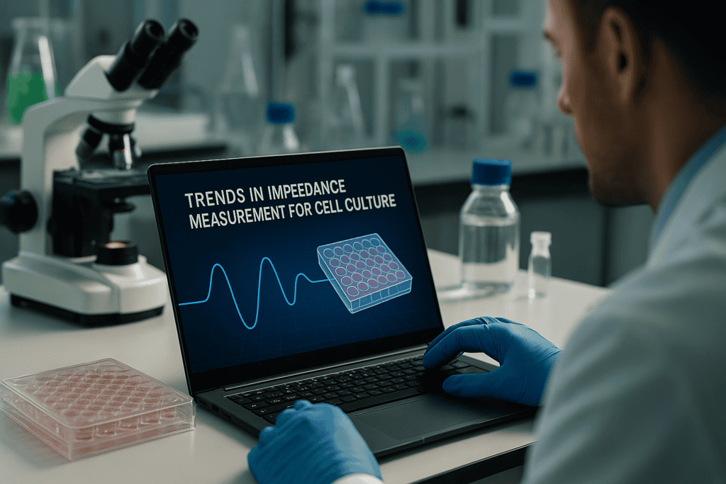
Trends in Impedance Measurement for Cell Culture
Impedance-based analysis is transforming how researchers monitor and quantify cellular behavior in real time. With increasing demand for non-invasive, label-free monitoring across biomedical research, drug discovery, and biotechnology development, electrical impedance spectroscopy (EIS) is receiving renewed attention. This article investigates the latest trends in impedance measurement for cell culture, explores the limitations of traditional methods, and outlines how integration with automated, incubator-based systems enhances reproducibility, throughput, and data richness.
Why Impedance Measurement Matters in Modern Cell Culture
Non-invasive, label-free monitoring for continuous data acquisition
Modern cell biology requires high-resolution, high-content data—with minimal interference to the cell microenvironment. Impedance measurement, particularly electrical impedance spectroscopy (EIS), offers a unique capability: monitoring living cells continuously without staining, washing, or optical systems. This technique is highly sensitive to cell attachment, proliferation, barrier function, and changes in morphology, making it ideal for real-time assessments of cell behavior in vitro.
- Continuous data acquisition over hours or days
- Compatible with various adherent cell types
- Ideal for assessing cell proliferation, migration, and cytotoxicity
- Minimal disruption to cell culture conditions
Increasingly, impedance-based readouts are being integrated into automated, high-throughput platforms, supporting complex assays such as wound healing models, barrier integrity tests (TEER), and 3D culture systems including organoids and spheroids.
Limitations of Conventional Methods in Live Cell Monitoring
Endpoint assays and manual workflows hinder reproducibility
For decades, optical microscopy, colorimetric assays (e.g., MTT, XTT), and fluorescence-based methods have been standard in cell culture laboratories. While effective for many applications, these systems introduce several limitations that impact high-throughput and longitudinal studies:
- Endpoint nature restricts temporal resolution
- Labeling or staining can influence cell physiology
- Manual workflows limit consistency and throughput
- Results often require cell lysis or fixation, ending the experiment
Furthermore, results can vary significantly depending on technician skill, reagent stability, and microscope calibration—factors that limit reproducibility, especially in multi-user or multi-site environments. In regulated sectors such as pharmaceutical development or diagnostics QA/QC, where lot-to-lot comparability and traceability are essential, these inconsistencies can impede assay validation and regulatory submission timelines.
Advances in Impedance-Based Technologies and Automation
From benchtop readers to integrated, incubator-compatible imaging systems
Contemporary impedance measurement technologies now support label-free, real-time monitoring with outputs that can be automated, digitized, and integrated into cloud-based workflows. Integrated systems such as incubator-compatible readers combine data acquisition and environmental control, reducing fluctuations that typically influence sensitive measurements.
An example is the zenCELL 貓頭鷹, a compact system designed to fit within standard incubators and to deliver continuous impedance-based cell monitoring under consistent temperature and humidity conditions. Such systems address key pain points in live-cell analysis by reducing the need to remove plates from CO₂ incubators, maintaining stable conditions and minimizing mechanical disturbances.
Core technical advances fueling the adoption of impedance systems include:
- Miniaturization of readout electronics, enabling multiwell integration (e.g., 24-, 96-, 384-well formats)
- Improved electrode manufacturing techniques for reproducible, low-noise signal acquisition
- Digital data handling, supporting scalable cloud storage and real-time analytics
- Compatibility with automation platforms for liquid handling and high-throughput screening
These developments have significantly advanced impedance applications beyond basic research, making them increasingly relevant in diagnostics development, biosensor validation, and pharmaceutical screening workflows.
Using Impedance Measurement with High-Content Workflows
Linking morphology, confluency, and viability to quantitative data
Modern cell culture research often integrates impedance measurements with live-cell imaging, enabling researchers to interpret complex cell behaviors more holistically. In such systems, impedance provides continuous quantification of cell attachment, proliferation, and confluency, while imaging captures morphological changes, organoid structure, and intercellular interactions.
Workflows combining impedance with high-content imaging support nuanced analysis in areas including:
- Cell differentiation and maturation (e.g., iPSC systems)
- Barrier function evaluation in endothelial or epithelial cell models
- Migration and wound healing assays through dynamic impedance mapping
- Drug sensitivity screening under physiologically relevant conditions
In HTS (high-throughput screening) settings, impedance readouts offer normalization capabilities for cell number variability and reduce the need for post-assay viability staining, expediting turnaround and minimizing material costs. By digitizing and timestamping each data point, these systems also enhance traceability during assay development and validation, a key requirement in GMP-compliant laboratory environments.
Benefits of Incubator-Based Impedance Systems
Improved reproducibility, sterility, and environmental consistency
Impedance systems integrated directly into incubators—rather than operated externally—offer crucial advantages for laboratories aiming to reduce variability and standardize workflows. As cell behavior is highly sensitive to environmental changes, even minor temperature fluctuations or mechanical disturbances can affect assay outcomes. By enabling true in situ monitoring, incubator-based systems provide:
- Stable CO₂, humidity, and temperature conditions throughout the experiment
- Reduced risk of contamination from plate handling or transport
- Higher data fidelity over extended culture periods
- Compatible setup with automated imaging and liquid handling systems
For facilities operating under Good Laboratory Practice (GLP) or transitioning into GMP workflows, these systems also offer advantages in traceability, as each monitored parameter is logged and time-stamped, enabling retrospective analysis and supporting audit readiness.
Key Applications of Impedance Measurement in Life Science Laboratories
Translational use cases across drug discovery and diagnostics
Impedance-based technologies support a wide range of biological analyses across preclinical research, translational biology, and quality control. Notable application fields include:
- Cell proliferation and cytotoxicity: Continuous monitoring of cell viability in response to compounds, without manual endpoint assays
- Barrier integrity and TEER: Real-time assessments of tight junction formation in epithelial and endothelial cell monolayers
- Migration and wound-healing assays: Dynamic impedance mapping following mechanical or chemical injury to the cell monolayer
- 3D culture models: Organoid growth assessed via impedance combined with microscopic imaging to track structural maturation
- Infectivity and pathogen assays: Host-pathogen interactions modeled through disruption in impedance profiles following viral or bacterial exposure
Use in diagnostic assay development is also growing, particularly in validating cellular responses to specific biomarkers or gene-editing strategies (e.g., CRISPR/Cas9). Because impedance systems offer quantifiable, label-free readouts, they are well-suited to early-stage screening as well as GMP-regulated validation phases, provided that system calibration and documentation standards are maintained.
继续阅读,以探索更深入的见解和策略。.
Optimizing Experimental Design with Impedance Parameters
Choosing the right frequency range and electrode setup for target assays
One of the most critical parameters influencing impedance measurements is the frequency range used for detection. Different frequencies probe specific electrical properties of cells and their surrounding matrix. Low frequencies (up to ~10 kHz) primarily assess extracellular ionic currents and barrier functions, while high frequencies (above 100 kHz) gauge intracellular dielectric properties. Therefore, selecting the appropriate impedance spectrum can tailor the analysis to specific biological behaviors—whether measuring tight junction formation during endothelial cell monolayer maturation or evaluating cytoplasmic changes during apoptosis.
In addition, electrode configuration—in terms of spacing, geometry, and coating—affects sensitivity and resolution. For instance, interdigitated electrodes with narrow gaps maximize surface area contact for adherent cells, enhancing signal quality. High-throughput systems often embed multiple electrode types within plates to support simultaneous analysis across conditions.
- Match frequency range to target readout: low (as low as 100 Hz) for barrier integrity, mid (10–100 kHz) for adhesion, high (>100 kHz) for intracellular changes.
Integrating Real-Time Impedance Data with AI-Based Analysis
Leveraging machine learning to detect subtle phenotypic shifts
With the proliferation of real-time impedance datasets, researchers are increasingly using machine learning (ML) algorithms to classify cell behavior patterns, detect anomalies, and predict outcomes. Modern impedance platforms often generate tens of thousands of data points per experiment, ideal for supervised learning approaches in phenotyping or toxicity prediction. Training ML models on labeled impedance profiles—for example, correlating characteristic patterns with apoptosis, senescence, or proliferation—can reveal subvisual physiological changes before morphology shifts are visibly apparent in imaging workflows.
One example is using convolutional neural networks (CNNs) to segment impedance data streams by pre-labeled profiles of cancer cell lines exposed to chemotherapeutic agents. This allows early identification of responder vs. non-responder populations in personalized oncology models.
- Use time-series clustering and ML classifiers to differentiate subtle phenotypes in high-throughput impedance datasets.
Case Study: Real-Time Drug Screening with Integrated Impedance Systems
High-throughput pharmacology in cancer cell lines using automated platforms
A pharmaceutical startup investigating kinase inhibitors adopted incubator-based impedance systems to accelerate their oncology pipeline. Using an integrated 96-well platform, they screened over 200 compounds across 10 cancer cell lines in a single week. The impedance system continuously monitored cytotoxicity and cell confluency in real time, eliminating the need for endpoint staining or plates withdrawal. Key advantages included early detection of acute toxicity, real-time EC50 curve generation, and reduced reagent costs.
Furthermore, integration with an automated liquid handler streamlined drug dilution and dispensing, producing fully reproducible conditions between replicates and across batches. Data export directly into cloud-based dashboards enabled pharmacokinetics teams to analyze curve shifts over time and correlate with imaging-derived morphology changes.
- Deploy impedance systems with automated liquid handling to dramatically reduce screening time while improving accuracy and replicability in compound libraries.
Combining Label-Free Impedance with Fluorescent Imaging
Multimodal workflows enhance mechanistic insight
While impedance gives excellent quantification of cellular status, combining it with fluorescence microscopy can enhance mechanistic investigations by pinpointing intracellular responses. Some impedance platforms support dual-modality analysis by synchronizing measurements with optical readouts in transparent-bottom well plates. This enables researchers to track cell membrane dynamics and nucleus organization alongside adhesion or proliferation indices.
Consider a wound healing assay using keratinocyte monolayers: impedance maps the closure of the wound in real time, while fluorescent tags such as phalloidin (F-actin regulator) reveal cytoskeletal alignment during migration. This dual approach allows a richer understanding of both macro (gap closure) and micro (migration directionality) dynamics.
- Use synchronized impedance and fluorescence imaging to explore both qualitative and quantitative dimensions of cell responses in one assay.
Reducing Reagent Costs and Error Potential with Label-Free Monitoring
Streamlining workflows while enhancing validity and reproducibility
Traditional live-cell assays often involve costly reagents, washes, and staining steps that increase variability and introduce user bias. Impedance-based systems require no labeling, significantly lowering consumables costs and minimizing potential for pipetting errors. The fact that experiments are monitored in real time also reduces the need for repeat runs due to missed time points or reagent instability.
In practical terms, shifting to a label-free impedance workflow saved one biotech firm over $25,000 annually in viability dye purchases during routine toxicity screens. Moreover, the switch freed up personnel from time-intensive tasks related to plate handling and endpoint preparation.
- Replace endpoint assays with impedance for cost-effective, high-throughput screening that minimizes user intervention and assay deviations.
Adoption in GMP and Regulated Workflows
Supporting documentation, traceability, and validation in compliant environments
As impedance platforms move into regulated environments such as biopharma QA/QC, diagnostic validation, and personalized medicine, they must meet standards for documentation and traceability. Leading systems now provide audit trails, exportable metadata, encrypted storage, and user access management—all essential for FDA 21 CFR Part 11 compliance. In biologics manufacturing, for instance, impedance readings are used to monitor cell growth in bioreactor-based systems, ensuring consistent lot-to-lot quality.
At a cell therapy manufacturer, impedance data are used to non-invasively evaluate stem cell expansion and differentiation, replacing destructive manual sampling. Historical datasets are then stored and compared to batch release criteria during regulatory reviews.
- Validate impedance measurement tools within compliant frameworks by using platforms equipped for auditability and GMP-ready reporting features.
Extending Impedance Applications to Co-Cultures and Organoids
Capturing complex biological dynamics in 3D and multi-cell models
With a growing emphasis on physiologically relevant models, impedance is now applied to 3D structures such as spheroids and organoids, as well as co-cultures modeling tissue interfaces. Impedance systems can measure collective adhesion forces, proliferation in dense matrices, or barrier dynamics in systems such as the blood-brain barrier (BBB). In these models, impedance can even help quantify lumen formation or detect necrotic core collapse in maturing spheroids—all without destructive sampling.
Researchers creating lung organoids to model COVID-19 used impedance as a readout of epithelial fusion, barrier tightness, and viral infectivity. Overlaying impedance data onto morphological reconstructions supported a better understanding of viral entry mechanics.
- Apply impedance to co-culture and 3D models to gain insight into multicellular dynamics, integrity, and differentiation in real time.
Cloud Connectivity and Remote Experiment Monitoring
Enabling flexible research environments and global collaboration
Cloud-connected impedance systems allow users to monitor experiments remotely, track data anomalies, or adjust protocols in real time. This capability has become especially relevant in hybrid research labs with offsite staff or global collaborative teams. Researchers can receive alerts about signal spikes, power interruptions, or threshold exceedances, ensuring minimal data loss. Shared dashboards allow real-time collaboration and troubleshooting across institutions.
During the COVID-19 pandemic, multiple academic centers reported that remote access to incubation-based impedance systems kept their drug screening workflows operational even under staffing restrictions. Dashboards enabled investigators to select hits, schedule follow-ups, or modify treatment protocols remotely without accessing the lab bench.
- Use cloud-based systems for real-time oversight and collaboration, ensuring productivity continuity across decentralized research teams.
接下来,我们将总结要点、指标和一个有力的结论。.
Future-Proofing Impedance Workflows with Modular Hardware
Scalable designs to support evolving assay demands
As experimental paradigms shift toward multiplexed, multi-organoid, and patient-derived models, impedance systems must be flexible enough to evolve. Modular impedance hardware—such as swappable electrode inserts, plate formats, and channel expansions—ensures compatibility with diverse applications, from cardiac spheroid beating assays to stem cell lineage tracking. Newer platforms now offer plug-and-play electrode arrays for microfluidic integration, allowing seamless incorporation into organ-on-chip setups.
This scalability means a single impedance reader can support both basic research and commercial screening simply by adjusting inserts or software parameters. For example, a startup developing gut-brain axis organoids migrated from planar 2D impedance plates to custom 3D well designs with integrated perfusion and real-time barrier monitoring—all while retaining the same analytic backend.
- Future-proof your lab by selecting impedance systems with modular hardware and cross-compatible accessories to support growing assay complexity.
Enhancing Interpretability with Integrated Metadata and Visual Dashboards
Making complex datasets actionable for diverse stakeholders
While impedance data is rich in temporal resolution, its interpretability depends heavily on context. Integrating metadata—such as cell type, well location, compound ID, exposure duration, and environmental conditions—ensures that patterns observed in impedance profiles can be interpreted and reused meaningfully across teams. Visualization tools now package this data into interactive dashboards, letting biologists explore signals alongside phenotypic annotations, and data scientists train AI models on standardized inputs.
One advanced approach overlays impedance traces with microscopy snapshots and drug identity, allowing real-time drill-down into anomalous wells or diverging phenotypes. For biopharma and translational teams, these dashboards facilitate data reviews without needing to parse raw signal files, enabling faster go/no-go decisions during early-stage development.
- Combine metadata integration and visual analytics to make impedance results accessible, reproducible, and actionable across interdisciplinary teams.
结论
As the life sciences field continues its shift toward high-information, physiologically relevant, and automation-compatible methodologies, impedance measurement stands out as a powerful, label-free modality capable of delivering real-time insights into cellular function. From optimizing electrode configurations to selecting frequency windows that align with biological endpoints, fine-tuning impedance parameters brings unmatched precision to experimental design.
By overlaying impedance maps with fluorescence imaging, or feeding continuous streams of data into machine learning models, researchers gain access to both qualitative and quantitative dimensions of cellular behavior. This multimodal synergy transforms standard assays—like wound healing or cytotoxicity screening—into dynamic platforms for mechanistic discovery and predictive insight. In co-culture and organoid settings, impedance excels by non-invasively tracking 3D dynamics, tissue integrity, and differentiation over time, providing a robust replacement or complement to endpoint-based techniques.
Moreover, the push toward digitized, remote-capable workflows has made cloud-connected impedance systems indispensable. Teams spanning continents can now collaborate in real time, adjusting protocols and making decisions without ever stepping into the lab. That flexibility isn’t just efficient—it’s transformative in a world where resilience, speed, and connectivity are essential to scientific progress.
As platforms grow increasingly modular and AI-integrated, and adoption rises across regulated environments like GMP and personalized medicine pipelines, impedance is no longer a niche technique—it is a core analytical pillar of modern cell biology, drug development, and biomanufacturing.
Whether you are optimizing a novel 3D assay, accelerating a drug screen, or building next-generation diagnostic models, impedance-based technologies offer the resolution, scalability, and insight needed to revolutionize your workflows. Now is the time to invest—not only in the hardware, but in the mindset shift toward dynamic, label-free, and data-rich experimentation. The future of cell culture analytics starts with an electric signal—and it’s already here.
High-Throughput Live-Cell Imaging: Scaling from 24 to 96-Well Monitoring
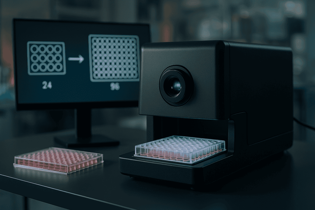
High-Throughput Live-Cell Imaging: Scaling from 24 to 96-Well Monitoring
Live-cell imaging technologies are redefining how researchers observe cellular behavior in real time. As laboratories move toward high-throughput, automated workflows, the demand for scalable, reproducible platforms for cell monitoring continues to grow. Transitioning from traditional 24-well plates to higher-density formats like 96-well plates introduces both technical challenges and significant advantages. This article explores the core principles guiding high-throughput live-cell imaging, practical considerations in scaling from 24 to 96-well formats, and the implications this has for assay development, data quality, and automation in modern laboratories. Key concepts such as optical consistency, environmental control, and equipment compatibility—especially in incubator-based systems like the zenCELL owl—will be examined in detail.
Why High-Throughput Live-Cell Imaging Matters
Real-Time Insights in Dynamic Cellular Systems
Live-cell imaging provides critical insights into cellular processes such as proliferation, migration, and differentiation. Unlike endpoint assays, it captures temporal changes, enhancing understanding of kinetics and morphological adaptations. Scaling live-cell imaging across multiple wells enables researchers to screen numerous conditions while minimizing variability—an essential feature for drug discovery, toxicology, and high-content analysis.
- Supports longitudinal studies under native conditions
- Reduces inter-experiment variability through continual imaging
- Compatible with assays requiring detailed kinetic profiling
Increasing Throughput Without Compromising Quality
Adapting live-cell imaging systems from 24-well to 96-well formats dramatically increases throughput while conserving reagents and cellular material. However, higher-density formats demand heightened optical precision, uniform environmental control, and robust imaging instrumentation capable of consistent, large-scale data acquisition without introducing artifacts or signal loss across wells.
- Enables simultaneous monitoring of 96 experimental conditions
- Paves the way for automated, parallelized experimentation
- Improves data richness per experiment while minimizing cost per condition
继续阅读,以探索更深入的见解和策略。.
Challenges in Scaling Live-Cell Imaging from 24 to 96-Well Formats
Optical and Physical Considerations in Multiwell Plate Design
High-throughput live-cell imaging requires plates with stringent optical and dimensional parameters. Standard 96-well plates feature smaller well diameters (approx. 6.4 mm) and lower working volumes compared to 24-well formats, which affects light path, depth of field, and signal intensity. Optical clarity and bottom thickness uniformity become critical in minimizing imaging inconsistencies.
- Uniform well geometry ensures consistent focal planes across wells
- Injection molding tolerances must maintain ±0.05 mm accuracy
- Selection of optical-grade polymers (e.g. polystyrene, COC) minimizes distortion
Culture Conditions and Evaporation Control
Smaller wells have higher surface area-to-volume ratios, increasing their susceptibility to evaporation and edge effects. For reproducible live-cell imaging, it is essential that environmental conditions such as humidity and CO2 levels remain tightly controlled within imaging-compatible incubators or chamber systems.
- Prevention of edge effects through plate design and sealing methodologies
- Stable temperature and humidity reduce experimental noise
- Plates designed with microclimates or perimeter wells for evaporation buffering
继续阅读,以探索更深入的见解和策略。.
Technological Advancements Enabling Scale-Up
Incubator-Compatible Imaging Systems
Traditionally, live-cell imaging required repeated manual intervention, exposing samples to environmental fluctuations. Modern systems such as the zenCELL owl integrate directly into standard CO2 incubators, enabling continuous, autonomous imaging of all wells in 24- and 96-well formats. These compact, modular platforms are optimized for minimal thermal footprint and extended in-incubator operation.
- Maintains physiological conditions throughout imaging sessions
- Removes handling-related variability in kinetic assays
- Supports remote and time-lapse imaging over multiple days
Automation and Image Analysis Integration
Coupling high-throughput imaging systems with intelligent image-processing software streamlines quantification of morphological features, growth rates, and phenotypic shifts across all wells. Data metadata tagging, segmentation algorithms, and machine learning tools now enable real-time analysis of thousands of data points per plate.
- Automated focus adjustment ensures clarity across well positions
- Built-in analysis pipelines reduce time-to-result
- Quantitative metrics such as confluence, velocity, and spreading can be extracted
继续阅读,以探索更深入的见解和策略。.
High-Throughput Live-Cell Imaging Applications
Migration and Wound Healing Assays in 96-Well Formats
Scratch or wound healing assays are widely used to study cell motility. When these assays are miniaturized in a 96-well plate, throughput is significantly increased, but precise confluence and visibility of the wound edge are essential. Live-cell imaging enables kinetic analysis of wound closure rate in each individual well without perturbation.
- Automated tracking of migration dynamics across all wells
- Optimized for screening compounds affecting cytoskeletal remodeling
- High reproducibility enabled by environmental stability during imaging
Organoid and Spheroid Monitoring
Three-dimensional culture models benefit from long-term real-time imaging to assess morphology and viability. Imaging systems scaled to 96-well plates with z-stack compatibility and sufficient focal depth allow for routine monitoring of organoid formation, aggregation, and response to treatment without frequent handling.
- Suitable for cancer biology, developmental biology, and toxicology research
- Time-lapse imaging tracks developmental trajectories non-invasively
- Small media volumes enable cost-efficient use of 3D culture reagents
Cell Proliferation and Kinetic Response Studies
Proliferation assays gain significant depth when converted from endpoint colorimetric readings to live-cell imaging of division events and morphological changes. Continuous imaging across 96 wells enables robust normalization across conditions and time points, supporting phenotype-driven drug screening.
- Enables calculation of doubling time and growth curves in real time
- Eliminates end-point reagent biases
- Data can be aligned with transcriptomic or metabolomic readouts
继续阅读,以探索更深入的见解和策略。.
Improvements in Reproducibility and Lab Efficiency
Minimizing Variation through Environmental Consistency
Integrating live-cell imaging devices directly into incubation environments removes a primary source of experimental noise—environmental fluctuations from door openings and transfers. Image acquisition without relocating cell culture plates supports higher consistency and minimizes osmotic and thermal stress across replicates.
- Maintains growth conditions throughout time-lapse imaging
- Useful for sensitive primary cell models or stem cell cultures
- Reduces stress-induced artifacts, especially in migration or cytotoxicity assays
Data-Driven Workflow Standardization
As live-cell imaging in high-density formats produces extensive quantitative datasets, laboratories can apply consistent data quality controls, calibration routines, and software-based normalization. Imaging-based workflows thus support reproducibility metrics mandated in preclinical validation and regulated lab documentation.
- Facilitates batch-to-batch comparability in regulated environments
- Links imaging data to LIMS or ELN systems through structured metadata
- Supports GLP or GMP-analogue documentation approaches in assay development pipelines
继续阅读,以探索更深入的见解和策略。.
Leveraging Machine Learning for High-Throughput Image Analysis
AI-Driven Pipelines Accelerate Discovery and Reduce Manual Bias
As high-throughput live-cell imaging produces thousands of images per experiment, manual quantification becomes impractical and subjective. Integrating machine learning (ML) algorithms allows automated interpretation of complex phenotypic data. Tools like CellProfiler Analyst, DeepCell, or custom TensorFlow-based models use supervised learning to distinguish cell types, track movement, or quantify morphological features such as nuclear size, sphericity, and clustering across all wells. Researchers can train models using annotated datasets and scale image classification efficiently, enabling real-time decisions on cell health, drug response, or toxicity.
- Use pretrained convolutional neural networks (CNNs) to accelerate segmentation accuracy
Combining Multiplexed Assays with Live-Cell Imaging
Parallel Phenotyping Enhances Experimental Depth
Live-cell imaging platforms can be used in conjunction with multiplexed fluorescent probes for real-time monitoring of cellular functions such as apoptosis, ROS activity, or mitochondrial integrity. Modern 96-well imaging systems support multiple fluorescence channels, enabling co-localization or temporal probe dynamics. For instance, using GFP-tagged biosensors alongside caspase-sensitive fluorophores allows simultaneous assessment of compound-induced cytotoxicity and pathway-specific activation. This multiplexing significantly increases the informational value of each well, especially in compound screens and pathway elucidation.
- Employ spectral unmixing algorithms to distinguish overlapping fluorophores in multiplexed readouts
Integrating Environmental Sensors for Closed-Loop Experiments
Adaptive Feedback Systems Enhance Experimental Control
In advanced live-cell imaging setups, environmental sensors (temperature, CO2, humidity) can be paired with imaging outputs to create closed-loop systems. For example, if a drop in confluency is detected during a toxicity screen, custom scripts can trigger alerts, initiate secondary assays, or even adjust incubation parameters. These feedback mechanisms are critical for long-term monitoring, particularly in stem cell or iPSC cultures that require tight condition control.
- Use programmable incubators and IOT-enabled sensors for real-time parameter adjustments
Real-Time Drug Screening at Scale
Accelerated Hit Identification with Continuous Monitoring
One of the biggest advantages of 96-well live-cell imaging is its application to high-throughput drug screening. Unlike traditional assays that rely on endpoint metabolic signals, real-time imaging provides kinetic insights into how drugs affect cell proliferation, death, or phenotypic changes. For example, anti-proliferative compounds can be assessed by monitoring changes in confluence curves or mitotic events within the first few hours. Some labs now complement live imaging with AI-curated phenotypic libraries for rapid compound triaging.
- Apply temporal normalization to account for initial seeding differences across plates
Advanced Plate Mapping and Metadata Management
Ensuring Accurate Data Attribution Across Complex Designs
As experimental layouts within 96-well plates grow more complex, rigorous plate mapping and metadata tracking become essential. Most live-cell imaging software now offers integrated design templates where experimental conditions are pre-assigned to specific wells. These templates are linked with experimental metadata, such as treatment concentration, cell line, and incubation time. Tools like PlateDesigner or proprietary LIMS integrations ensure traceability and reduce errors during data preprocessing or result reporting.
- Leverage barcoded plates and automated loggers to reduce manual error in metadata capture
Temporal Resolution Strategy for Imaging Optimization
Balancing Image Frequency with Data Volume and Biological Relevance
Determining an optimal image acquisition frequency is crucial for data richness without overwhelming storage systems. For fast-changing dynamics like mitosis or cytoskeletal rearrangement, imaging intervals of 10–15 minutes per well may be necessary. Conversely, for slow processes like differentiation, hourly or even daily acquisition suffices. Adaptive scheduling algorithms embedded in zenCELL owl and similar systems can automatically regulate imaging frequency based on observed changes in cellular phenotype—maximizing efficiency while safeguarding important transitions.
- Use pilot runs to determine the minimal temporal resolution required for your biological endpoint
Remote Monitoring and Collaborative Experimentation
Virtual Access Enables Real-Time Collaboration and Rapid Troubleshooting
Many incubator-based imaging systems now include remote access features, allowing users to monitor experiments from anywhere via secure web portals. This supports globally distributed teams and reduces the need for repeated lab entry. For example, researchers studying patient-derived organoids can grant access to collaborators or CRO partners in real time. Remote monitoring also supports rapid troubleshooting—if early apoptosis is detected in one condition, adjustments can be made mid-experiment without interruption.
- Use cloud-based storage and encryption protocols for secure, scalable data access
Case Study: Accelerated Antiviral Compound Screening Using Live-Cell Imaging
Real World Application of High-Content Screening in 96-Well Format
During a recent outbreak response study, a virology laboratory used the zenCELL owl 96-well imaging platform to screen over 300 antiviral candidates for cytopathic effect reduction. By employing confluency and cell death quantification metrics derived from time-lapse imaging, the team rapidly identified 12 promising candidates within 72 hours. Each compound’s kinetic profile was linked to its mechanism of action, verified by multiplexed fluorescent labeling of viral load and host viability. The imaging system operated autonomously over four days inside a controlled incubator, minimizing contamination risk and maximizing data fidelity.
- Combine morphological imaging with biosafety-compliant enclosure systems in infectious disease research
接下来,我们将总结要点、指标和一个有力的结论。.
Automated Data Analysis Pipelines
From Raw Images to Actionable Insights
As high-throughput imaging generates terabytes of data per experiment, scalable and automated data analysis pipelines are essential. Image preprocessing, segmentation, feature extraction, and classification must occur with minimal manual intervention. Platforms that utilize Python-based workflows—integrating OpenCV, scikit-image, or deep learning models—enable streamlined data flow from image acquisition to quantified results. These pipelines can be configured to operate in parallel across computational clusters or GPU-enabled environments, drastically reducing turnaround time from days to hours. Downstream, results export directly into statistical visualization tools or cloud dashboards for rapid interpretation.
- Use modular analysis pipelines that can be adapted across assay types and cell models
Scalability and Future-Proofing Experimental Design
Designing for Flexibility, Speed, and Reproducibility
One of the most powerful aspects of 96-well live-cell imaging is its ability to scale. From pilot screens with a handful of compounds to full-deck evaluations, well-aligned hardware and software infrastructures ensure that assays remain flexible yet reproducible. Standardizing protocol templates, creating reusable imaging schemas, and storing versioned model checkpoints allows teams to replicate and iteratively improve experiments with confidence. As future imaging platforms integrate higher resolution, broader spectral windows, or AI-based real-time control, labs prepared today with structured, data-centric workflows will adapt seamlessly without redesigning processes from scratch.
- Version-control all experimental parameters to ensure reproducibility across time and teams
Ethical Data Stewardship and FAIR Principles
Building Sustainable and Shareable Bioimage Repositories
In an era of increasing data volumes, ensuring ethical image data management is both a responsibility and an opportunity. Applying the FAIR (Findable, Accessible, Interoperable, Reusable) data principles to live-cell imaging projects facilitates knowledge dissemination, reproducibility, and multi-lab collaboration. Rich metadata annotation, standardized file formats (e.g., OME-TIFF), and integration with public or institutional image databases support long-term utility of datasets. Moreover, transparent usage of AI models—alongside mechanisms for bias detection—builds trust in analytical outcomes and strengthens the interpretive power of image-derived biological knowledge.
- Adopt community standards like OME-NGFF and maintain detailed provenance logs for images and annotations
结论
High-throughput live-cell imaging in 96-well format has redefined the pace and precision of modern cell biology. Through the integration of machine learning algorithms, multiplexed probe strategies, environmental feedback systems, and cloud-enabled remote monitoring, researchers can now perform deeper, broader, and more dynamic investigations with unprecedented efficiency. From real-time drug response tracking to long-term stem cell differentiation assays, each well becomes a window into complex cellular behaviors across time.
This technological synergy not only minimizes manual burden and subjectivity but also unlocks avenues for scaling up discovery pipelines. By incorporating advanced metadata frameworks, automated analysis pipelines, and FAIR data principles, labs ensure their work remains reproducible, shareable, and impactful. Systems like the zenCELL owl showcase how seamless instrumentation, rich data capture, and intelligent automation make it feasible to screen hundreds of conditions, track phenotypic changes in real-time, and unveil subtle cellular trends that traditional assays might overlook.
As the demand for real-world, high-content cellular analysis continues to rise—in contexts ranging from infectious disease surveillance to precision oncology—the role of modular, scalable, and intelligent 96-well imaging platforms will only grow stronger. Researchers equipped with these tools are at the forefront of a new era—where every experiment can be digitized, analyzed in real-time, and translated rapidly into actionable insights that drive therapy, innovation, and impact.
Whether you’re optimizing a new assay, evaluating a lead compound, or exploring stem cell phenotypes, the convergence of high-throughput live-cell imaging with AI, IoT, and cloud technologies ensures that your experiments are not only faster—but smarter. Embrace this transformative workflow, and turn your next imaging study into a data-rich, discovery-driven journey.
基于AI的细胞计数与汇聚度分析:从手动误差到自动化精度
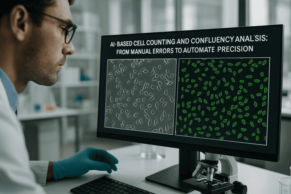
基于AI的细胞计数与汇聚度分析:从手动误差到自动化精度
In the fast-evolving landscape of cell biology and biotechnology, accuracy and reproducibility have become indispensable. Traditional cell counting and confluency assessment methods, reliant on human interpretation, are increasingly viewed as bottlenecks in modern research workflows. With advancements in artificial intelligence and live-cell imaging, laboratories can now shift from subjective manual techniques to objective, automated systems.
This article dives into how AI-based cell counting and confluency analysis are redefining precision in cell culture research. We’ll explore the limitations of manual approaches, examine the rise of automation technologies, and provide real-world lab workflows demonstrating how AI-powered tools such as incubator-based imaging systems are transforming experimental consistency and throughput.
Whether you are a cell culture specialist, a lab manager aiming to optimize resources, or a biotech professional scaling up assays, understanding these innovations is essential to maintaining competitiveness and scientific rigor.
传统方法的常见挑战和局限性
The Subjectivity Problem in Manual Cell Counting
Cell counting is foundational in cell biology, yet the standard procedures using hemocytometers or manual microscope observations are surprisingly prone to error. Despite being long-established, these techniques depend heavily on user experience, consistency in sample preparation, and visual interpretation, leading to variable outcomes between operators and even across time in the same experiment.
- High intra- and inter-operator variability
- Manual fatigue, especially in large-scale or time-lapse experiments
- Difficulty distinguishing overlapping, dead, or clustered cells
Limitations in Conventional Confluency Estimation
Confluency assessment—crucial for cell passage timing or treatment administration—is often visually approximated, using phrases like “70% confluent.” This introduces ambiguity and subjectivity, making it difficult to reproduce decisions across labs or replicate published findings. Furthermore, intermittent sampling risks missing critical morphological changes or growth milestones.
- Lack of real-time tracking of cell growth trends
- Variability from phase-contrast image interpretation
- Disruption of cell culture conditions during analysis
Together, these challenges highlight the pressing need for more reliable, automated solutions that can deliver quantifiable and reproducible data, especially in high-throughput or time-sensitive research environments.
技术进步与自动化趋势
How AI Is Reshaping Quantitative Cell Analysis
Artificial intelligence, specifically machine learning and computer vision algorithms, has significantly improved the accuracy and consistency of image-based cell analysis. AI-based cell counting and confluency analysis platforms leverage trained image recognition models to segment, count, and classify cells with levels of precision far beyond manual techniques.
Unlike traditional thresholding or morphological filters, AI systems can:
- Adapt to varied imaging conditions and cell types
- Distinguish overlapping cells and differentiate cell health states
- Continuously learn and improve through dataset refinement
Automation Across the Cell Culture Workflow
Automation has evolved from pipetting robots and media handlers to encompass real-time image acquisition and analysis. When combined with AI-driven software, these systems support closed-loop feedback mechanisms—allowing labs to monitor metrics like growth rates or cell viability and make dynamic adjustments without disrupting incubated cultures.
Key automation capabilities include:
- Uninterrupted data capture over extended periods
- Automated image analysis for kinetic studies
- Cloud-based data storage for collaborative review
Such technologies align with the growing emphasis on Good Laboratory Practice (GLP), data integrity, and heightening throughput demands in fields like regenerative medicine, cancer biology, and pharmacological testing.
活细胞成像的实用示例和工作流程
Continuous Monitoring without Culture Disruption
Live-cell imaging systems housed within incubators enable uninterrupted observation of cellular behavior from seeding through proliferation or differentiation. Rather than removing plates from the incubator for periodic inspection—risking temperature and CO2 fluctuations—these systems image cultures under consistent physiological conditions, preserving the natural state of cell populations.
For example, using a compact, incubator-compatible platform such as the zenCELL owl, researchers can automatically acquire high-frequency images across multiple wells in standard formats. This facilitates longitudinal studies that yield far more granular data than single time-point evaluations.
Automated Cell Counting Workflow in Practice
A typical automated workflow leveraging AI-based cell counting may include the following steps:
- Plate seeding with predefined cell density
- Image acquisition at intervals (e.g., every 30 minutes over 72 hours)
- Real-time image analysis providing cell number, confluency, and morphology statistics
- Data export in standardized formats for downstream analysis
Researchers can easily monitor population doubling time or assess the impact of a compound on cell proliferation dynamics, all while increasing experimental reproducibility and reducing hands-on time.
Integration with Other Automated Systems
Advanced systems can be integrated into broader automation pipelines, including robotic liquid handlers, environmental monitoring systems, and laboratory information management systems (LIMS). This bridges imaging and quantification directly with treatment applications or logistical scheduling in high-throughput screening (HTS) environments.
- Minimized human intervention and error rates
- Streamlined data flow across experimental modalities
- Support for 24/7 operation in drug discovery or production labs
继续阅读,以探索更深入的见解和策略。.
Enhancing Experimental Reproducibility with Quantitative AI Metrics
From qualitative observations to reproducible datasets
One of the most transformative advantages of AI-based cell analysis is the shift from qualitative, user-dependent results to quantitative, standardized metrics. Traditional annotations like “moderate proliferation” or “good viability” are replaced by precise, time-stamped numerical data—such as confluency percentages, cell counts per field, migration rate, and doubling time—generated automatically at each imaging cycle.
This objectivity not only improves internal consistency but also facilitates cross-study comparisons, meta-analyses, and regulatory reporting. For example, in stem cell expansion for cell therapy, consistent monitoring and documentation of proliferation metrics are critical for meeting Good Manufacturing Practice (GMP) standards.
- Use consistent, AI-generated numerical outputs to enable auditable and reproducible experiment logs.
AI-Powered Morphological Classification and Cell Health Assessment
Detecting subtle variations beyond human perception
Modern AI algorithms go beyond simple counting—they’re now capable of segmenting individual cells and classifying them based on morphological features. This allows researchers to distinguish between healthy, apoptotic, necrotic, and mitotic cells in culture without the need for staining or labeling.
For instance, AI-enabled software can analyze nuclear condensation, blebbing, or cytoplasmic granularity to flag early signs of apoptosis. In cancer research, such fine-grained discrimination supports dynamic cytotoxicity assays without disrupting cell viability, enabling longitudinal tracking of drug efficacy.
- Train AI models on specific image sets to tailor morphological classifications for your unique research goals.
Adapting AI Workflows to Diverse Cell Types and Assay Conditions
Flexibility of deep learning models across research disciplines
One of the barriers to broad AI adoption in life sciences has been the diversity of cell phenotypes—fibroblasts, neurons, spheroids, T-cells—each presenting unique morphology. However, AI solutions now incorporate convolutional neural networks (CNNs) capable of learning from varied datasets, adapting to both adherent and suspension cultures, as well as 2D and 3D systems.
Leading platforms allow researchers to curate their own training datasets or utilize pre-trained models optimized for specific assays, such as wound healing, neurite outgrowth, or spheroid growth inhibition studies. This flexibility dramatically shortens setup time and increases out-of-the-box accuracy.
- Select AI tools with customizable training pipelines to handle new or rare cell models.
Accelerating Decision Making with Real-Time Alerts and Dashboards
Enabling timely intervention with automated notifications
With integrated dashboards and remote-access platforms, AI-enabled systems can send real-time alerts when specific thresholds are crossed—such as reaching 80% confluency or detecting sudden declines in cell health. This capability minimizes lag between observations and interventions, which is particularly crucial when managing time-sensitive tasks like transfection or induction of differentiation.
For example, production-scale labs using CHO cells for biopharmaceutical manufacturing can rely on such alerts to optimize feeding schedules or harvest timing, improving yield while conserving resources.
- Configure dynamic alerts based on custom metrics (e.g., doubling time deviation or peak proliferation rate).
Optimizing High-Content Screening for Drug Discovery Pipelines
From image capture to actionable insight—at scale
AI-powered imaging platforms have revolutionized high-content screening (HCS) by automating not only image acquisition but also multiparametric analysis. In pharmacological testing, this means simultaneously assessing proliferation, viability, morphology, and response markers across thousands of compounds, dramatically accelerating the lead identification process.
Large pharmaceutical firms deploy systems such as the Incucyte® or ImageXpress linked with neural networks trained on cytotoxicity endpoints. Integration with LIMS enables auto-tagging of positive hits, reducing days of manual effort to hours of automated processing.
- Integrate AI-based image analysis directly into compound screening pipelines to reduce false positives and accelerate validation.
Minimizing Bias through Blind, AI-Based Analysis
Combatting confirmation bias and user influence
Conventional manual analysis is inherently vulnerable to cognitive bias. Whether consciously or subconsciously, researchers may interpret borderline results in favor of their hypothesis. AI systems, by contrast, apply the same analytical criteria across all samples, blind to experimental groups or desired outcomes.
This objectivity is particularly valuable in blinded studies or preclinical trials where regulatory bodies demand unbiased, statistically robust data. By eliminating observer bias, AI enhances transparency and reinforces data credibility in grant applications, publications, and audits.
- Standardize analysis protocols across team members and time points using predefined AI analytic templates.
Case Study: Streamlining QA in a Biotech Manufacturing Environment
How one biotech optimized quality assurance using live-cell AI tools
A mid-sized biotech firm producing stem cell-derived cardiac cells faced issues related to variability in cell differentiation and contractility. Manual inspections led to subjective judgments and inconsistent batch quality. After implementing an AI-based live-cell imaging system inside the QA incubator, the team began acquiring hourly microscopy images across cloned production flasks.
AI counted cells, measured confluency, and evaluated pre-trained beat-pattern algorithms to monitor coordinated contractions. Insights from early differentiation stages now allow the team to calibrate media changes proactively. The result: a 40% reduction in failed batches and a 30% improvement in downstream consistency.
- Use AI-generated insights to standardize criteria for batch release and reduce manual QC bottlenecks.
Leveraging Cloud Integration for Multi-Site Collaboration
Real-time data access empowers distributed research teams
As collaborations expand across academic and industrial sites, cloud-integrated imaging systems allow real-time access to AI-analyzed cell culture data from anywhere in the world. Labs can now compare culture confluency, proliferation trends, and endpoint results without shipping samples or scheduling virtual microscopy sessions.
Such centralized access streamlines remote troubleshooting, enhances transparency for cross-institutional studies, and ensures faster feedback loops in contract research or CRO settings. Teams using platforms like Axion Biosystems, Sartorius IncuCyte, or zenCELL owl can jointly annotate or flag anomalies during the culture period, reducing decision delays.
- Choose systems with open APIs or cloud support to unify remote data access and analysis pipelines.
接下来,我们将总结要点、指标和一个有力的结论。.
Scaling AI-Enabled Workflows with Automation and Robotics
Bridging digital image analysis with physical lab automation
The next step in transforming experimental reproducibility lies in integrating AI-powered image analysis with robotic handling systems and automated incubators. By pairing real-time confluency data or health metrics with programmable robotic protocols, workflows such as passaging, media exchange, or compound dosing can be fully automated based on objective criteria, not time-based approximations.
For example, an AI-monitored culture can signal when proliferation slows—automatically triggering a robotic pipetting sequence for replenishing growth media or initiating differentiation protocols. This closed-loop interaction between digital analysis and physical action reduces operator variability and allows true 24/7 lab automation, essential for high-throughput screening and regenerative medicine production pipelines.
- Link AI analysis outputs with lab robotics to enable conditional, event-driven process automation.
Future Horizons: Incorporating Predictive Modeling in Cell Culture Analytics
Beyond observation—toward anticipation and optimization
The frontier of AI in cell culture is moving from descriptive to predictive analytics. By leveraging historical culture data, environmental parameters, and morphological trends, machine learning models can anticipate outcomes such as culture failure, peak efficiency points, or optimal harvest windows. This evolution transforms AI from a monitoring tool into a proactive forecasting engine.
In long-term organoid cultures or perfusion bioreactors, time-series analyses can forecast necrotic core formation or nutrient depletion events before visible signs occur. Early warnings empower lab teams to adjust protocols preemptively—shifting from reactive troubleshooting to proactive optimization.
- Incorporate historical datasets into training pipelines to enhance predictive power and preempt failure points.
结论
The integration of AI-based cell analysis is fundamentally redefining how labs conduct, monitor, and interpret biological experiments. From eliminating subjective assessments to enabling predictive insight, these technologies form the bedrock of a more reproducible, efficient, and scalable research environment. Whether you’re navigating early-stage discovery or managing GMP-compliant production, the objectivity and precision afforded by AI can elevate both the rigor and speed of your workflows.
Key takeaways include the ability to generate consistent, quantitative metrics that enhance both internal validity and cross-lab comparisons; the capability to detect subtle morphological variations invisible to the human eye; and the adaptability of AI models across diverse cell types and assay formats. As AI tools continue to evolve, features like real-time alerts, cloud-based collaboration, and predictive modeling further bridge the gap between experimentation and actionable decision-making.
Moreover, as these platforms become increasingly interoperable—with APIs, LIMS integration, and robotics compatibility—labs can design fully automated, closed-loop workflows that are not only reproducible but also scalable for industrial applications. This democratization of high-content imaging and analysis ensures that teams of all sizes can harness the power of AI without extensive computational infrastructure.
Now is the time to shift from fragmented, manual analysis to a unified, AI-powered strategy that boosts transparency, accelerates discovery, and minimizes bias. Whether you’re striving for publication-grade data, regulatory readiness, or operational excellence, AI-based image analysis offers the clarity and consistency modern science demands.
Invest in these tools not just for automation or convenience—but to future-proof your science. By embracing AI today, you’re laying the foundation for a more reliable, reproducible, and insightful tomorrow.
自动化伤口愈合和迁移检测:如何实现可重复的结果

自动化伤口愈合和迁移检测:如何实现可重复的结果
Cell migration and wound healing assays are essential tools in cell biology, oncology, regenerative medicine, and pharmacological research. Traditional scratch assays, while widely used for studying collective cell movement and regeneration, often suffer from inconsistencies and subjective data interpretation. With the increasing need for high-throughput screening, real-time observation, and reproducibility in life science applications, automated wound healing and migration assays have emerged as a robust solution.
This article explores the scientific and technical considerations for achieving reproducible results in automated assays, covering validation strategies, live-cell imaging technologies, and trends in scalable labware development. Researchers, lab managers, and biotech developers will gain a deep technical understanding of the methods and materials that support the reliability of automated wound healing workflows under regulated conditions.
Challenges in Traditional Wound Healing Assays
Technical Limitations of Manual Scratch Methods
The classic wound healing assay involves manually creating a cell-free zone (“wound”) in a confluent cell monolayer using pipette tips or blades. Despite its simplicity, this method introduces significant bias across time points and replicates due to mechanical inconsistencies and human error. These technical variabilities limit assay reproducibility and reduce confidence in comparative data.
- Manual scratches vary in width, edge shape, and cell detachment effects.
- Edge damage can release intracellular contents, altering local microenvironments.
- Subjective imaging and endpoint analyses hinder standardization in multi-well formats.
Environmental and Workflow Inconsistencies
Reliance on traditional microscopes outside of incubators introduces temperature and CO₂ fluctuations that disturb cell physiology. Moreover, inconsistent assay timing and imaging delays further impair reproducibility, especially in time-sensitive applications such as drug screening or migration kinetics.
- Movement of plates between incubators and imaging stations creates environmental shocks.
- Manual imaging scheduling leads to uneven observation intervals.
- Data quality suffers from off-incubator imaging due to focus drift and condensation.
Technology Advancements Driving Automation
Automated Live-Cell Imaging Platforms
To ensure consistent observation and quantitative data generation, many laboratories are adopting incubator-compatible imaging systems. Continuous monitoring using compact, automated devices—such as the zenCELL owl—enables real-time data acquisition without removing cells from optimal culture conditions.
- Real-time kinetic data of cell migration and gap closure.
- Imaging within a standard incubator reduces environmental variability.
- Multichannel and time-lapse images support comprehensive, unbiased analysis.
Precision Labware for Assay Standardization
Lab plastics tailored for migration assays, such as pre-defined inserts and wound field designs in multiwell formats, offer mechanical consistency and improve performance metrics across experiments. These precision-molded formats eliminate edge variability and are compatible with automated liquid handling systems, crucial for scalable workflows.
- Custom-designed wells ensure consistent scratch width and geometry.
- Transparent, optically clear plastics (e.g. polystyrene, COC) support high-resolution imaging.
- Surface functionalization (e.g. TC treatment) promotes even cell adhesion and growth.
Implementing Automated Wound Healing Assays
Workflow Integration in Regulated Lab Environments
Transitioning to automated wound healing and migration assays involves synchronizing hardware, consumables, and software within a validated quality control framework. Especially in GMP or cGMP-compliant labs, every aspect from assay design to data output must adhere to robust documentation and reproducibility standards.
Key considerations include:
- Use of validated, traceable labware and imaging instruments.
- Implementation of audit trails and data storage compliant with 21 CFR Part 11.
- Standard operating procedures (SOPs) for gap creation, cell seeding, media change, and imaging.
Examples of Optimized Automated Assay Protocols
The use of precision multiwell plates combined with real-time imaging allows for reproducible assay designs. For instance, combining a 24-well plate with embedded cell exclusion zones and the zenCELL owl system allows a continuous 72-hour migration monitoring without manual intervention. Such workflows are particularly valuable in kinetic drug response studies or testing growth factor effects on cell mobility.
Benefits include:
- Simultaneous real-time monitoring across multiple wells or conditions.
- Reduction in assay-to-assay variability through standardized plate formats and protocols.
- Minimized operator time while maximizing data resolution and analysis consistency.
Enhanced Reproducibility with Incubator-Based Imaging
Environmental Stability Improves Cellular Fidelity
Maintaining cells within controlled incubator conditions during imaging preserves metabolic activity and cellular behavior, especially important for sensitive cell types. Incubator-compatible systems like the zenCELL owl eliminate the need for sensor recalibration and refocusing between observations, reducing variability introduced by manual microscopy sessions.
- Sustained 37 °C, 5% CO₂ eliminates thermal and pH shifts during time-lapse studies.
- High-frequency imaging captures transient events and accelerates migration rate calculations.
- Time-resolved imaging enables statistical analysis of wound closure kinetics across biological replicates.
Automated Image Acquisition and Analysis
Advanced software algorithms quantify wound area and cell movement automatically, reducing observer bias. Integration of tailored software workflows allows users to standardize analysis endpoints and minimize data handling errors. These systems also enable batch processing for screening applications requiring high-throughput assay formats such as 96-well plates.
- Image segmentation algorithms ensure consistent wound edge detection.
- Metadata tagging ensures traceability for GMP record-keeping requirements.
- Analysis modules support quantitative kinetics for migration speed and proliferation indices.
Applications Beyond Classical Wound Healing
Cell Migration, Organoids, Proliferation, and Drug Screening
Automated wound healing assays form the basis for several related in vitro assessments. Researchers apply similar protocols for evaluating fibroblast, endothelial, or cancer cell invasion under defined gradients. Furthermore, organoid-based migration assays and barrier integrity models are expanding the scope of these techniques by integrating 3D formats and co-culture systems.
- Migratory behavior in cancer models to assess metastasis potential.
- Barrier reformation in epithelial monolayers to study tight junction recovery.
- Proliferation tracking alongside migration for combined mechanistic investigations.
High-Throughput Screening (HTS) and Multiplexed Studies
Automated imaging and labware compatibility with robotic pipetting platforms support high-throughput settings where multiple drug candidates or treatment conditions must be evaluated simultaneously. Optically clear injection-molded lab plastics in 96- or 384-well plates allow for scalable adoption of migration and wound healing assays while preserving imaging fidelity.
- HTS-compatible plate formats reduce reagent volumes and increase parallelism.
- Data consistency ensures reliable lead identification in early drug discovery.
- Integrated assay automation supports streamlined workflows across R&D and quality labs.
继续阅读,以探索更深入的见解和策略。.
Advanced Assay Calibration for Quantitative Accuracy
Optimizing Imaging Parameters and Reference Controls
Achieving consistent, high-fidelity results in automated wound healing assays requires calibration of imaging parameters—especially when using time-lapse systems and multichannel microscopy. Factors such as exposure time, focus depth, and pixel resolution must be precisely defined during assay development and kept constant throughout the experiment. The use of internal reference controls and calibration beads enables normalization across different imaging sessions or assay runs, improving inter-experimental repeatability.
- Perform flat-field correction and illumination uniformity tests to avoid uneven signal intensity.
- Include wells with known cell migration rates or migration-inhibited controls for internal benchmarking.
Optimizing Cell Density and Seeding Uniformity
Consistent Monolayer Confluence Enhances Assay Comparability
Uneven or low initial cell densities lead to variability in wound closure dynamics. For accurate wound healing measurement, it’s critical to standardize the seeding process across wells and experiments. Automated liquid handlers or multi-channel pipettes ensure reproducible delivery, while pre-coating plates with extracellular matrix components like fibronectin or collagen enhances uniform cell attachment and spreading. In high-throughput formats, vortex mixing followed by automated dispensing prevents cell clumping and supports monolayer homogeneity.
- Validate optimal seeding densities for each cell type to reach 90–100% confluence before wound initiation.
- Use robotic plate fillers or cell dispensers to minimize pipetting-driven variation during multicondition assays.
Chemical and Mechanical Gap Creation Strategies
Consistent Exclusion Zones Enable Standardized Kinetics
To eliminate the inconsistency of manual scratches, many labs have transitioned to mechanical inserts and hydrogel-based stencils for wound generation. These devices create reproducible gaps in monolayers without damaging surrounding cells. For example, silicone insert systems or removable polymeric stoppers allow users to lift predefined barriers after cell adhesion, enabling sharp, repeatable exclusion zones. Alternatively, enzymatic methods using dispase or non-cytotoxic peeling films can detach cells precisely from designated regions, facilitating gentle wounding in sensitive cultures.
- Use wound inserts sized to fit your specific multiwell plate and application format.
- Evaluate enzymatic or mechanical approaches based on target cell sensitivity and assay duration.
Automated Data Management for Regulatory Compliance
Scalable, Audit-Ready Workflows for GxP Environments
In regulated lab settings, automated wound healing platforms must support traceability, data integrity, and compliance with global standards such as 21 CFR Part 11 or EU GMP Annex 11. Integration of imaging systems with laboratory information management systems (LIMS) ensures secure data storage, retrieval, and auditability. Real-time tagging of metadata—including incubation parameters, imaging intervals, and treatment conditions—further enhances downstream data mining and reproducibility.
- Implement secure cloud-based storage or encrypted servers with digital access control verification.
- Use SOP-defined filename conventions and version control for image and analysis documentation.
Custom Software for Tracking Cell Behavior Over Time
Quantitative Analysis Algorithms Enhance Biological Insights
Modern imaging platforms deploy machine learning (ML) and AI-powered software to track individual cell movements, collective migration patterns, and proliferation events. These advanced tools allow researchers to differentiate between random cell motility and directed migration or chemotaxis. For example, software can calculate velocity vectors, persistence time, and path tortuosity, providing deeper biological meaning to mere wound area reduction metrics.
Several systems incorporate automated segmentation for cell tracking using DIC, fluorescence, or phase-contrast imaging. Users can define dynamic thresholds for wound area clearance, confluence index, and morphological parameters, enabling high-content screening directly from the wound healing assay.
- Use AI-assisted tracking to distinguish between contact inhibition, mitotic activity, and true migration.
- Apply morphokinetic metrics such as circularity and aspect ratio to evaluate epithelial-to-mesenchymal transitions (EMTs).
Case Study: Real-Time Drug Response Profiling
Automated Wound Healing as a Phenotypic Screening Tool
In one applied example, a pharmaceutical R&D team utilized a zenCELL owl system combined with barrier-based 24-well migration plates to analyze the effect of kinase inhibitors on breast cancer cell motility. Cells were seeded into the plates with removable stoppers forming 500-micron wounds. After a 24-hour treatment with varying drug concentrations, cell migration was tracked hourly for 48 hours. Software automatically quantified wound closure rates, providing EC₅₀ values correlated with cell viability and morphological changes.
This workflow eliminated manual analysis steps, reduced turnaround time by 67%, and increased reproducibility by 35% compared to traditional microscopy and hand-drawn ROI analysis. Integration with a LIMS system allowed the same workflow to be reused for other cancer cell lines and therapeutic candidates.
- Automated systems support reproducible, high-resolution phenotypic profiling in early-stage drug selection.
- Time-course migration tracking allows insight into both onset and durability of drug responses.
Multiparametric Analysis: Migration Meets Proliferation
Dissecting Cellular Contributions Using Combined Readouts
Distinguishing between cell migration and proliferation is critical for interpreting wound healing data, particularly in cancer models or regenerative medicine. Advanced assays incorporate dual-channel analysis, where a proliferation marker like BrdU or EdU is added in tandem with live-cell imaging. This approach allows researchers to decouple the effect of treatment on cytostasis versus directional movement. Furthermore, overlaying cell cycle reporters such as FUCCI enables a cell-by-cell phase analysis within the migrating population.
Some commercial assay platforms now integrate fluorescence overlays directly into their imaging timelines, providing seamless correlation of cell division markers with positional data. This dual profiling enhances mechanistic understanding and leads to more targeted therapy optimization.
- Use cytostatic controls alongside migration inhibitors to benchmark assay outputs and avoid data misinterpretation.
- Integrate nuclear and cytoplasmic markers for real-time proliferation tracking within wound edges.
Strategies for Time-Efficient Optimization of Assay Conditions
Reducing Setup Time Without Compromising Data Quality
To streamline assay setup across multiple conditions or cell lines, labs can adopt modular optimization strategies. This includes miniaturized pilot runs in 12- or 24-well formats using automation-compatible inserts and imaging loops to quickly assess optimal seeding density, confluence timing, and treatment start times. Imaging software presets can then be programmed for batch acquisition and stitched image compilation where required.
Instituting a Design of Experiments (DoE) approach across temperature, serum levels, and coating conditions accelerates parameter tuning while maintaining scientific rigor. With automated cuvette or plate washer compatibility, solutions used for washing or media change are made more uniform, further boosting inter-assay comparability.
- Implement DoE-based pilot studies for rapid optimization of cell and media variables.
- Maintain matched biochemical conditions across wells using automated liquid handling protocols.
接下来,我们将总结要点、指标和一个有力的结论。.
Quality Control Checkpoints Across the Workflow
Ensuring Consistency from Reagent Prep to Data Output
Maintaining consistency across multiple runs of automated wound healing assays depends on well-defined quality control (QC) checkpoints. Each step—from reagent handling to image acquisition—can introduce variability if not properly standardized. Including both technical replicates and biological controls ensures that assay robustness remains high despite inevitable experimental shifts. For instance, preparing master mixes of media or inhibitors reduces reagent batch effects, while validating cell health using viability stains like calcein-AM or PI provides an upstream QC trigger.
Image pre-processing QC is often overlooked; however, verifying focus stability, drift correction, and stitching accuracy is essential when dealing with multiday, time-lapse imaging. Automated software platforms increasingly include preconfigured validation protocols that can flag anomalies in image acquisition or well-level inconsistencies.
- Design QC gates based on biological endpoints (e.g., confluence threshold) and technical parameters (e.g., image illumination profile).
- Create dashboards within your LIMS to track cell line passage numbers, reagent expiry, and system calibration dates.
Scalable Deployment Across Screens and Teams
From Discovery to Preclinical Development
As laboratories scale their wound healing workflows beyond single-user research setups into multiuser or interdepartmental pipelines, harmonizing protocols and data interpretation becomes imperative. Automated wound healing systems support scalability by enabling protocol preset sharing, remote access for data review, and standardization via machine-readable metadata. These benefits are especially valuable for pharmaceutical teams operating in dispersed preclinical ecosystems or organizations performing global concurrent screening studies.
To aid reproducibility, many setups utilize shared protocol libraries that ensure consistency in assay composition, imaging schedules, segmentation algorithms, and analysis parameters. Furthermore, multi-site teams can implement collaborative QC matrices that monitor assay fidelity across operator, site, and run timeline, creating a robust knowledge base built on consistent and well-annotated data.
- Standardize workflows using interchangeable run templates and centralized data labeling taxonomies.
- Use shared LIMS or cloud-based ELN platforms to propagate validated protocols across teams and programs.
结论
Automated wound healing and migration assays have evolved into high-precision, reproducible platforms capable of delivering deep phenotypic insights across cell biology, oncology, and regenerative research. By embracing advancements in imaging calibration, cell seeding practices, and gap generation strategies, researchers can significantly minimize inter-assay variability while capturing rich, biologically relevant data.
Through integration with LIMS systems and application of custom software analytics, these platforms now support not only robust migration tracking but also time-resolved proliferation analysis and mechanistic dissection of cellular behaviors. The inclusion of dual readouts and multiparametric overlays allows for a comprehensive view of wound closure dynamics, improving the fidelity of conclusions drawn in both discovery and translational settings.
Key success factors include adopting standardized imaging protocols, consistent reagent preparation, and automated data handling practices to conform with regulatory requirements. As demonstrated in the case study and execution strategies, the shift to scalable, automation-driven workflows doesn’t just save time—it elevates the entire assay strategy, accelerating the path from cellular insight to actionable outcome.
Whether you’re optimizing for high-throughput compound screening, unraveling the underpinnings of migration in disease models, or validating therapeutic interventions, mastering these advanced wound healing assay techniques will put you ahead. By aligning precise assay architectures with flexible software and hardware integration, your lab can scale discoveries confidently and reproducibly.
Now is the time to rethink your approach to in vitro migration studies. Invest in automation, apply rigorous standardization, and let modern imaging technologies work for you. The future of wound healing assay performance lies in reproducibility, resolution, and real-world scalability—embrace it fully, and transform your research outcomes one experiment at a time.
What is human serum and how i use it in cell culture applications?

What is human serum and how i use it in cell culture applications?
Understanding Human Serum: Definition and Biological Role
What is Human Serum?
Human serum is the cell-free, coagulated fraction of human blood. It is derived by allowing whole blood to clot and then removing the clot and cellular components through centrifugation. The resulting fluid contains a complex mixture of proteins, electrolytes, hormones, and growth factors, but lacks fibrinogen and other clotting factors present in plasma. The absence of clotting components can reduce variability in certain assays and supports applications where antibodies or cytokines in the native serum matrix are critical.
- Contains immunoglobulins, albumin, electrolytes, and various metabolic regulators
- Lacks fibrinogen and clotting cascade proteins found in plasma
- Harvested under standardized, traceable conditions to ensure biosafety
继续阅读,以探索更深入的见解和策略。.
Scientific Applications of Human Serum in Cell Culture
Use in Primary Human Cell Cultures
Primary cells derived from human tissues often perform optimally in media supplemented with human serum due to species-specific compatibility. For example, human mesenchymal stem cells (hMSCs), peripheral blood mononuclear cells (PBMCs), and dendritic cells commonly show improved viability and differentiation when cultured in human serum compared to fetal bovine serum (FBS). The aligned cytokine and growth factor profiles support physiological cell behavior and reduce immunogenic artifacts.
- Supports functional maturation in immune cell assays
- Minimizes xenogeneic immune responses in model development
- Enhances translational relevance in personalized medicine research
Immunology and Antibody Research Applications
In immunology workflows, human serum provides an authentic matrix for testing antibody-antigen interactions, complement activation, and cytokine responses. Its endogenous immunoglobulins and complement proteins are particularly relevant when modeling immune mechanisms in vitro. Laboratory workflows such as antibody screening and flow cytometry often require serum batch testing to avoid interference or nonspecific binding.
- Enables study of native Fc receptor interactions
- Supports complement-dependent cytotoxicity (CDC) assays
- Preserves in vivo-like conditions for diagnostic development
继续阅读,以探索更深入的见解和策略。.
Addressing Variability and Quality Control in Human Serum
Donor Variability and Batch Consistency
Due to its human origin, human serum demonstrates inherent donor variability in protein concentration, hormone levels, and immunoglobulin content. This variability can influence reproducibility across experiments unless appropriately managed. Sourcing strategies, such as using pooled human serum from multiple donors, help mitigate this issue. Additionally, each batch should be tested in the target cell system to verify performance consistency.
- Pre-screening batches in relevant cell lines is advisable
- Pooled serum reduces individual donor outliers
- Traceability and documented donor screening support ethical compliance
Documentation and Regulatory Considerations
Human-derived reagents must comply with strict ethical, biosafety, and documentation standards. Sera for research use are typically collected under informed consent and subject to infectious disease screening, including HIV, HBV, HCV, and syphilis. Technical documentation, typically available from providers such as shop.seamlessbio.de, should include certificate of origin, donor eligibility criteria, and testing methods.
- Certificates of analysis support GLP and GMP-aligned workflows
- Lot traceability reduces compliance and reproducibility risk
- Alignment with region-specific ethical guidelines (e.g., EU Tissues Directive)
继续阅读,以探索更深入的见解和策略。.
Best Practices for Using Human Serum in the Laboratory
Handling and Storage Guidelines
To preserve the functional integrity of human serum, proper storage and handling are essential. Serum should be stored at -20°C or lower to avoid degradation of labile components. Before use, it should be thawed slowly at 2–8°C and gently inverted to ensure uniform mixing. Repeated freeze-thaw cycles should be avoided to maintain bioactivity and minimize protein denaturation.
- Single-use aliquots minimize freeze-thaw artifacts
- Transition to cell culture flasks or plates should be done under sterile conditions
- Compatible with standard plasticware from sources such as shop.innome.de
Serum Qualification in Specific Assays
Experimental design often necessitates serum batch qualification, especially in sensitive downstream assays. For example, in monoclonal antibody screening, the presence of endogenous IgG in human serum might confound measurements if not accounted for. Live-cell imaging platforms, such as the incubator-compatible system described at zencellowl.com, may assist in monitoring how specific serum lots affect cell morphology and behavior in real-time, aiding the selection of optimal batches.
- Consider testing multiple batches in parallel experimental setups
- Incorporate documentation of serum lot into laboratory records
- Use live-cell imaging to evaluate growth kinetics and morphology dynamically
继续阅读,以探索更深入的见解和策略。.
Strategic Integration of Human Serum in Workflow Design
Long-Term Project Support and Risk Management
In longitudinal studies or large development programs, variability in biological materials can compromise reproducibility. To mitigate this, many laboratories implement custom batch reservation, qualification testing, and lot documentation support services. These approaches are particularly critical in workflows involving antibody development, where consistent cellular responses and matrix backgrounds are vital for screening fidelity.
- Reserve characterized serum batches for long-term studies
- Use custom testing services to qualify sera under target assay conditions
- Document donor origin, protein content, and immunoglobulin levels to maintain traceability
Cultural and Ethical Considerations
Use of human biological materials must adhere not only to scientific standards but also to ethical and legal frameworks. Human serum products intended for research are typically sourced from screened, consenting blood donors. Researchers must ensure compliance with local governance bodies and institutional review boards and consider regional variations in sourcing guidelines and donor screening practices.
- Check donor consent protocols and legal sourcing documentation
- Align usage with institutional biosafety and ethics guidelines
- Review technical data sheets for comprehensive testing panels
继续阅读,以探索更深入的见解和策略。.
Streamlining Serum Lot Selection for Experimental Reproducibility
Implementing an Evidence-Based Qualification Workflow
Selecting the right human serum lot can significantly impact experimental outcomes, especially for high-sensitivity assays or regulatory-stage workflows. A rational approach to serum qualification involves screening multiple lots side-by-side using standard operating protocols (SOPs) to compare cell viability, proliferation, morphological changes, and biomarker expression. Incorporating performance metrics, such as population doubling time or immunophenotyping outcomes, allows researchers to choose lots that align with assay-specific requirements.
- Develop a scoring system for batch comparison based on relevant assay metrics
- Use benchmarked cell lines or donor cells to standardize responses
- Record all experimental parameters in laboratory data management systems (e.g., ELN or LIMS)
Utilizing Human Serum in 3D and Organoid Culture Systems
Enhancing Physiological Relevance in Advanced Cell Models
Human serum plays a pivotal role in supporting 3D cell culture models and organoid systems by better mimicking in vivo conditions than animal-derived supplements. In models such as liver organoids or tumor spheroids, human serum provides human-specific growth stimulators and cytokines that support more accurate tissue-like behavior. Studies have shown increased functional expression of epithelial markers and metabolic enzymes in organoid cultures exposed to human serum compared to those raised on FBS-supplemented media.
- Precondition medium with human serum to promote uniform cell aggregation
- Monitor specific tissue markers like albumin in hepatic organoids as functional readouts
- Combine with hydrogel matrices for tissue-like architecture
Supporting Serum-Free to Human Serum Transitions
Engineering Media for Hybrid Feeding Strategies
Transitioning from serum-free or defined media to human serum-supplemented conditions can be challenging due to differences in osmolarity, nutrient concentrations, and signaling molecule profiles. A hybrid conditioning approach—where cells are gradually exposed to increasing concentrations of human serum—helps mitigate stress responses and maintain phenotypic consistency. For example, clinical-grade stem cell expansion protocols often incorporate a stepwise adaptation from xeno-free media to human serum-enriched media to preserve differentiation potential without inducing shock or apoptosis.
- Introduce human serum in 10-20% increments every 24–48 hours
- Track cell morphology, confluency, and doubling time after each transition
- Validate pathway activation using flow cytometry or qPCR markers
Custom Supplementation and Reconstitution Approaches
Tailoring Human Serum for Targeted Applications
For specific research demands, custom supplementation of human serum is often employed to enhance or suppress targeted pathways. For instance, supplementation with recombinant growth factors like EGF or IL-2 can boost proliferation or immune activation on particular platforms. Some researchers also use immunoglobulin-depleted or heat-inactivated variants of serum to tune the impact on signaling cascades or complement activity. Providers often offer customized processing services for batch-specific modification upon request.
- Use cytokine-spiked human serum for T cell activation or NK cell assays
- Heat-inactivate serum at 56°C for 30 minutes to eliminate complement activity where undesired
- Consider delipidated or charcoal-stripped variants for hormone-sensitive assays
Integrating Human Serum into Automated High-Throughput Systems
Ensuring Compatibility with Robotics and Screening Pipelines
Automated liquid handling and high-throughput screening (HTS) platforms demand consistency and stability in reagent composition. Human serum can be fully integrated into these systems with careful preparation—such as pre-filtering and aliquoting—to avoid clumping or pipetting inconsistencies. In HTS drug discovery pipelines, human serum adds critical relevance to pharmacokinetic and cytotoxicity modeling by providing a protein-binding environment closer to human plasma.
- Use 0.22 µm sterile filtration to reduce particle formation before robot loading
- Test inter-assay and intra-assay CV for serum-containing wells in 96- or 384-well plates
- Analyze serum-induced background signals in luminescence or absorbance-based assays
Case Study: Enhancing PBMC-Based Assays with Human Serum
Real-World Example from an Immuno-Oncology Laboratory
A Brussels-based biotechnology group developing bispecific antibodies for T cell redirection encountered variability in PBMC-based cytotoxicity assays using FBS. Upon transitioning to pooled human AB serum, they observed increased reproducibility in inter-donor responses and improved cytokine signatures reflective of in vivo conditions. Importantly, the presence of functional complement proteins in the human serum allowed evaluation of both complement-dependent cytotoxicity (CDC) and antibody-dependent cellular cytotoxicity (ADCC) in parallel systems.
- Switched from FBS to pooled AB serum to reduce xenogeneic immune impact
- Validated cytotoxicity using IFN-γ ELISA and CD107a degranulation markers
- Incorporated live-cell imaging (via Zencell Owl) to confirm target-directed lysis events
Data-Driven Documentation to Support Regulatory Submissions
Capturing Complete Audit Trails and Performance Logs
When research progresses toward therapeutic product development, regulators require full traceability of all raw materials including reagents like human serum. Documentation should log batch numbers, donor eligibility summaries, processing methods, storage conditions, and all pre-use qualification data. Tools such as digital laboratory notebooks (DLNs) and laboratory information management systems (LIMS) allow seamless linking of cell culture data, serum lot details, and experimental observations, simplifying regulatory filings and inspection processes.
- Digitally archive each serum lot’s Certificate of Analysis (CoA)
- Assign QR-coded vials or barcoded aliquots for inventory tracking
- Integrate documentation platforms (e.g., Benchling or Labguru) with experimental planning tools
Advanced Batch Pooling Strategy for Multi-Phase Studies
Mitigating Batch-to-Batch Variability Over Time
In projects spanning several quarters or involving multiple study phases, a risk mitigation strategy involves creating a large pooled batch at project inception. Collaborating closely with suppliers, researchers can draw from multiple donor lots to create a homogenized, well-characterized master lot of serum. This can either be cryogenically preserved in aliquots or distributed across project-specific workgroups. This approach helps safeguard against lot-to-lot deviations that could compromise longitudinal data sets.
- Work with suppliers for batch pooling and pre-release functional testing
- Establish quality acceptance criteria prior to pooling (protein levels, cytokine activity)
- Cryostorage at -80°C supports year-long usability without degradation
接下来,我们将总结要点、指标和一个有力的结论。.
Collaborating with Suppliers for Consistency and Traceability
Establishing Long-Term Partnerships for Reagent Reliability
Maintaining consistent experimental performance increasingly demands close collaboration between research teams and serum suppliers. Working collaboratively allows researchers to receive advance notifications about lot availability, secure reserved inventory, or even co-develop custom processing pipelines for specific applications. Long-standing partnerships also enable access to more detailed donor demographics or health screening data—factors that can be critical when modeling specific disease states or regulatory-dependent cellular therapies.
- Communicate forecasting needs early to ensure uninterrupted access to preferred lots
- Request donor-level or demographic granularity for precision medicine models
- Leverage supplier expertise in clinical-grade serum sourcing and compliance pathways
Training Teams and Standardizing Protocols
Empowering Users for Serum Handling Excellence
Even with top-tier materials, improper serum handling can introduce avoidable variability. Standardizing how lab personnel thaw, aliquot, store, and use human serum is critical to preserving integrity and ensuring consistent outcomes. Implementing internal training programs, SOP adherence audits, and deviation tracking forms safeguard experiment quality at scale. Additionally, clear labeling protocols—such as freeze/thaw count indicators or barcode-based traceability—help large teams manage serum resources efficiently across multi-user platforms.
- Develop and distribute serum handling SOPs for new users and collaborators
- Include serum QC checkpoints in onboarding plans for technical staff
- Track freeze/thaw cycles visually or digitally to prevent performance drift
结论
The strategic integration of human serum into cell culture methodologies offers transformative enhancements across a wide spectrum of biomedical research and development activities. From standard monolayer assays to advanced 3D organoid platforms, human serum contributes crucial biochemical cues that improve physiological relevance, reproducibility, and translational fidelity. This article has outlined the multifaceted best practices for selecting, qualifying, customizing, and documenting the use of human serum to empower both basic research and clinical-stage workflows.
Whether navigating early-stage assay optimization, transitioning from serum-free conditions, integrating into automated systems, or preparing for regulatory submission, a data-informed and protocol-driven approach is essential. The implementation of evidence-based qualification workflows—underscored by lot comparison metrics, cell phenotyping, and assay-specific benchmarks—supports confident serum selection that aligns with experimental objectives. Furthermore, adopting pooling strategies, establishing supplier partnerships, and utilizing digital inventory tools helps mitigate lot variability risks and ensure long-term consistency across multi-phase studies.
Crucially, as advanced cellular platforms like organoids, tumor spheroids, and immunotherapy models gain prominence, tailoring serum inputs for those specific systems—whether by heat inactivation, cytokine enrichment, or donor profiling—has become a best-in-class standard. The versatility of human serum, when approached deliberately, serves to support robust modeling of tissue physiology, immune interaction, and therapeutic responsiveness with higher fidelity. As demonstrated by real-world applications such as PBMC-based cytotoxicity studies, well-qualified human serum enables researchers to recapitulate key immunological and cellular processes that are often underrepresented in traditional serum systems.
Ultimately, investing time into proper serum management—from sourcing and qualification to handling and documentation—pays dividends in reproducibility, data integrity, and regulatory readiness. For laboratories working on cutting-edge projects where accuracy and compliance are paramount, human serum is not merely a supplement, but a strategic component of experimental design. Scientists, lab managers, and quality teams alike should view serum optimization as a collaborative cross-disciplinary endeavor that supports scientific credibility and innovation at every level.
Now is the time to revisit your current serum practices and explore how a more structured, human-focused approach can elevate your cell culture outcomes. Partner with trusted vendors, empower your personnel through protocol harmonization, and commit to continuous optimization. Exceptional science begins with exceptional inputs—let human serum, curated and correctly applied, be part of your laboratory’s success story.
