
Monitor spheroid growth, morphology, drug response and outgrowth dynamics continuously — brightfield, label-free, 24 wells simultaneously. No incubator disruption. No manual timepoints. Up to 14 days unattended.
Book Free Demo All Applications →Spheroids — three-dimensional multicellular aggregates — better mimic the cell-cell and cell-matrix interactions of living tissue. They are increasingly used in drug development, toxicology, oncology, and stem cell research as a bridge between simple 2D assays and complex animal models.
Spheroids form gradients of oxygen, nutrients, and metabolites — mimicking the microenvironment of tumors and tissue. Drug penetration and response differ fundamentally from 2D cultures.
IC50 values from 3D spheroid assays are significantly more predictive of in vivo drug response than standard 2D cytotoxicity assays. Increasingly required in cancer drug development.
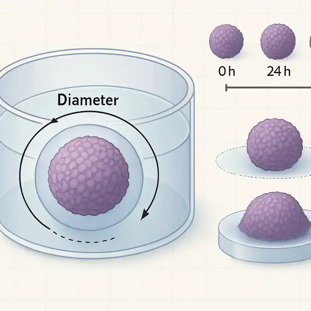
Spheroid size, morphology, compactness and growth rate are all measurable in brightfield without fluorescent dyes — no protocol changes, no phototoxicity, no endpoint fixation.
Continuous monitoring captures growth curves, outgrowth dynamics, and drug response onset in real time — not just a before/after comparison at a single endpoint.
Hepatotoxicity, nephrotoxicity, cardiotoxicity — 3D spheroid models from primary cells or organotypic cultures provide more accurate safety assessment than 2D monolayers.
Embryoid bodies, neurospheres, and early-stage organoids — all visible and quantifiable in brightfield time-lapse without disrupting the culture environment.
Published in collaboration with the University of Regensburg and Fraunhofer EMFT Munich. Both experiments run fully automated inside the incubator with zenCELL owl.
Non-adhesive surface · MCF-7 breast cancer cells
Treated surface · 7-day-old MCF-7 spheroids

All metrics calculated automatically from brightfield images — no staining, no manual measurement, no operator variability.
Continuous growth curves — diameter and projected area at every imaging interval. Full kinetic growth profile from T0 to endpoint.
Compact vs. irregular spheroid morphology quantified as roundness score. Drug-induced morphology changes detected automatically.
Orthogonal dimensions tracked over time — useful for spheroids with non-circular cross-sections or directional growth patterns.
For adhesion assays: migrating cell monolayer area quantified separately from the spheroid core — outgrowth rate per timepoint.
Rate of diameter increase per hour — comparable across 24 conditions. Drug response onset: exact timepoint when growth slows or reverses.
AVI time-lapse per well, PNG image stack for ImageJ, CSV kinetic data for GraphPad Prism or R. All export formats publication-ready.
Works with agarose-coated plates, ULA plates, Matrigel domes, and standard TC-treated plates. No protocol modifications required.
Form spheroids using your preferred method — agarose-coated plates, ULA 96-well, or hanging drop. Transfer to 24-well format at desired size (typically after 3–7 days of formation).
Standard spheroid protocolPlace zenCELL owl on the incubator shelf. Connect USB-C to PC outside the incubator. Setup takes under 2 minutes — no engineers, no dedicated incubator required.
⏱ Under 2 minutesPlace 24-well plate on zenCELL owl. Select wells, set imaging interval (10 minutes recommended for spheroids), and experiment duration (up to 14 days). AI autofocus calibrates per well.
⏱ Under 5 minuteszenCELL owl captures brightfield images of all selected wells at every interval. Monitor remotely from any PC. Receive automatic alerts at defined size thresholds.
Fully automated · 24/7zenCELL owl software automatically calculates diameter, area, roundness and outgrowth per well over time. Export CSV kinetic data, AVI time-lapse, PNG stack. Full ImageJ compatibility.
CSV · AVI · PNG · ImageJ96-well spheroid screening is possible with the zenCELL owl XYZ SCAN stage add-on — automated multi-position imaging across a full 96-well ULA plate in under 90 seconds per scan. Contact us for specifications.
| 功能 | zenCELL 貓頭鷹 | Manual Microscopy | Others |
|---|---|---|---|
| 持续监控 | ✓ Every 10 min · 14 days | ✗ Manual timepoints only | ✓ 是 |
| Inside incubator | ✓ Any CO₂ incubator | ✗ Plate removed each time | Dedicated incubator needed |
| Wells simultaneously | 24 (96 with XYZ) | 1 at a time | Up to 6 positions |
| 荧光 | No — brightfield only | 是的 | Yes — 3 channels |
| Label-free | ✓ No dyes needed | Depends on assay | ✓ Brightfield available |
| Automated analysis | ✓ Diameter · area · roundness | ✗ Manual ImageJ | ✓ Spheroid module |
| 价格 | from €14,000 · no annual fee | ~€0 (existing microscope) | ~€220,000 + annual fee |
| 安装 | Any incubator · <10 min | Standard microscope setup | Dedicated incubator required |
zenCELL owl spheroid monitoring has been validated and published in collaboration with two of Germany's leading life science research institutions.
Joint application note: timelapse imaging of MCF-7 spheroids — 96-hour growth monitoring, morphological quantification, adhesion and outgrowth dynamics. Published with support from distribution partner Omni Life Science. Available as free PDF download.
zenCELL owl is compatible with standard 24-well plates — including agarose-coated plates, tissue culture-treated plates, and standard flat-bottom plates with ULA coating. For 96-well ULA plates, the XYZ SCAN stage add-on enables full-plate automated imaging. Matrigel dome cultures in 24-well format are also compatible. Contact us for plate-specific recommendations.
No. For growth monitoring, morphology tracking, size quantification, and drug response kinetics — brightfield is sufficient. Fluorescence is required for specific viability dyes (calcein AM, propidium iodide), GFP reporter lines, or multiplexed readouts. For brightfield-compatible spheroid assays, zenCELL owl provides a complete label-free solution at significantly lower cost than fluorescence systems.
zenCELL owl reliably detects spheroids from approximately 100–150 µm diameter upwards. Early-stage aggregates below 100 µm may have lower contrast in brightfield. For very small aggregates, contact us to discuss your specific application — imaging parameters can be adjusted to optimize contrast for your cell line and plate format.
Yes — this is one of the most common applications. Add compound to spheroid wells after formation, and monitor the effect on growth, morphology, and viability continuously over 24–96 hours across 24 conditions simultaneously. The continuous kinetic data reveals onset of drug effect, growth arrest, and recovery — not just a before/after comparison.
For brightfield spheroid growth monitoring, zenCELL owl covers the same core assay at over 90% lower cost. Key differences: zenCELL owl fits inside any existing CO₂ incubator (Others requires a dedicated incubator), has no annual licence fee, and includes free ImageJ integration. Others adds fluorescence channels and a larger validated assay library. For label-free spheroid growth monitoring, zenCELL owl is the cost-effective alternative.
Free 20-minute remote demo — real spheroids, real incubator, full software. Twice a week via MS Teams.
Book Free Demo Download Application Note → English English
 Deutsch
 Français
 Español
 简体中文
简体中文