Monitor growth, morphology and drug response continuously across 24 wells. Brightfield, label-free, timelapse — directly inside your CO₂ incubator. From €14,000. No annual fee.
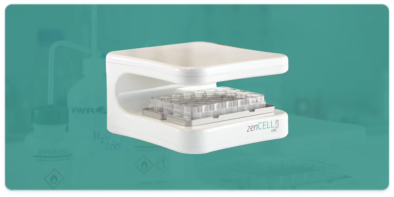
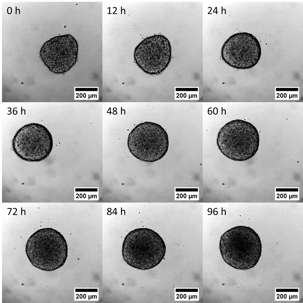
Request a free demo Product details →Left: the zenCELL owl system inside your incubator — always on, always imaging. Right: a cellular spheroid with migrating monolayer captured by brightfield timelapse.
Organoids grow, differentiate and respond to treatment — and all of it happens while no one is watching. Manual spot-checks mean removing plates from the incubator, breaking environmental equilibrium and losing the kinetic data that makes your results meaningful.
Day 0: embed. Day 3: quick check. Day 7: treat. Day 14: endpoint. What happened in between? Without continuous imaging, you cannot know.
Was Batch A growing at the same rate as Batch B before treatment? Without growth traces, you are comparing unknowns.
A well already necrotic before treatment. A batch that never grew. Without imaging, you only see the outcome — never the cause.
When did the treatment take effect? Was it reversible? How fast? Endpoint assays answer none of these questions.
Setup in under 10 minutes. No engineers, no dedicated incubator, no protocol changes. Works with your existing plates, media and workflows.
Compatible with all standard 24-well plates, Matrigel domes, suspension culture, ULA plates and 3D scaffolds
The compact device fits inside any standard CO₂ incubator. Route the USB-C cable through the incubator's cable port to your PC.
⏱ ~2 minutesSoftware detects the device automatically. No calibration needed. Create a new project and name your experiment.
⏱ ~2 minutesPlace the 24-well plate directly on the zenCELL owl. Select the wells you want to monitor (up to 24 simultaneously). Adjust focus height per well if needed.
⏱ ~3 minutesRecommended interval for organoids: every 2–4 hours. For fast-moving assays (drug response): every 30–60 minutes. Press Start — the system runs fully automatically.
⏱ ~1 minuteCheck images and growth data from your desk, without entering the lab. All data is saved automatically to your PC or network drive.
24/7 automatedThe software automatically calculates area, roundness, diameter and confluence per well. Export to CSV for statistics, or as PNG/AVI timelapse videos. Compatible with ImageJ for further morphological analysis.
Export: CSV · PNG · AVIzenCELL owl is not a replacement for your confocal microscope. It covers the continuous monitoring phase that makes everything before the endpoint more reliable.
Track aggregate formation, size consistency and timing across all wells simultaneously. QC your starting conditions before investing further resources.
zenCELL owl · idealContinuous brightfield timelapse across all 24 wells in parallel. No plate removal, no temperature drops, no interruptions to your culture.
zenCELL owl · idealConfirm that organoids across all conditions are morphologically comparable before adding compound. Catch outlier wells before they skew your results.
zenCELL owl · idealWatch treatment effects unfold in real time — size reduction, morphology shifts, growth arrest, lysis onset. Full kinetics, not just the endpoint.
zenCELL owl · idealHigh-resolution fluorescence-based endpoint analysis — viability dyes, GFP reporters, immunostaining. A different tool for a different question.
Confocal / Core facilityUse zenCELL owl for daily workflow and growth kinetics. Use a core facility confocal for fluorescence endpoints. Many groups run both — at a fraction of the total Incucyte cost.
Any organoid or 3D structure visible in brightfield can be monitored continuously. No dyes, no labels, no modifications to your existing protocol.
Growth kinetics, morphology QC, batch comparison — continuous monitoring of iPSC-based cerebral organoid cultures without disruption.
Size, morphology and — in mature cardiomyocyte aggregates — rhythmic contraction visible as motion in timelapse brightfield.
Drug screening kinetics, growth curves, lysis timepoint detection across multi-day toxicity assays.
Budding patterns, cystic vs. solid morphology, drug response — automatically documented across all wells without sampling.
Continuous drug screening kinetics, growth inhibition curves, morphology changes — IC50 kinetics over days, not a single endpoint.
HTS-compatible in 96-well ULA plates. Size, roundness, growth rate — fully automated across all wells simultaneously.
Two brightfield monitoring systems — one at ~90% lower cost than the other. Here is where they differ and how to choose.
| Feature | zenCELL owl | Incucyte S3 |
|---|---|---|
| Purchase price | €14,000 (one-time) | ~€220,000 (one-time) |
| Annual licence fee | None — software included | Mandatory — system locked without it |
| Fluorescence channels | No (brightfield only) | Yes (3 channels) |
| Simultaneous wells | 24 | Up to 6 scan positions |
| Installation | USB-C, any CO₂ incubator, <10 min | Dedicated incubator required |
| Maintenance | None — no moving parts | Service contract recommended |
| Organoid analysis module | Included in base system | Separate paid software module |
| Price transparency | Published openly | Published openly |
| DACH reference labs | Uni Regensburg, Fraunhofer EMFT Munich | Widely used globally |
When Incucyte is the right choice: if your assays require GFP reporters, fluorescence multiplexing or a large validated assay library. For organoid growth monitoring, batch QC and brightfield drug response kinetics, zenCELL owl covers the full workflow at approximately 90% lower cost — with no annual lock-in.
Also comparing against other systems? Axion Omni vs. zenCELL owl → | CYTENA CellCyte X vs. zenCELL owl → | Full comparison overview →
zenCELL owl has been validated for 3D spheroid and organoid timelapse imaging in a joint project between the University of Regensburg and Fraunhofer EMFT Munich, published as a peer-reviewed application note.
Institute of Analytical Chemistry, Chemo- & Biosensors. Reference site for spheroid timelapse imaging and morphological analysis with zenCELL owl.
Joint application note for 3D cell culture timelapse monitoring. Primary DACH industrial reference site for zenCELL owl.
Toxicology, vascular biology, immunotherapy, 3D cell culture — validated across a broad range of cell models and research applications.
University of Regensburg & Fraunhofer EMFT Munich — in partnership with OMNI Life Science. MCF-7 spheroids, 96-hour timelapse, morphological quantification.
Reproducibility, high-content data, minimal perturbation — how continuous imaging is changing the field.
Migration assays, organoid development, HTS — how incubator-based imaging transforms workflows.
MCF-7 spheroid growth, adhesion dynamics, morphological analysis — full experimental results.
For organoid-specific culture media, chemically defined and xeno-free formulations, FBS and GMP-grade media — our distribution partner SeamlessBio specialises in exactly this. ISO 9001 and ISO 13485 certified, EU-produced, full regulatory documentation.
Yes. zenCELL owl is compact and fits inside any standard CO₂ incubator. It connects via USB-C and takes under 10 minutes to install — no modifications, no dedicated incubator, no service engineer required. The system works alongside your existing cultures without disruption.
Yes. Organoids in Matrigel domes, suspension culture, ULA plates and other 3D culture formats are all visible by brightfield. For very thick gels, imaging depth may be limited depending on the organoid position within the gel layer. For defined, animal-origin-free media alternatives to Matrigel, our partner SeamlessBio can help.
For growth kinetics, morphology tracking, batch QC and drug response monitoring — no. Brightfield is sufficient for the majority of organoid workflows. Fluorescence is required for specific viability dyes (propidium iodide, Calcein AM) or GFP-based reporters. For those endpoints, a fluorescence system or core facility confocal is the right tool. zenCELL owl and a core facility confocal complement each other well.
zenCELL owl costs €14,000 (one-time purchase). There is no annual licence fee, no software subscription and no mandatory service contract. All software updates are included in the purchase price. The price is published openly — unlike most competitor systems which require a quote.
The standard field of view is 1.08 mm² — well-suited for individual organoid and spheroid monitoring in standard well formats. Organoids typically range from 0.1 to 2 mm in diameter, fitting comfortably within the imaging field. Expanded imaging capability is currently in development. Contact us for the latest specifications and what is coming next.
Up to 24 wells simultaneously — a full 24-well plate or selected wells across different formats. The minimum capture interval across all 24 wells is 30 seconds. Multiple zenCELL owl units can operate in the same incubator at the same time for higher throughput.
Yes. We offer a free 30-minute remote live demo — book your slot here. You will see real cells, real data and a full software walkthrough directly inside a running incubator. Available twice per week via MS Teams.
Images export as PNG, JPG or BMP. Timelapse videos export as AVI. Quantitative analysis data (area, roundness, confluence per well over time) exports as CSV — compatible with Excel, GraphPad Prism and R. Raw images are also compatible with ImageJ for custom morphological analysis.
Free 30-minute remote demo — real cells, real data, inside a real incubator. No commitment required.
Book your free demo Product page →