ScratchMaker scratch assay plates feature photosensitizer-coated bottoms for standardized, reproducible wound healing assays. Available in 6-, 24-, and 96-well format. Requires a fluorescence microscope with blue/violet excitation (~385-408 nm, DAPI channel) – 3 cm distance, 60 sec per well. No washing step or medium change required.
For new customers: we create a personal offer based on your format and quantity needs.
The method you use to create a wound determines what biology you can study. Not all scratch assay plates are equal — the wound type matters. Studies on cell migration assays consistently show that physiological relevance is the critical differentiator between insert-based and photochemical wound models.


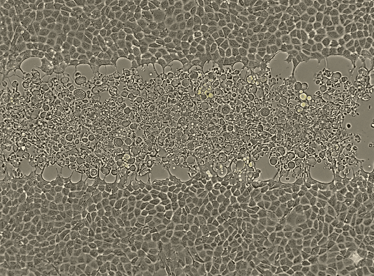
Most labs still create scratch wounds by dragging a pipette tip across the monolayer by hand. This mechanically ruptures cells at the wound edge — width, angle, and depth vary with every pass, every operator, every plate. ScratchMaker scratch assay plates replace this manual step entirely with a precisely defined, reproducible photochemical wound.

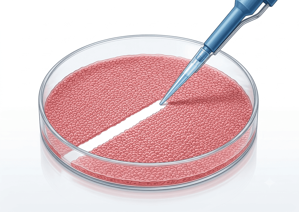
"The cells, when they move towards the wound, they not only close the wound, but also digest the cell bodies which are on the way. So we believe that this closely mimics the physiological situation, the real-time situation, what's happening at a biological level."
— Researcher feedback, zenCELL owl live demo · June 2026UV-A/visible light (wavelength typically ~340–408 nm). Standard fluorescence microscopes with Hg-vapour lamp (DAPI filter cube) or 405 nm laser are fully compatible. Photosensitizer absorption maximum: ~395 nm. No dedicated UV lamp or laser required — exposure time 20–60 seconds per well.
Around 12,000 PubMed entries involve scratch assays – most performed manually with pipette tips. Inter-assay variability is the most cited limitation in wound healing assay publications.
Manual pipette scratches vary significantly depending on operator pressure, angle, and tip size – making quantification unreliable.
Results cannot be directly compared between experiments, time points, or operators – destroying statistical power.
Lack of standardization is a frequent reason for rejection in peer review of migration and wound healing studies.
96 manual scratches per plate is time-consuming and error-prone – high-throughput screening is not realistic.
The pipette tip physically tears cells at the wound edge, generating uncontrolled debris and stress responses that confound migration readouts.
Each well is scratched at a slightly different moment and with a slightly different hand motion – timing and technique drift accumulate across a plate.
Most labs start with the scratch assay plates alone and grow into the full ScratchMaker workflow step by step.
You're generating reproducible, photochemical scratch assay wounds – manually imaging each plate under your fluorescence microscope.
Your ScratchMaker plate sits inside the zenCELL owl, capturing every well automatically over 24–72 h – no manual imaging needed.
Standard quantification not enough? We can build a custom analysis algorithm tailored to your cell type, wound geometry, or readout – tell us about your project.
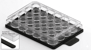
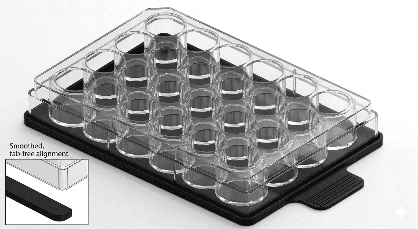
Available in 6-, 24-, and 96-well format. Order plates and masks separately or as a complete Starter Kit. Individual sample offers available for new customers.

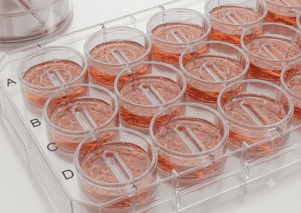
Photosensitizer-coated glass bottom plates for standardized, reproducible wound healing assays. Available in 6-, 24-, and 96-well format. No washing step or medium change required after light exposure.

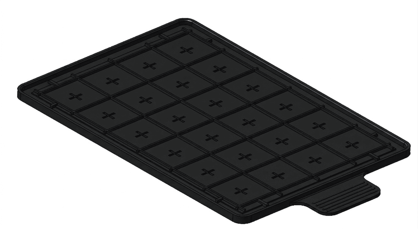
The light mask clips under the ScratchMaker plate and defines exactly where the photosensitizer is activated. Ensures identical wound geometry across all wells, all experiments, all operators. One mask per format — reusable.

Everything you need to run your first standardized scratch assay. Includes ScratchMaker plates in your chosen format, the matching light mask, and a step-by-step protocol. Up to 50% off for new customers.
Ideal for larger cell populations, higher cell input, or applications requiring more image area per well. Compatible with most brightfield and fluorescence microscopes.
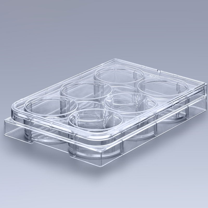
The standard format for scratch assay experiments. Fits directly into the zenCELL owl 24-channel imager. Best balance between throughput and image quality.
For high-throughput screening applications. Compatible with zenCELL owl XYZ SCAN stage add-on. Screen up to 96 conditions in a single experiment.
All ScratchMaker plates and masks are available for individual sample offers. Contact us at info@innome.de with your format, quantity, and institution — we will create a personal quote within 24 hours. New customers receive up to 50% off their first Starter Kit order.
ScratchMaker plates with integrated Microelectrode Array (MEA) compatibility — precisely defined wound position relative to the electrode for electrical cell migration measurements. Contact us for early access.
From confluent monolayer to defined photochemical wound – a standardized workflow in four steps. Compatible with 6-, 24-, and 96-well formats.
Seed cells, incubate to ≥95% confluency
Mask clips under the plate, defines exposure zone
Blue/violet light (~408 nm, DAPI channel), 3 cm distance, per well
No washing, no medium change – ready to go
Seed your cells onto the ScratchMaker scratch assay plate at the appropriate density. Incubate until ≥95% confluency is reached – typically 16–24 h depending on cell line.
Clip the ScratchMaker light mask onto the underside of the well plate. The mask acts as holder and light guide – it defines exactly which area of each well will be exposed.
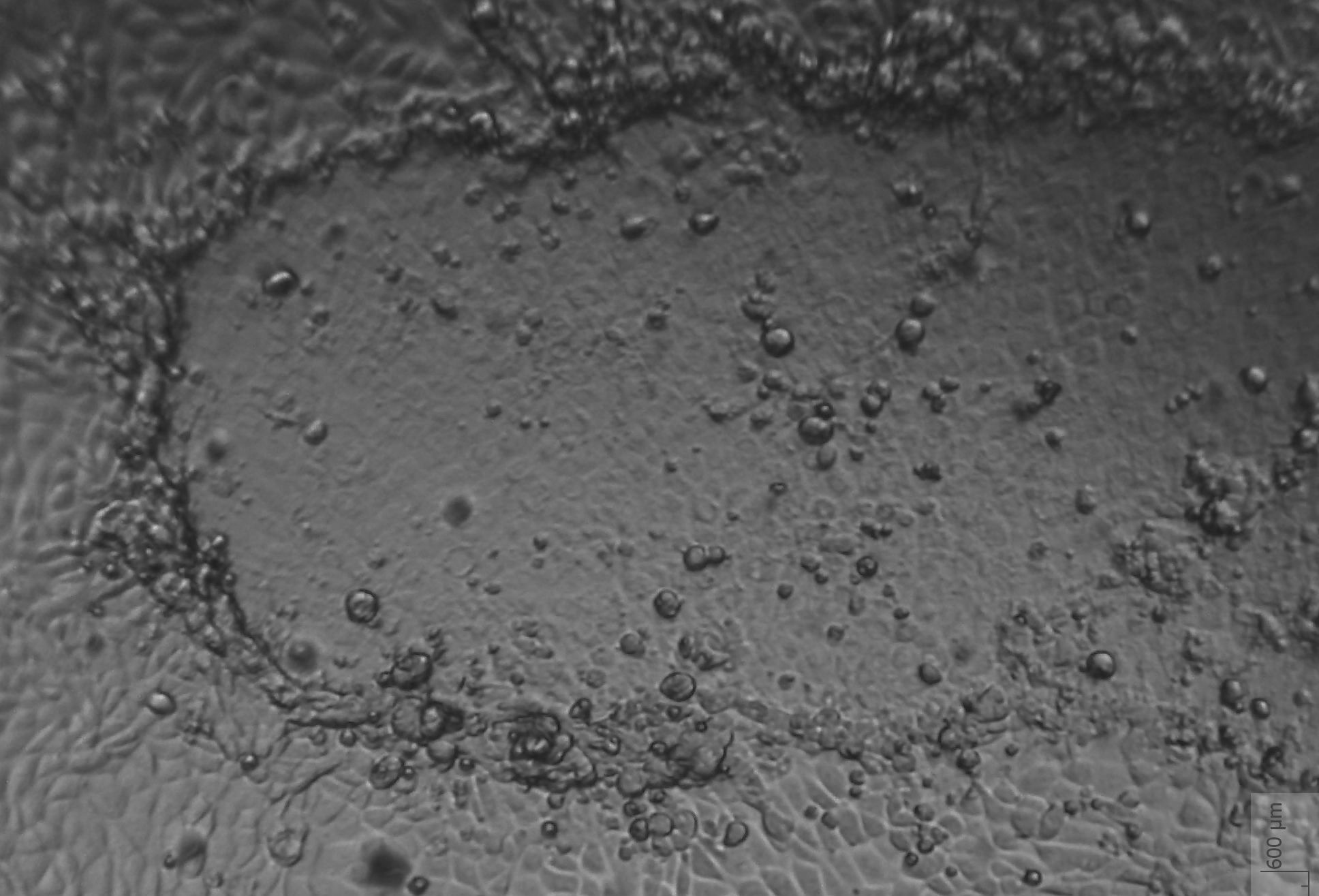
Place each well under your fluorescence microscope light source for 60 seconds at a distance of 3 cm. The blue/violet excitation light resonantly activates the photosensitizer embedded in the glass coating, generating singlet oxygen (¹O₂) precisely within the illuminated area. This induces localized cell death only where the light hits – cells just outside the exposed zone remain completely unaffected.
The scratch assay plate is ready immediately after light exposure. No washing or medium change is required – cells remain in their original medium. Place directly under your microscope or into the zenCELL owl and start live-cell monitoring.

Works with 6-Well, 24-Well, and 96-Well ScratchMaker scratch assay plates. The same light mask system applies to all formats – clip on, expose, start imaging.
A direct comparison across the three most common wound healing assay approaches – and why photochemical scratch assay plates produce the most physiologically relevant data.
| Feature | ScratchMaker Plates | Pipette Tip | Ibidi Insert |
|---|---|---|---|
| Wound type | Photochemical cell death | Mechanical rupture | Gap – no cell death |
| Physiological relevance | High – mimics in vivo | Moderate | Low – no debris clearance |
| Cell debris clearance | Yes, defined zone | Yes, uncontrolled | No |
| Insert detachment risk | No insert | No insert | Yes – after 2+ hours |
| Scratch reproducibility | High | Low (±30–60%) | High |
| 6-Well | Yes | Yes | Yes |
| 24-Well | Yes | Yes | Yes |
| 96-Well | Yes | Laborious | Yes |
| Microscope requirement | Fluorescence microscope (min.) · 3 cm distance | Any | Any |
| Partial plate use | Yes – expose wells step by step (sterile) | Yes | No |
| Washing after wound | Not required | Required | Required |
| Cost per plate | from €59 | ~€0 | €50–80 |
| Microelectrode Array (MEA) | Compatible | Alternative method | Alternative method |
| Publication suitability | Yes | Limited | Yes |
ScratchMaker plates are compatible with Microelectrode Array (MEA) technology, offering a precise alternative to insert-based or manual scratch methods for electrical migration measurements. Because the wound position is precisely defined by the light mask, the MEA electrode is always located at the same position relative to the wound – making electrical migration measurements significantly more reproducible and valid.
ScratchMaker scratch assay plates are designed to work directly with the zenCELL owl incubator imager. Place the plate inside, press start – the system captures every well, every hour, automatically.
Fully automated photochemical wound generation for our scratch assay plates – 96 identical wounds in under 60 seconds. Currently in prototype development.

Illuminate all wells simultaneously in one step using UV-A/visible light (~385–408 nm) illumination. Compatible with all ScratchMaker scratch assay plates – 24-well and 96-well. Early access on request.
Answers to the most common questions about light sources, wound geometry, plate usage, and compatibility.
A washing step is not strictly required — cells remain in their original medium and the plate is ready for imaging immediately after light exposure.
However, many researchers perform a medium change before and/or after illumination to remove floating dead cells and improve image quality, as well as to wash out any potentially generated by-products.
When adding reagents such as migration inhibitors or stimulants, a medium change is recommended to introduce them at a defined time point relative to wound creation — either before or after illumination depending on your experimental design.
Protocol guidance validated in collaboration with Stefanie Michaelis, Fraunhofer EMFT / University of Regensburg.
Price list, protocol PDF, and sample order for ScratchMaker scratch assay plates – contact us directly.
Contact Us
In return, we ask for a short application note, video clip, or written statement within 90 days — your experience helps other researchers discover ScratchMaker.