Predicting Failure: Detecting Cell Stress and Early Apoptosis in Real-Time

“`html
Predicting Failure: Detecting Cell Stress and Early Apoptosis in Real-Time
Cell stress and early apoptosis are crucial phenomena in cell biology, underpinning various cellular responses to environmental changes. As scientific methods evolve, the necessity to predict and monitor these processes in real-time has never been more imperative. This capability is vital in areas such as drug development, toxicology, and cancer research. In this article, researchers, lab managers, and biotech professionals will explore the challenges of traditional methods, the impact of new technologies, and practical strategies using advanced tools like live-cell imaging systems to improve research outcomes.
Common Challenges and Limitations of Traditional Approaches
Understanding Traditional Cell Stress Detection
Traditionally, detecting cell stress and early apoptosis involved endpoint assays that provide static snapshots of cellular events. Techniques such as flow cytometry and Western blotting, while informative, often miss dynamic processes that occur rapidly or intermittently. These methods also introduce variability due to manual handling, thus compromising reproducibility and accuracy.
- Static nature of endpoint assays
- Manual handling increases variability
- Potential to miss transient cellular events
Technological Advances and Automation Trends
The Rise of Live-Cell Imaging Systems
Technological breakthroughs have ushered in new methods for continuous and non-invasive monitoring of live cells. Live-cell imaging systems provide invaluable insights into cell dynamics, allowing researchers to monitor cell health in real-time. Automation within these systems plays a pivotal role by minimizing manual intervention, thus enhancing data reproducibility and reliability.
- Non-invasive real-time monitoring
- Reduction in manual handling
- Enhanced reproducibility of results
Practical Examples and Workflows Using Live-Cell Imaging
Leveraging Live-Cell Imaging for Real-Time Monitoring
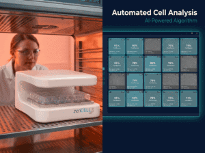
Implementing live-cell imaging into lab workflows transforms traditional approaches. For example, researchers can establish baseline cell health metrics, detect stress markers before traditional assays show change, and observe apoptotic events as they unfold. By integrating devices such as the zenCELL owl — known for its compact and incubator-compatible design — institutions can maintain environmental controls essential for accurate live-cell analysis.
- Real-time data capture of cellular changes
- In situ monitoring within incubators
- Efficient integration with existing workflows
How Incubator-Based Imaging Improves Reproducibility and Data Quality
Insights Into Enhanced Experimental Design
Incubator-based imaging systems like the zenCELL owl allow for continuous observation under optimal conditions. Such systems ensure that cells are undisturbed, maintaining physiological relevance. This leads to higher data fidelity and reduces the chance of experimental errors that occur during sample handling.
- Increased physiological relevance during analysis
- Minimized sample disturbance
- Consistency across experimental runs
Summary and Outlook for Future Lab Workflows
Embracing the Future of Cell Culture Research
By investing in and integrating advanced imaging and real-time monitoring technologies, labs can significantly enhance their research capabilities. These innovations promise more accurate predictions of cellular responses, ultimately leading to more reliable diagnostics and therapeutic developments. As technology progresses, the potential for more sophisticated applications in organoid studies, proliferation assays, and high-throughput screening continues to grow. Embracing these trends will propel laboratory research into a new era of precision and efficacy.
- Adoption of cutting-edge imaging technologies
- Improved diagnostics and therapeutic research outcomes
- Potential for innovative applications in multiple fields
Continue reading to explore more advanced insights and strategies.
“`
“`html
Integrating Data Analytics with Live-Cell Imaging
Harnessing Data for Predictive Modeling
The synergy of data analytics and live-cell imaging has paved the way for predictive modeling—an innovation boosting the accuracy of forecasting cellular behaviors. By leveraging advanced data analytics platforms, researchers can integrate massive data sets captured from live-cell imaging to construct predictive models. These models facilitate understanding of how cells respond to stress and initiate apoptosis, leading to proactive rather than reactive research approaches.
- Use data analytics to identify patterns and trends in cell behavior
- Develop predictive models to forecast cellular responses to treatments
Employing Machine Learning for Enhanced Decision-Making
Artificial Intelligence and Cell Biology: A Powerful Combination
Machine learning algorithms have revolutionized our understanding of complex biological systems. By incorporating these algorithms into the data interpretation phase of live-cell imaging, scientists can obtain quantitative insights into cell health and behavior. For instance, AI-driven analysis of imaging data can help discern subtle early signs of apoptosis, streamlining decision-making in both research and clinical settings.
- Implement AI algorithms to refine data analysis and interpretation
- Enhance decision-making processes with quantitative insights
Combining Live-Cell Imaging with High-Throughput Screening
Maximizing Efficiency and Discovery in Research
The integration of live-cell imaging into high-throughput screening (HTS) workflows represents a powerful strategy to accelerate the discovery of potential drug candidates. By enabling real-time monitoring of cellular responses in HTS setups, researchers can rapidly identify promising compounds and observe their effects on cell viability and stress levels. This approach significantly reduces the time and cost associated with drug discovery pipelines.
- Incorporate live-cell imaging systems in high-throughput environments
- Streamline discovery processes to identify effective compounds
Case Study: Real-World Application of Live-Cell Imaging
Pioneering Research in Oncology
A pharmaceutical company recently integrated live-cell imaging systems into their cancer research laboratories, aiming to better understand drug resistance mechanisms. By allowing scientists to monitor tumor cell dynamics in real-time, the company identified novel apoptosis pathways that standard assays overlooked. This technological integration not only elucidated new therapeutic targets but also enhanced the predictive power of preclinical studies, resulting in more robust drug candidates entering clinical trials.
- Optimize drug discovery by understanding tumor cell dynamics
- Identify previously unknown pathways for potential therapeutic targeting
Algorithmic Approaches to Improve Imaging Workflows
Streamlining Data Processing with Computational Tools
The workflows for live-cell imaging can be greatly improved through the use of algorithmic methodologies that automate image capture and processing. By combining imaging systems with computational software, labs can achieve heightened levels of efficiency and accuracy. This automation reduces the workload associated with manual data analysis, allowing research staff to focus more on experimental design and less on routine data processing tasks.
- Employ computational tools to automate and streamline imaging workflows
- Shift focus from data processing to experimental innovation
Cloud-Based Platforms for Collaborative Research
Redefining Lab Collaboration Through Virtual Means
The adoption of cloud-based platforms is transforming how researchers collaborate and share data in cell biology. These platforms facilitate seamless data sharing and collective analysis, thus enabling cross-disciplinary teams to work together regardless of location. The integration with live-cell imaging data promotes a collaborative research environment where insights are rapidly exchanged, accelerating the pace of discovery.
- Utilize cloud technologies to foster collaboration and data sharing
- Enhance cross-disciplinary research through virtual data platforms
Enhancing Workflow Efficiency with Automated Image Processing
Utilizing Software Tools for Real-Time Data Analysis
Advanced software tools are essential for the automated processing of live-cell imaging data. These tools can segment images, quantify cell morphology, and detect changes over time, providing immediate feedback. By automating image processing, labs can increase throughput and reduce errors associated with manual interpretation, ensuring results are both timely and precise.
- Adopt software solutions for automated and precise image processing
- Increase data throughput while minimizing human error
Strategic Insights into Future Innovations
Positioning Labs for Next-Generation Cellular Research
The landscape of cell biology research is rapidly evolving, with live-cell imaging at the forefront of innovation. Labs must stay agile, continuously adopting new technologies and methodologies to remain competitive. By doing so, they will be well-positioned to tackle upcoming challenges, explore next-generation applications such as personalized medicine, and contribute meaningfully to the broader scientific community.
- Continuously integrate emerging technologies into research workflows
- Prepare for next-generation applications in cell biology research
Next, we’ll wrap up with key takeaways, metrics, and a powerful conclusion.
“`
“`html
Key Metrics in Evaluating Live-Cell Imaging Systems
Measuring Impact with Quantitative Benchmarks
In the rapidly advancing field of live-cell imaging, establishing quantitative benchmarks is vital for assessing system performance and impact on research outputs. Metrics such as resolution, sensitivity, throughput, and image acquisition speed are crucial in determining the efficacy of imaging systems. Regular evaluation of these metrics ensures that labs maintain high standards and can effectively leverage their imaging setups to drive groundbreaking discoveries.
- Assess imaging systems based on key metrics like resolution and throughput
- Optimize imaging performance to support high-quality research outcomes
Innovative Collaborations in Live-Cell Imaging
Building Synergies for a Brighter Future
Collaboration across multiple disciplines forms the backbone of innovation in live-cell imaging. By engaging in partnerships with academic, clinical, and industrial entities, labs can pool expertise and resources to address complex biological questions. These initiatives amplify the reach and potential of live-cell imaging, drawing on a diverse range of perspectives to unlock new insights into cellular dynamics and disease mechanisms.
- Forge strategic partnerships with diverse research entities
- Leverage interdisciplinary approaches to tackle complex biological challenges
Exploring the Role of Live-Cell Imaging in Personalized Medicine
Individualized Insights for Enhanced Therapies
The application of live-cell imaging in personalized medicine represents a significant leap towards individualized healthcare solutions. By providing real-time insights into patient-specific cellular responses, live-cell imaging enables the development of bespoke treatment plans that reflect the unique biological makeup of each patient. This approach promises to enhance treatment efficacy and minimize adverse effects, marking a paradigm shift in medical practice.
- Utilize live-cell imaging to tailor personalized healthcare solutions
- Improve treatment outcomes by understanding patient-specific cellular responses
Conclusion
The integration of live-cell imaging with advanced data analytics, machine learning, and high-throughput screening is revolutionizing the field of cell biology. This synergy offers unparalleled insights into cellular mechanisms, significantly enhancing research accuracy and efficiency. The fusion of artificial intelligence with imaging technology provides precision in understanding and predicting cellular responses, accelerating drug discovery and facilitating breakthroughs in understanding cancer, neurodegenerative diseases, and myriad other biological phenomena.
Live-cell imaging empowers researchers with the tools to monitor cellular activities in real time, effectively bridging the gap between observation and action. Strategic utilization of cloud-based platforms fosters global collaborations, breaking down geographical barriers to innovation. The seamless sharing of data and expertise heralds a new era of scientific discovery characterized by speed, efficiency, and unparalleled depth of insight.
The article underscores the imperative for laboratories to remain agile, continuously incorporating emerging technologies to stay at the forefront of cellular research. As research evolves towards personalized medicine, live-cell imaging serves as a cornerstone for developing patient-specific treatment plans, reflecting a holistic progression towards individualized healthcare solutions.
Looking to the future, the commitment to integrating these advanced technologies into research practices will position labs to surpass existing boundaries in cell biology. Researchers are encouraged to adopt forward-thinking strategies, thereby shaping the future of scientific exploration and innovation. Embrace these tools and methodologies as we endeavor to unlock the vast potential of live-cell imaging and drive transformative changes in health care and beyond.
Stay inspired, stay innovative, and harness the power of these cutting-edge technologies to continue pushing the boundaries of what is possible in the fascinating world of cell biology.
“`