The short answer is: for most applications, no — you do not need fluorescence. Here is how to determine which category your experiment falls into.
The Decision: Brightfield or Fluorescence?
Brightfield is sufficient when:
- Your readout is cell morphology or density
- You are measuring wound closure or gap area
- You are monitoring confluency over time
- You are tracking neurite outgrowth (PC12, iPSC)
- You are measuring cytotoxic morphological changes
- You are monitoring spheroid diameter and compactness
- You are checking barrier integrity (MDCK, Caco-2)
- Your experiment runs 24–72 hours or longer
Fluorescence IS required when:
- Imaging GFP/RFP-tagged proteins or reporters
- Measuring specific ion concentrations (Ca²⁺ sensors)
- Distinguishing cell populations by fluorescent label
- Sub-cellular localisation studies
- FRET-based signalling reporters
- Immunofluorescence of specific structures
- Tracking individual cells by fluorescent marker
What Brightfield Actually Shows You
Brightfield live cell imaging uses transmitted white light to visualize cells without any labelling. The resulting images show cell morphology, boundaries, density, and movement with remarkable clarity for most common applications. Here is what is directly quantifiable:
- Confluency — percentage of well surface covered by cells, calculated automatically from image texture
- Wound area — cell-free gap area in wound healing assays, measured in µm²
- Wound closure rate — µm²/hour, calculated from consecutive timepoints
- Neurite length — clearly visible PC12 and iPSC-neuron extensions in brightfield
- Spheroid diameter — 3D aggregate size and compactness from transmitted light
- Cell morphology changes — rounding, blebbing, retraction — visible before cell death
- Contamination — background texture changes visible 6–12h before medium turbidity
The Hidden Cost of Unnecessary Fluorescence
Choosing fluorescence when brightfield would suffice has real costs beyond the price of the microscope:
| Factor | Champ clair | Fluorescence (when not needed) |
|---|---|---|
| Phototoxicity | None — low-intensity white light | Risk of cellular damage affecting results |
| Photobleaching | None — no fluorophore | Signal fades — limits multi-day imaging |
| Labelling protocol | Pas requis | Additional time, cost, potential interference |
| Dye cytotoxicity | Zero risk | Dyes can affect cell behavior at working concentrations |
| Equipment cost | Significantly lower | Fluorescence optics multiply system cost |
| Experiment duration | Days to weeks — no signal degradation | Limited by photobleaching and phototoxicity accumulation |
Real Applications — Brightfield Is Enough
Wound healing and scratch assays
The cell-free gap in a wound healing assay is clearly visible in brightfield — the wound zone appears lighter than the surrounding monolayer. Automated image analysis quantifies wound area at every timepoint without any staining. This is the standard approach in the field and sufficient for publication-quality data.
PC12 neuronal differentiation
Neurites extending from PC12 cell bodies are clearly visible as thin processes in brightfield. Length, branching angle, and network formation can all be quantified without fluorescent labelling — eliminating phototoxicity risk over 24–72 hour differentiation experiments.
Cytotoxicity assays
Morphological changes preceding cell death — rounding, blebbing, detachment — are visible in brightfield hours before the cell actually dies. Confluency decline after compound addition is a direct proxy for cell loss, without requiring any metabolic assay reagent.
Spheroid growth monitoring
Spheroid diameter, compactness, and necrotic core formation are all quantifiable from brightfield images. The dense aggregate is clearly visible against the surrounding medium without any labelling.
Brightfield live cell imaging — free demo
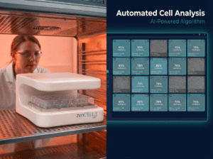
See zenCELL owl imaging 24 wells simultaneously in brightfield. No fluorescence needed.
Foire aux questions
Can I switch from fluorescence to brightfield for my wound healing assay?
Yes — and for most wound healing assays, brightfield gives equivalent or better results. The gap is clearly visible without labelling, phototoxicity does not alter migration kinetics, and the experiment can run continuously for 24–48 hours without signal degradation. The only reason to use fluorescence for a wound healing assay is if you are simultaneously tracking a fluorescent reporter in the same cells.
Is brightfield quantitative enough for publication?
Yes. Brightfield-based wound closure quantification (% closure, wound closure rate, t½ gap closure time) is accepted by reviewers across cell biology journals. The Wound Healing Size Tool for ImageJ/Fiji (Suarez-Arnedo et al., PLoS ONE 2020, 900+ citations) provides standardized brightfield analysis. zenCELL owl automated analysis generates the same metrics automatically.
What resolution does brightfield give compared to fluorescence?
For the applications described above — confluency, wound closure, morphology, spheroid size — brightfield resolution is entirely sufficient. zenCELL owl uses a 5MP sensor, providing detailed images of cell morphology, neurite structure, and monolayer integrity. Sub-cellular resolution (organelles, cytoskeletal structures) requires fluorescence or high-NA oil immersion objectives, but is not needed for any of the applications discussed here.