“`html
From Plate to Paper: Streamlining Image Analysis for Faster Documentation
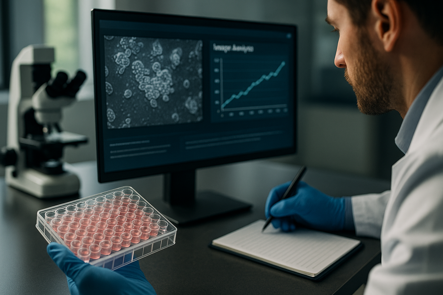
In the swiftly evolving landscape of modern cell culture research, the need for efficient and accurate image analysis is more critical than ever. As scientific inquiries grow in complexity, researchers, lab managers, and biotech professionals are tasked with not only cultivating their samples but also interpreting vast amounts of data. This article delves into the significant topic of streamlining image analysis — from plate to paper — and discusses how advancements in live-cell imaging and laboratory automation are crucial for faster documentation.
Challenges in Traditional Image Analysis Methods
Traditional image analysis methods in cell culture research have often been labor-intensive, requiring significant manual input and time investment. Scientists may face a myriad of challenges that impede their ability to promptly interpret and document their findings.
Manual Processes and Human Error
Relying on manual imaging techniques introduces a high potential for human error. Variability in capturing images, subjective assessments, and disparate interpretations can all lead to inconsistent data. This inconsistency hampers reproducibility — a cornerstone of scientific research.
- Time-consuming manual imaging hampers efficiency.
- Inconsistencies arise from subjective interpretation.
- High potential for human error affects data quality.
Limited Throughput
The limitations of traditional approaches extend to throughput capabilities. Manual strategies struggle to keep up with the demands of high-throughput screenings (HTS) necessary for comprehensive studies, such as drug discovery or genetic analysis.
- Traditional approaches have low throughput.
- Lack of automation limits scalability of experiments.
技术进步与自动化趋势
Advancements in technology offer promising solutions to the pitfalls of traditional image analysis. The integration of automation and enhanced imaging techniques allows for a more streamlined workflow — taking researchers from plate to paper with increased efficiency and accuracy.
The Rise of Automated Imaging Systems
Automated imaging systems alleviate many burdens inherent to manual processes. These systems not only enhance the precision of image capture but also facilitate consistent documentation of cell behaviors over time. The zenCELL owl, for example, exemplifies a compact, incubator-compatible imaging model that offers uninterrupted monitoring and analysis within the optimal environment for cell culture.
- Automated systems improve image consistency.
- Continuous monitoring ensures data accuracy and robustness.
Data Management and Analysis Software
Modern software solutions complement automated hardware by streamlining data management and analysis. These platforms offer powerful tools designed to handle vast quantities of data, providing researchers with swift insights and analysis. Improved software interfaces allow for easy integration with laboratory information management systems (LIMS), enhancing workflow efficiency.
- Advanced software facilitates large data set management.
- Integration with LIMS enhances workflow efficiency.
Practical Workflows Using Live-Cell Imaging
Integration of Continuous Monitoring
Live-cell imaging offers a real-time glimpse into cellular dynamics, significantly enriching experimental outcomes. Its integration transforms the data collection process from simple snapshots to continuous monitoring, offering rich datasets that provide deeper insights into cellular processes.
- Continuous monitoring enhances dataset richness.
- Real-time insights into cellular processes aid in precise analyses.
Application in Various Cell Models
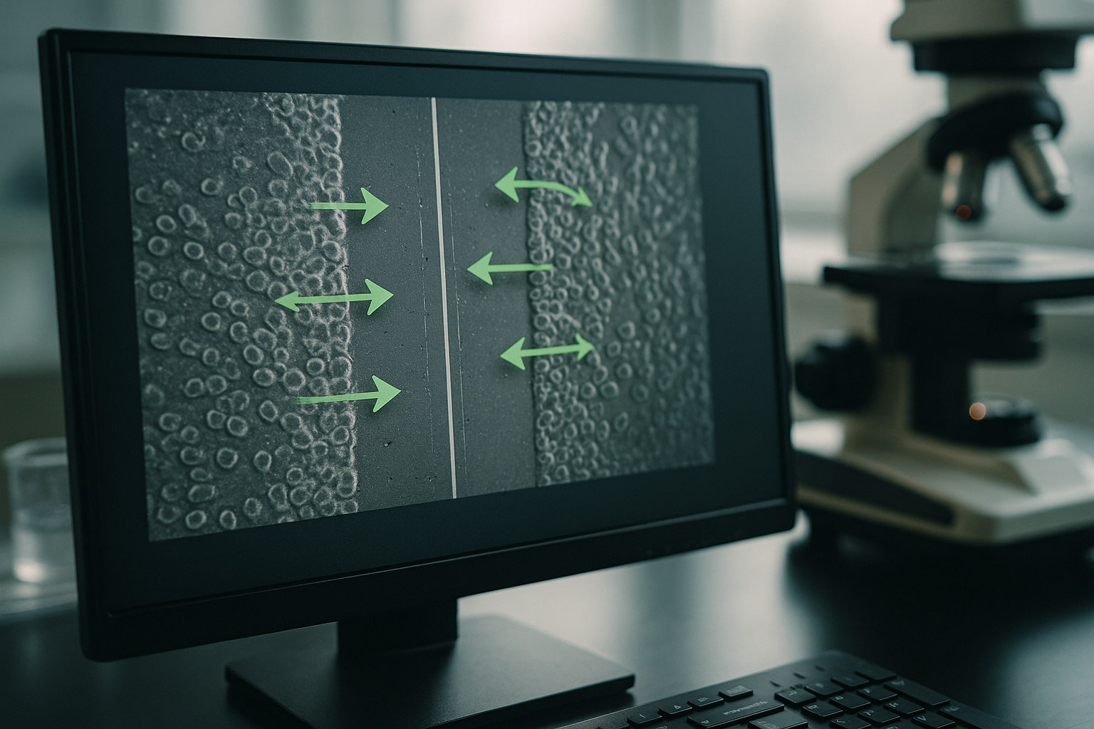
Researchers using live-cell imaging can apply it across various cell models with applications expanding to include migration assays, organoid studies, and proliferation analyses. This flexibility supports diverse research objectives and encourages thorough experimental exploration.
- Live-cell imaging is applicable to varied cell models.
- Supports applications such as migration assays and organoid studies.
继续阅读,以探索更深入的见解和策略。.
“`
“`html
Leveraging AI in Image Analysis
Smart Algorithms for Enhanced Precision
The integration of artificial intelligence (AI) in image analysis has transformed the accuracy and speed with which researchers can interpret complex data sets. AI algorithms are capable of recognizing patterns and structures in cell imagery that may be overlooked by the human eye. By employing machine learning models, researchers can automate the detection and classification of cellular events, drastically shortening the time from observation to documentation. A study by Harvard University highlighted the use of AI-driven systems to achieve a 95% accuracy rate in distinguishing between different cell types, a task that traditionally required significant manual labor.
- Employ AI solutions to enhance pattern recognition accuracy.
- Integrate machine learning for dynamic data analysis.
Enhancing Data Reproducibility
Standardization Across Laboratories
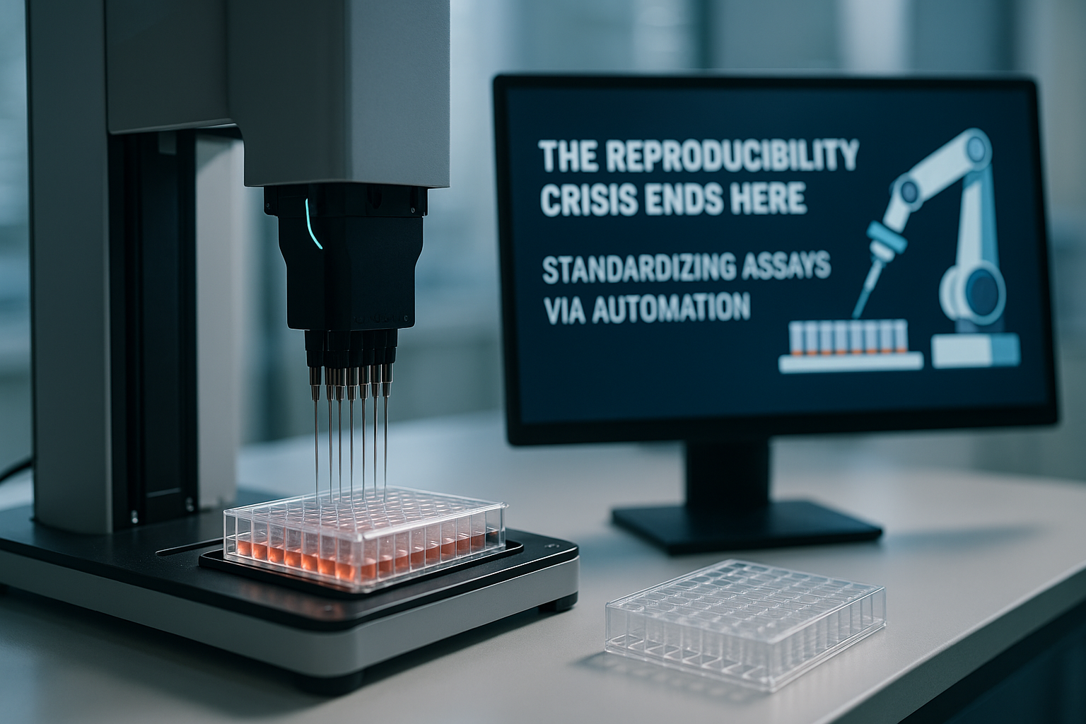
Data reproducibility is a fundamental challenge in scientific research. With automated image analysis, standardization across laboratory practices becomes more achievable. Platforms like the zenCELL owl ensure that all images are captured under uniform conditions, minimizing variability. Additionally, software-driven data logging ensures that experimental conditions are meticulously recorded and easily retrievable, making it easier to replicate studies. In a comparative study, laboratories that employed standardized automated imaging protocols reported a 30% improvement in experiment reproducibility.
- Implement uniform imaging protocols for all laboratory environments.
- Utilize automated data logging to enhance replicability.
Advanced Imaging Techniques in Action
Case Study: Drug Efficiency Testing
A leading pharmaceutical company adopted automated and live-cell imaging to expedite its drug testing process. By integrating high-content screening with real-time data analysis, they reduced the drug candidate selection phase from 12 months to 8 months. The company’s success hinged on their ability to monitor drug-cell interactions continuously, allowing for robust, data-driven decision-making. This automation also enabled parallel testing of multiple compounds, significantly increasing throughput.
- Use live-cell imaging to quickly identify potential drug candidates.
- Parallel testing with automation increases throughput and efficiency.
Interfacing with IoT for Remote Monitoring
Wireless Technology and Laboratory Efficiency
The Internet of Things (IoT) continues to revolutionize laboratory operations. With new IoT-enabled devices, researchers can remotely monitor live-cell imaging setups and access data in real time on any smart device. This not only promotes flexibility in where and how researchers work but also ensures that data collection is not disrupted by physical constraints. Research institutions implementing IoT solutions have reported increased productivity, with a 40% reduction in on-site monitoring requirements.
- Incorporate IoT solutions for seamless remote data monitoring.
- Utilize smart devices to access real-time insights from anywhere.
Adapting Laboratory Automation for Sustainability
Eco-Friendly Innovations
Modern laboratories are also faced with the challenge of reducing their environmental footprint while maintaining high levels of productivity. Automated imaging technologies contribute to sustainability by reducing waste associated with manual processes, such as excessive reagent use and energy consumption. For instance, automated systems can precisely control the amount of reagents applied in assays, minimizing waste. The University of Leicester’s pilot project demonstrated a 25% reduction in energy consumption after implementing automated imaging systems.
- Opt for automated systems to minimize reagent waste.
- Implement energy-efficient hardware to support sustainability goals.
Integrating Virtual Reality for Immersive Analysis
Beyond Traditional Viewing
The application of virtual reality (VR) in cell culture research offers a multi-dimensional approach to image analysis. By visualizing images in a VR environment, researchers can better understand the spatial relationships and interactions within cell models. This approach is particularly beneficial for complex studies involving 3D cell cultures or organoids. VR platforms allow for the manipulation of 3D cellular maps, offering an unparalleled depth of analysis and understanding that enhances research conclusions.
- Utilize VR to explore cellular interactions in a 3D environment.
- Enhance visual comprehension with interactive, immersive tools.
Boosting Collaboration through Cloud Technologies
Seamless Teamwork with Cloud Sharing
Cloud-based platforms facilitate global collaboration by allowing researchers to share image data and analyses swiftly and securely. Collaborative tools enable multiple users across different locations to contribute to image interpretation, fostering interdisciplinary cooperation and accelerating innovation. The University of Tokyo’s research team successfully used cloud technologies to collaborate on stem cell research with international partners, significantly reducing project timelines.
- Embrace cloud platforms for convenient data sharing and collaboration.
- Encourage interdisciplinary teamwork through shared digital workspaces.
接下来,我们将总结要点、指标和一个有力的结论。.
“`
“`html
Data Security in Digital Imaging
Safeguarding Sensitive Information
As laboratories increasingly leverage cloud and IoT technologies, ensuring data security becomes paramount. Protecting sensitive research data from unauthorized access and breaches involves implementing robust cybersecurity measures. Encrypted data transfers and access control protocols are essential to safeguarding digital image repositories. Research institutions such as Stanford University have set benchmarks in data security, reducing breach incidences by 60% after adopting enhanced security frameworks for their imaging databases.
- Encrypt data communications to prevent unauthorized access.
- Implement stringent user authentication and access controls.
Machine Learning and Predictive Modelling
Exploring Future Research Opportunities
The evolution of machine learning opens new avenues for predictive modelling in image analysis. By training algorithms with vast datasets, AI can forecast cellular behaviours and treatment outcomes, potentially revolutionizing early diagnostics and personalized medicine. This predictive capability empowers researchers to identify trends and anomalies early, optimizing experimental designs. The Mayo Clinic has pioneered using predictive models to guide treatment plans, with preliminary results demonstrating improved patient outcomes by 20%.
- Leverage machine learning for early anomaly detection in cellular data.
- Develop predictive models to tailor personalized treatment strategies.
Ethical Considerations in Automated Imaging
平衡创新与责任
The rapid adoption of automated imaging technologies poses ethical questions about data privacy and algorithmic bias. As researchers harness these tools, it is crucial to maintain transparency in AI processes and ensure equitable representation in data used for training algorithms. Ethical review boards play a pivotal role in guiding institutions to adopt fair practices that uphold the integrity of scientific inquiry. The Royal Society emphasizes the significance of ethical guidelines, encouraging ethical audits and inclusive datasets to foster responsible innovation.
- Adopt transparent AI practices to build trust and accountability.
- Conduct ethical audits to ensure fairness in algorithmic applications.
结论
The integration of advanced technologies in image analysis is undeniably transformative, offering unprecedented precision, reproducibility, and scalability in scientific research. Through the strategic use of AI, IoT, VR, and cloud-based platforms, laboratories can now operate with enhanced efficiencies and insights far beyond traditional methods. These advancements not only streamline workflows but also enable researchers to unlock new levels of understanding in complex biological systems, paving the way for groundbreaking discoveries.
The significance of these technologies extends beyond mere efficiency; they play a crucial role in fostering collaboration, sustainability, and ethical responsibility within the scientific community. By adopting automated and digital solutions, laboratories can reduce their environmental footprint, standardize processes across global teams, and ensure data security and ethical compliance. The journey from plate to paper no longer needs to traverse through tedious manual processes but can transform into a seamless digital experience with infinite possibilities.
This article underscores the transformative power of technology in scientific research, presenting a compelling case for embracing these innovations to accelerate progress and ensure the highest quality of results. By integrating these advanced imaging solutions, researchers can not only safeguard scientific rigor but also inspire a new era of investigation powered by technology and collaboration.
As we continue to explore the future of automated imaging, I urge researchers and institutions to engage proactively with these advancements, investing time and resources to leverage their full potential. The future of research is here, and it invites us to explore, innovate, and elevate our understanding of the natural world. Embrace these tools and witness the profound impact they can have on your scientific quest.
“`