Au-delà de l'instantané : pourquoi la microscopie de point d'extrémité freine votre recherche
“`html
Au-delà de l'instantané : pourquoi la microscopie de point d'extrémité freine votre recherche
Le monde de la recherche en culture cellulaire évolue rapidement. Avec l'avènement de technologies innovantes, les chercheurs sont désormais mieux équipés que jamais pour dévoiler les couches de complexité cellulaire. Cependant, la dépendance continue à la microscopie de point final, une approche traditionnelle où les cellules sont fixées et imagées à des points de temps spécifiques, présente des limites importantes. Cette méthode agit souvent comme un goulot d'étranglement, empêchant les chercheurs de capturer la nature dynamique des cellules vivantes. Dans cet article, nous examinons les limites de la microscopie de point final, explorons les avancées technologiques en imagerie de cellules vivantes et discutons des applications pratiques qui transforment les flux de travail de laboratoire standards.
Défis et limites courants des approches traditionnelles
La nature statique de la microscopie des points d'extrémité
La microscopie des points terminaux, bien qu'étant une pierre angulaire de l'imagerie cellulaire, est intrinsèquement limitée par sa nature statique. Cette technique consiste à capturer des images à intervalles fixes, souvent après une fixation chimique qui interrompt les processus cellulaires. Par conséquent, les chercheurs passent à côté d'interactions dynamiques vitales et d'événements transitoires se produisant à l'intérieur des cellules vivantes. Les images statiques ne fournissent qu'un ‘ instantané ’, conduisant à une compréhension fragmentée du comportement et des interactions cellulaires. Cette limitation est particulièrement évidente dans les études nécessitant une surveillance en temps réel, telles que la progression mitotique, le réarrangement du cytosquelette et la réponse cellulaire aux stimuli.
- Perte d'information cellulaire dynamique.
- Potentiel d'artefacts dû aux processus de fixation.
- Limitations de la résolution temporelle.
Avancées technologiques et tendances d'automatisation
Imagerie de cellules vivantes : un tournant décisif pour la recherche cellulaire
Le passage à l'imagerie de cellules vivantes représente un changement de paradigme dans la recherche en culture cellulaire. Contrairement à la microscopie à point final, les techniques d'imagerie de cellules vivantes permettent l'observation continue des processus cellulaires en temps réel. Cela a été facilité par les progrès des systèmes optiques, des marqueurs fluorescents et des logiciels d'imagerie qui offrent une résolution spatiale et temporelle améliorée. En permettant l'étude des cellules dans leur environnement naturel, l'imagerie de cellules vivantes permet aux chercheurs de capturer des réponses cellulaires subtiles et des processus physiologiques dynamiques qui étaient auparavant indétectables.
- Visualisation en temps réel des processus cellulaires.
- Résolution temporelle et spatiale améliorée.
- Meilleure compréhension des comportements cellulaires dynamiques.
Exemples pratiques et flux de travail utilisant l'imagerie de cellules vivantes
Améliorer la recherche avec des données en temps réel
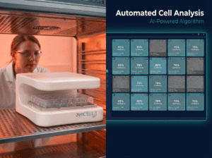
L'imagerie des cellules vivantes révolutionne les flux de travail en fournissant des informations sur la dynamique cellulaire qui étaient auparavant inaccessibles avec les méthodes d'analyse par points finaux. Un exemple typique inclut la microscopie à balayage temporel, où les systèmes d'imagerie des cellules vivantes tels que le zenCELL owl peuvent capturer des images de haute qualité à haute fréquence, fournissant des informations précieuses sur la division cellulaire, la migration et les changements morphologiques. Cette surveillance continue offre un ensemble de données robuste qui contribue à une analyse quantitative précise, conduisant à des résultats expérimentaux reproductibles et hautement fiables.
- Microscopie en accéléré pour l'analyse de processus dynamiques.
- La surveillance continue améliore la reproductibilité des données.
- Permet une analyse quantitative robuste.
Continuez votre lecture pour explorer des perspectives et des stratégies plus avancées.
“`
“`html
Le pouvoir des marqueurs fluorescents en imagerie cellulaire
Illuminer l'invisible
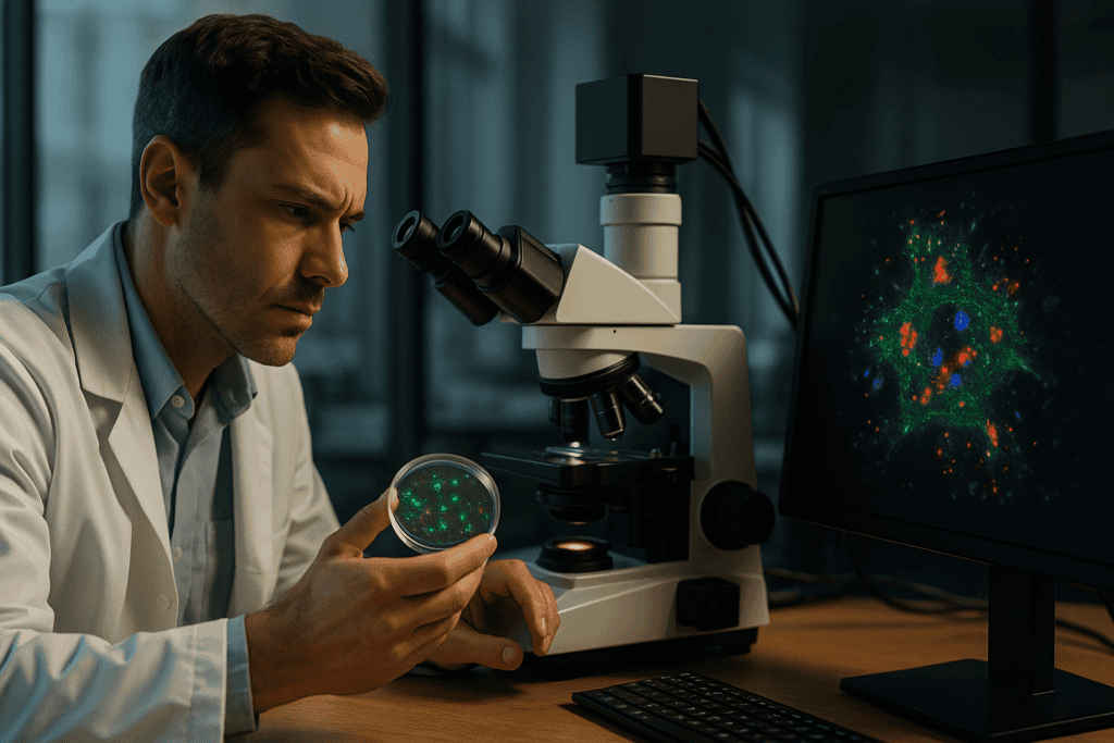
Les marqueurs fluorescents ont révolutionné le domaine de l'imagerie cellulaire, rendant visible l'invisible grâce à l'utilisation de colorants et de protéines fluorescents qui se lient à des composants cellulaires spécifiques. Ces marqueurs sont essentiels pour permettre le suivi des cellules vivantes, fournissant des informations sur l'architecture cellulaire, les voies de signalisation et les interactions protéiques. Par exemple, l'application de protéines marquées par la GFP permet aux chercheurs de suivre la localisation et le mouvement des protéines dans les cellules vivantes, dévoilant des processus qui étaient auparavant obscurcis par des méthodologies d'analyse en fin de vie.
- Identifier et valider des marqueurs spécifiques à vos objectifs de recherche.
Intégration de l'IA et de l'apprentissage automatique dans l'imagerie
Transformer les données en informations
Avec l'explosion des données générées par l'imagerie de cellules vivantes, l'IA et l'apprentissage automatique sont devenus essentiels pour extraire des informations significatives d'énormes ensembles de données. Ces technologies aident à analyser des données d'imagerie complexes en reconnaissant des modèles et des anomalies souvent manqués par les observateurs humains. Par exemple, des logiciels avancés comme CellProfiler utilisent des algorithmes d'apprentissage automatique pour segmenter les cellules, quantifier les phénotypes cellulaires et même prédire les réponses cellulaires. En automatisant ces analyses, les chercheurs peuvent améliorer la précision, réduire les biais et augmenter le débit.
- Intégrer des outils d'IA pour rationaliser les flux de travail d'analyse de données.
Cryoconservation : Maintenir la qualité des cellules vivantes
Préservation de l'intégrité cellulaire dans le temps
La cryopréservation joue un rôle crucial dans les études d'imagerie de cellules vivantes, permettant aux chercheurs de maintenir la viabilité des cellules sur de longues périodes. Cette méthode garantit que les échantillons de cellules vivantes conservent leur fonctionnalité et leur réactivité, essentielles pour les études longitudinales. Des techniques telles que la congélation à vitesse contrôlée et la vitrification atténuent la formation de cristaux de glace, qui peuvent endommager les structures cellulaires. La capacité de préserver les cellules à des taux de viabilité élevés permet des configurations expérimentales cohérentes, réduisant la variabilité qui peut fausser les données d'imagerie de cellules vivantes.
- Mettre en œuvre des protocoles de cryopréservation appropriés pour garantir la viabilité cellulaire.
Le rôle des microscopes d'incubateur dans l'efficacité des flux de travail
Surveillance continue sans interruption
Les microscopes incubateurs offrent un environnement idéal pour l'imagerie de cellules vivantes en permettant une observation continue sans perturber les conditions de culture. Ces systèmes intègrent des contrôles environnementaux (température, CO2, humidité) directement dans l'installation d'imagerie, facilitant ainsi les études à long terme nécessaires pour capturer les changements cellulaires progressifs. Des appareils tels que l'Olympus IXplore Live aident les chercheurs à mener des imageries en temps réel tout en maintenant les conditions physiologiques cruciales pour la santé et les fonctions normales des cellules.
- Utiliser des microscopes d'incubateur pour maintenir les conditions expérimentales.
Technologies d'imagerie 3D haute résolution
Élargir les perspectives en recherche cellulaire
Les technologies d'imagerie 3D telles que la microscopie confocale et multiphotonique offrent une profondeur et une résolution inégalées, permettant aux chercheurs de visualiser les structures cellulaires en trois dimensions. Cette avancée est essentielle pour les études impliquant des tissus complexes ou des structures multicellulaires, où les interactions se produisent dans toutes les dimensions spatiales. Par exemple, son application dans la recherche sur le microenvironnement tumoral éclaire les complexités de la progression du cancer, dévoilant des interactions auparavant cachées au sein des matrices tissulaires denses.
- Envisagez l'imagerie 3D pour une compréhension complète de l'architecture tissulaire.
Maximiser l'efficacité avec des pipelines d'imagerie automatisés
Optimisation des processus pour une productivité élevée
Les pipelines d'imagerie automatisés simplifient le flux de travail en gérant l'acquisition, le traitement et l'analyse des images avec une intervention minimale de l'utilisateur. Cette automatisation réduit les erreurs, augmente la répétabilité et permet de gagner un temps de recherche précieux. Des plateformes telles que NIS-Elements de Nikon rationalisent ces processus en s'intégrant de manière transparente à l'équipement robotique, permettant ainsi des applications de criblage à haut débit qui accélèrent le processus de découverte de médicaments.
- Exploitez les systèmes automatisés pour augmenter le débit et la cohérence des données.
Stratégies de recherche collaborative et de partage de données
Amplifier l'impact de la recherche par la connectivité
La collaboration au sein de la communauté scientifique est renforcée par des plateformes de partage de données qui facilitent l'échange de jeux de données d'imagerie et de méthodologies. Les plateformes open-source, y compris l'Image Data Resource (IDR), permettent un accès multi-institutionnel à des données d'imagerie de haute qualité, favorisant la collaboration et l'innovation. Ces ressources permettent aux chercheurs de s'appuyer sur les travaux existants, d'éviter les redondances et de maximiser l'utilisation des ressources.
- Participez au partage de données pour améliorer les collaborations de recherche.
Ensuite, nous conclurons avec les points clés à retenir, les métriques et une conclusion percutante.
“`
“`html
Réalité virtuelle et augmentée en imagerie cellulaire
Au-delà de la visualisation traditionnelle
Alors que les limites de l'imagerie cellulaire s'étendent, l'intégration de la réalité virtuelle (RV) et de la réalité augmentée (RA) présente de nouvelles façons d'interagir avec des ensembles de données cellulaires complexes. Les technologies RV et RA permettent aux chercheurs de visualiser et de manipuler des données biologiques tridimensionnelles dans des environnements immersifs, offrant des perspectives profondes sur les relations spatiales et la dynamique. Cette capacité améliore les résultats éducatifs et ouvre la voie à une meilleure compréhension de phénomènes tels que la connectivité neuronale et le développement des tissus, difficiles à appréhender dans des formats bidimensionnels. En utilisant des plateformes telles que le CAVE Automatic Virtual Environment, les scientifiques peuvent simuler des processus cellulaires à une échelle et une perspective inégalées par les méthodes conventionnelles.
- Explorer la RA/RV pour une approche interactive de l'interprétation des données.
Relever les défis de l'imagerie des cellules vivantes
Surmonter les contraintes pour libérer le potentiel
Malgré des avancées remarquables, l'imagerie de cellules vivantes présente des défis qui doivent être abordés pour exploiter pleinement son potentiel. L'un des principaux défis est la phototoxicité, qui résulte d'une exposition prolongée à la lumière, pouvant altérer le comportement cellulaire et compromettre l'intégrité des données. Des stratégies telles que l'optimisation des concentrations de colorants, l'utilisation de marqueurs photostables et l'intégration des avancées dans les technologies à faible photon sont essentielles pour réduire les photodommages. De plus, le volume considérable de données peut submerger les infrastructures de stockage et de traitement traditionnelles, soulignant la nécessité de solutions évolutives et de ressources informatiques avancées pour gérer efficacement les données volumineuses.
- Adoptez des techniques pour atténuer la phototoxicité afin d'obtenir une imagerie précise.
Considérations éthiques dans la recherche en imagerie
Naviguer dans le paysage moral
Alors que les techniques d'imagerie cellulaire continuent d'évoluer, les implications éthiques entourant leur utilisation se font de plus en plus sentir. Il est essentiel de veiller à ce que les études d'imagerie respectent l'intégrité et la confidentialité cellulaires, en particulier lors de la recherche sur des modèles cellulaires sensibles ou propriétaires. Aborder les préoccupations éthiques de manière transparente et adhérer à des directives strictes favorise la confiance au sein de la communauté scientifique et du grand public. En établissant des cadres éthiques solides, les chercheurs peuvent équilibrer la quête de connaissances avec l'impératif de mener des recherches de manière responsable.
- S'engager avec des cadres éthiques pour maintenir l'intégrité de la recherche.
Conclusion
Dans le parcours “ Au-delà du cliché ”, l'imagerie de cellules vivantes s'impose comme une force de transformation, redéfinissant ce qui est possible dans la recherche cellulaire. L'intégration de techniques et de technologies telles que l'IA, l'apprentissage automatique, l'imagerie 3D et même la VR/AR remodèle nos approches scientifiques, offrant des perspectives plus dynamiques, précises et perspicaces sur les comportements et les interactions cellulaires. Cette révolution élargit non seulement notre compréhension, mais ouvre également de nouvelles voies pour les innovations dans le traitement des maladies et les applications de bio-ingénierie.
Au milieu de ces avancées technologiques, on ne saurait trop insister sur l'importance de maintenir des normes scientifiques rigoureuses et des considérations éthiques. Alors que nous sommes au bord d'avancées sans précédent, il incombe aux chercheurs de réfléchir continuellement à l'impact de nos méthodologies et de nos protocoles de gestion des données. Les obstacles de la phototoxicité, de la gestion des données et de l'intégrité éthique soulignent un parcours marqué par la collaboration, l'innovation et un engagement indéfectible envers la rigueur scientifique.
En fin de compte, la valeur de l'adoption des avancées de l'imagerie des cellules vivantes ne réside pas seulement dans l'atteinte de l'excellence académique et professionnelle, mais aussi dans la contribution significative à l'effort scientifique collectif. Les chercheurs sont encouragés à adopter ces outils et stratégies de pointe, à favoriser un esprit de connectivité et de collaboration, et à participer aux efforts qui repoussent les limites de la recherche biologique plus loin que jamais auparavant. Alors que nous illuminons l'invisible et décodons les complexités des structures cellulaires, allons de l'avant avec courage et une quête inébranlable de savoir, sachant que nos découvertes d'aujourd'hui jettent les bases des innovations de demain.
“`