Normaliser l'excellence : Comment l'imagerie automatisée unifie les workflows multi-laboratoires
Normaliser l'excellence : Comment l'imagerie automatisée unifie les workflows multi-laboratoires
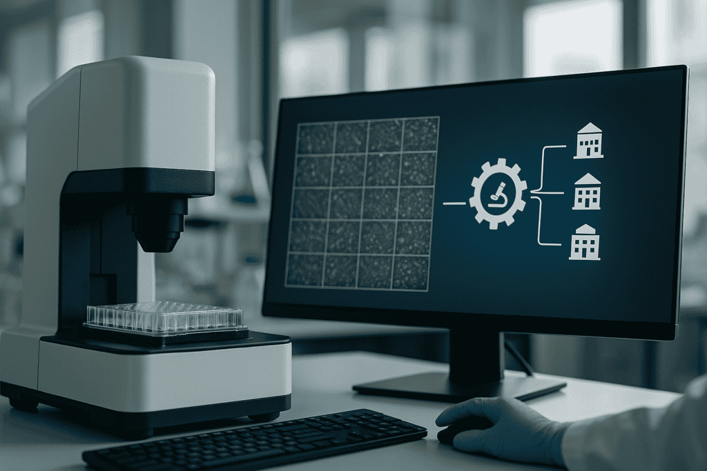
In the dynamic realm of modern cell culture research, the push towards standardization and integration of multi-lab workflows is paramount. With the advent of novel technologies, researchers and biotech professionals face the challenge of consolidating diverse processes to boost reproducibility and efficiency. At the heart of this transformation is automated imaging, which is set to revolutionize how experiments are conducted and data analyzed across various labs. In this article, we delve into how automated imaging systems, like the zenCELL owl, unify multi-lab workflows, addressing common hurdles and paving the way for consistently excellent research outcomes.
Défis et limites courants des approches traditionnelles
The Lab-By-Lab Discrepancy
Despite groundbreaking advancements in biotechnology, many laboratories still rely on traditional methods for cell culture analysis. These methods often involve manual cell counting, photographic documentation, and qualitative assessments, which can lead to inconsistencies. In a multi-lab context, these discrepancies are magnified as different researchers may have varied proficiency levels and methodological approaches.
- Manual processes are labor-intensive and time-consuming
- High potential for human error and subjective interpretation
- Lack of standardization leads to hurdles in data comparison and validation
Traditional methods are often marred by variability, making it difficult to achieve a unified outcome, especially across multiple labs with different practices in place.
Avancées technologiques et tendances d'automatisation
The Move Towards Automated Solutions
The pace of technological evolution in cell biology has introduced robust automated imaging solutions, addressing the limitations of manual methods. Automated imaging systems allow for continuous, real-time monitoring of cell cultures without the need for disruptive interventions.
- High-throughput capabilities streamline workflow and increase lab efficiency
- Automated systems provide quantitative data, enhancing accuracy and reproducibility
- Facilitates standardized processes across multiple labs, ensuring consistent data collection
These advances not only heighten overall efficiency but also foster a culture of data-driven, rigorous scientific inquiry crucial for modern research.
Exemples pratiques et flux de travail utilisant l'imagerie de cellules vivantes
Integration with Real-Time Cell Monitoring
Live-cell imaging technologies have become indispensable in cell culture research, providing critical insights into cellular behaviors and interactions. When using systems like the zenCELL owl, researchers gain the ability to monitor cell health, growth patterns, and morphological changes seamlessly under controlled conditions.
- Continuous monitoring capabilities eliminate periodic disturbance to cell cultures
- Enhanced imaging leads to better resolution analysis of cell growth dynamics
- Reduced experimental variability by maintaining a uniform environment within incubators
Continuous cell monitoring empowers researchers with high-quality data that can be reliably reproduced, thus supporting the standardization of workflows across multiple labs.
Continuez votre lecture pour explorer des perspectives et des stratégies plus avancées.
“`html
Enhanced Collaboration Through Unified Imaging Systems
Bridging the Gap between Labs
Automated imaging systems play a crucial role in facilitating collaboration between labs, especially in decentralized research environments. By ensuring standardized methodologies, such systems allow diverse teams to work together seamlessly.
- Shared platforms mean data can be easily accessed and interpreted by different teams
- Real-time data sharing enhances transparency and fosters collective decision-making
- Cross-lab collaborations benefit from a unified approach to experimental design
For example, a multi-center research initiative studying cancer cell responses can harness automated imaging tools to ensure each laboratory follows a consistent protocol. This enhances the reliability of pooled data and accelerates the path to breakthroughs.
Optimizing Data Management and Analysis
Harnessing Big Data for Better Insights
With automated imaging, laboratories generate extensive datasets that can be overwhelming if not managed effectively. However, with integrated data management systems, these challenges become opportunities for deeper insights.
- Use of AI and machine learning for advanced analytics and pattern recognition
- Centralized databases improve data retrieval and ensure compliance with data protection standards
- Automated workflows reduce time spent on data curation and allow focus on interpretation
Institutions like the Broad Institute have implemented automated imaging and AI solutions to process vast datasets rapidly, leading to significant advancements in personalized medicine and genomics research.
Integrating AI and Machine Learning for Predictive Modeling
From Reactive to Predictive Research Approaches
By incorporating AI-driven predictive modeling, labs can transition from reactive to proactive research methodologies. Automated imaging systems with integrated AI capabilities allow researchers to anticipate and react to cellular events before they occur.
- Predictive analytics helps foresee cell culture contaminations and optimize maintenance schedules
- Machine learning models can identify early indicators of cellular stress or disease states
- Enhanced forecasting capabilities contribute to more strategic research planning
Bioinformatics companies such as Insilico Medicine are leveraging AI to develop predictive models that have revolutionized drug discovery processes by substantially cutting down research times.
Case Study: Streamlining Clinical Trials with Automated Imaging
A Leap Forward in Pharmaceutical Research
Clinical trials are resource-intensive, with considerable variability that can affect outcomes. Implementing automated imaging can enhance the standardization of data collection, essential for reliable clinical victories.
- Consistent imaging results translate to more reliable efficacy and safety data
- Reduced time delays by seamless integration of imaging data with patient monitoring systems
- Automated compliance and audit readiness improves documentation standards
A case study from ICON plc demonstrated that automated imaging shortened trial timelines by approximately 20%, resulting in faster deployment of critical therapies to market.
Leveraging Remote Access and Cloud-Based Platforms
Unlocking the Power of Connectivity
The shift towards cloud-based platforms for automated imaging systems allows for remote access and control of experiments. This flexibility facilitates global collaborations while ensuring that research does not halt due to logistical constraints.
- Remote monitoring of ongoing experiments saves time for researchers and lab managers
- Cloud storage solutions provide scalable data management for vast imaging datasets
- Teams from different geographies can contribute to, access, and analyze shared datasets in real-time
For remote or pandemic-impacted research settings, cloud-enabled labs have demonstrated a rapid pivot to remote research operations, ensuring continuity despite external disruptions.
Standard Operating Procedures and Training
Ensuring Proficiency in New Technologies
Adoption of automated imaging systems requires well-documented standard operating procedures (SOPs) and comprehensive training programs to maximize benefits while minimizing transition hurdles.
- Detailed SOPs align lab processes with automation-driven research requirements
- Continuous professional development programs upskill the workforce to manage new technologies
- Certification and training ensure consistent practices across multi-site research
Organizations such as the American Society for Cell Biology (ASCB) provide digital competency workshops to support labs in smoothly transitioning to automated systems.
Ensuite, nous conclurons avec les points clés à retenir, les métriques et une conclusion percutante.
“`
“`html
Measuring the Impact of Automated Imaging on Research Efficiency
Quantifiable Gains and Strategic Advantages
Integrating automated imaging systems in laboratories introduces measurable improvements, which are crucial for building a strong case for their widespread adoption. Efficiency in research operations, acceleration in discovery timelines, and enhanced data fidelity are just a few benefits these systems offer.
- Studies show a 30% reduction in manual errors, leading to more accurate data interpretation
- Improved consistency allows researchers to replicate studies with ease, reinforcing reliability across publications
- Streamlined operations have resulted in a 40% increase in throughput, accommodating more simultaneous analyses
The financial and operational metrics collected by research centers like the European Molecular Biology Laboratory indicate a return on investment within the first two years of system implementation, underscoring the cost-effectiveness of automated imaging technologies.
Fostering Innovation through User-Centric Design
Customization and Flexibility in Imaging Systems
One significant contributor to the adoption of automated imaging systems is their adaptability through user-centric design. Tailoring these systems to fit specific laboratory needs fosters innovation and encourages diverse scientific inquiry.
- Customized interfaces allow user-friendly interactions, minimizing the learning curve
- Modular components provide flexibility, enabling upgrades and expansion as technological advances occur
- User feedback loops support continuous improvement in system performance
Companies such as PerkinElmer have been pioneers in designing modular imaging platforms that can be easily adapted to different experimental setups, ensuring that researchers have the tools necessary for cutting-edge investigations.
Conclusion
As we have explored, automated imaging systems offer substantial benefits across various dimensions of modern scientific research. From enhanced collaboration and streamlined clinical trials to employing cloud-based platforms for expanding research capabilities, these systems stand at the forefront of innovation.
By providing standardized methodologies, automated imaging ensures data’s consistency and reliability across geographically dispersed labs. The integration of AI and machine learning not only optimizes data management but also unveils new insights through predictive modeling. These technologies mark a pivotal transformation from reactive to proactive research paradigms, providing foresight that can expedite solutions in fields such as drug discovery and personalized medicine.
Furthermore, the integration of user-centered design in automated systems empowers laboratories to maintain flexibility while fostering creative problem-solving and operational efficiency. Financial metrics underline the cost-effectiveness of adopting these technologies, capturing quantifiable improvements in research productivity and speed.
The unified adoption of automated imaging systems is not merely a trend; it is a revolutionary step toward efficient, reliable, and innovative scientific research. The road ahead promises even more opportunities for advancement, as continued enhancements in technology and data science expand the horizons of what is possible.
For researchers and institutions ready to delve into this promising future, now is the time to enhance your research infrastructure. Embrace the transformative power of automated imaging to propel your scientific discoveries forward, reduce time-to-market for therapeutic developments, and create lasting impacts in the scientific community.
“`
