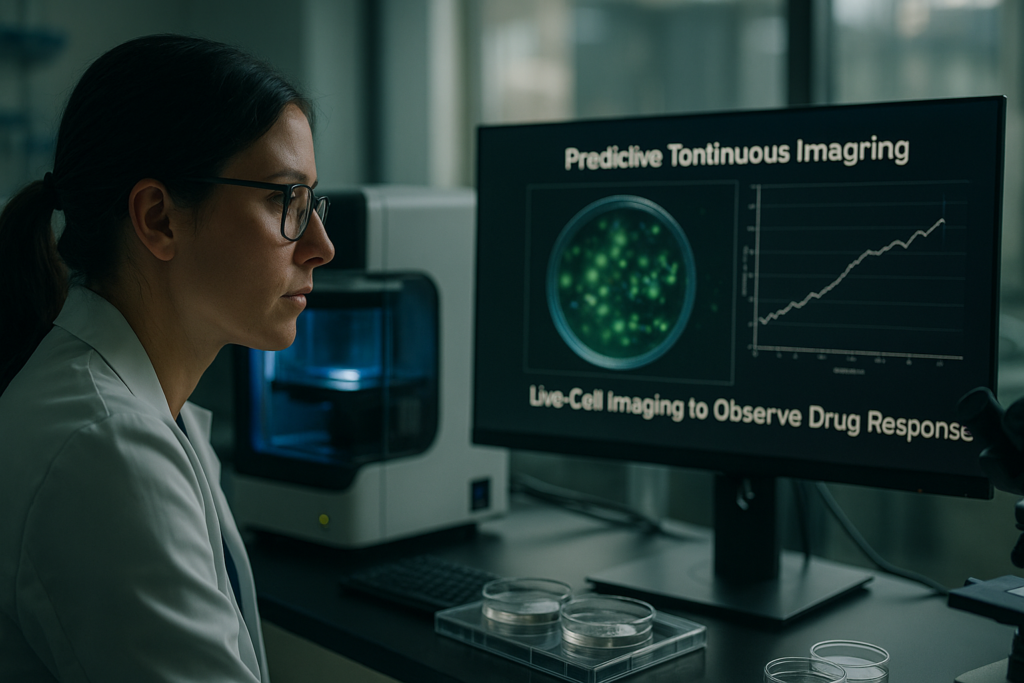
Predictive Toxicology: Using Continuous Imaging to Observe Drug Response
Predictive Toxicology: Using Continuous Imaging to Observe Drug Response
In the ever-evolving field of biomedical research, predictive toxicology is gaining significant traction as a key investigative approach. With its focus on forecasting the toxic effects of new pharmaceutical compounds, this discipline is crucial in preclinical trial phases. Continuous live-cell imaging provides an invaluable tool in this process, offering researchers a more dynamic view of how cells respond to potential drugs over time. This article dives into why continuous imaging is transforming predictive toxicology, and how it promises enhanced accuracy and efficiency in cell culture research.
Défis et limites des approches traditionnelles
The Static Nature of Conventional Methods
Traditional toxicology approaches typically rely on endpoint assays, where observations are made at a single point in time. This method inherently overlooks dynamic changes in cell behavior, potentially missing critical transient responses. Moreover, these techniques often require destructive sample processing, thereby limiting the ability to collect longitudinal data from the same sample.
- Endpoint assays fail to capture dynamic responses.
- Destructive methods limit longitudinal observations.
Without the capacity to observe continuous cellular responses, researchers are faced with a significant gap in understanding real-time drug interactions. A shift towards techniques that allow for ongoing observation could rectify these limitations.
Avancées technologiques et tendances d'automatisation
The Rise of Real-Time Imaging Technologies
Recent technological advancements, such as live-cell imaging systems, have revolutionized predictive toxicology. These systems enable the real-time observation of cellular responses to compounds. Automation trends, particularly in cell culture technologies, are enhancing data accuracy and throughput, providing a more comprehensive view of cellular dynamics.
- Enhanced accuracy through real-time monitoring.
- Improved throughput with automated systems.
Implementing continuous imaging provides a constant stream of data, which is particularly crucial in high-throughput screening (HTS) environments, ensuring robust and reproducible results that reflect true cell behavior.
Exemples pratiques et flux de travail utilisant l'imagerie de cellules vivantes
Integrating Live-Cell Imaging in Lab Workflows
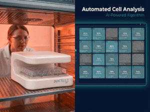
Incorporating continuous imaging into laboratory workflows begins with selecting the appropriate technology that suits experimental designs. Devices like the zenCELL owl, an incubator-compatible imaging system, allow seamless integration with existing lab setups. This compact system automates the imaging process, maintaining optimal conditions within an incubator while capturing high-quality data.
- Automated imaging increases consistency and reduces manual error.
- Seamless integration into current lab infrastructures.
Utilizing these tools enables scientists to closely monitor cellular changes over time, adapting experimental designs in real-time, thus bolstering the reliability of predictive toxicology studies.
Comment l'imagerie basée sur incubateur améliore la reproductibilité et la qualité des données
Maintaining Optimal Conditions for Consistent Results
One of the most significant advantages of incubator-based imaging systems is their ability to maintain stable conditions, minimizing fluctuations that could lead to altered cell behavior. By keeping the environment constant—in terms of temperature, humidity, and gas concentration—high data quality and reproducibility are achievable, which are critical for toxicology studies.
- Reduced environmental variation ensures data consistency.
- Enhanced reliability of continuous imaging data.
Such consistency is invaluable when assessing the toxicological profile of compounds, as it allows researchers to accurately trace how environmental changes can influence cellular responses to drugs.
Continuez votre lecture pour explorer des perspectives et des stratégies plus avancées.
Bridging In Vitro and In Vivo with Predictive Models
Leveraging Predictive Models for Better Drug Assessment
With the help of continuous imaging, the gap between in vitro cell cultures and in vivo responses can be increasingly bridged through sophisticated predictive models. These models utilize data garnered from real-time imaging to simulate how drugs might behave in a living organism. By doing so, researchers can foresee potential adverse effects and efficacy without the need for early-stage animal testing, saving both time and resources in drug development.
- Develop and refine predictive models to enhance correlation with in vivo data.
For example, machine learning algorithms can be employed to analyze time-lapse imaging data, recognizing patterns that correlate with known toxicological outcomes. This integration of technological advancements with biological research enhances the predictive power and reliability of preclinical assessments.
Quantifying Cellular Changes with Advanced Metrics
Utilizing Quantitative Imaging Techniques
Continuous imaging not only visualizes cellular processes but also quantifies them. Advanced metrics, such as cell proliferation rates, morphological changes, and signal transduction pathways, offer precise data that can be analyzed to understand drug interactions at a granular level. This form of quantification helps in discerning subtle changes in cellular behavior, which might otherwise be missed through traditional endpoint analysis.
- Employ image analysis software to extract and interpret quantitative data efficiently.
Transitioning from qualitative to quantitative imaging has allowed researchers to develop more refined metrics that better predict a compound’s potential for toxicity, thereby driving more informed decision-making processes in drug development.
Harnessing High-Content Screening for Comprehensive Analysis
Streamlining Drug Discovery with High-Content Approaches
High-Content Screening (HCS) is an invaluable advancement in predictive toxicology. By combining continuous imaging with high-throughput technologies, researchers can perform large-scale screening of compounds more efficiently. HCS permits the simultaneous analysis of multiple cellular parameters, yielding comprehensive insights into drug effects across various pathways and models.
- Leverage multi-parametric analysis to maximize dataset richness.
For instance, pharmaceutical companies have adopted HCS to expedite their drug discovery pipelines, allowing them to screen thousands of compounds for toxic effects in a fraction of the time required by traditional methods. This has resulted in faster, more cost-effective drug development cycles.
Designing Adaptive Experimental Protocols
Creating Flexible Methodologies to Capture Dynamic Responses
Continuous imaging empowers researchers to develop adaptive experimental protocols that can evolve based on real-time data feedback. This dynamic approach allows scientists to adjust variables mid-experiment to better understand the temporal nature of drug responses, refining the focus of the study as new patterns emerge.
- Implement adaptive protocols to respond immediately to data trends.
Practically, this means employing real-time data analytics to modify or halt experiments when unexpected toxicological changes are observed, thus preventing resource waste and time spent on non-viable drug candidates early on.
Enhancing Collaboration and Data Sharing
Facilitating Open Science through Shared Imaging Data
Advancements in continuous imaging have paved the way for enhanced collaboration and data sharing across scientific communities. Open databases containing imaging data allow for cross-institutional comparisons and validations, fostering a more collaborative environment in predictive toxicology research.
- Engage in collaborative networks to enrich research outcomes.
For example, initiatives like the Open Microscopy Environment (OME) provide platforms for the free exchange of imaging data and analytical tools, enhancing transparency and enabling researchers worldwide to synergize their efforts in combating toxicological challenges.
Exploring Case Studies of Successful Implementations
Real-World Applications Demonstrating Imaging Power
Several case studies highlight the transformative impact of continuous imaging in predictive toxicology. One notable example involves a biotech firm utilizing zenCELL owl imaging technology to optimize their lead compound’s safety profile. Through continuous monitoring, researchers could precisely adjust dosages in cell lines indicative of human physiology, resulting in a drug candidate with significantly reduced adverse effects when preclinically trialed.
- Analyze case studies to derive best practices and guide future research.
These real-world implementations showcase how embracing continuous imaging leads to improved predictive accuracy and better-informed decisions regarding drug safety assessments.
Future Directions and Emerging Innovations
Anticipating the Next Wave of Technological Progress
The field of predictive toxicology is constantly evolving, with emerging technologies poised to further enhance continuous imaging capabilities. Innovations such as artificial intelligence and CRISPR-based gene editing are likely to be integrated with live-cell imaging, paving the way for even more personalized and precise toxicological assessments.
- Stay informed about cutting-edge innovations to maintain research competitiveness.
Future advances are expected to facilitate a deeper understanding of multi-cellular interactions in heterogeneous environments, paving the way for breakthroughs in personalized medicine and targeted therapies.
Ensuite, nous conclurons avec les points clés à retenir, les métriques et une conclusion percutante.
“`html
Integrating Patient-Derived Models
Personalizing Drug Assessments with Human-Derived Cells
Incorporating patient-derived cells into continuous imaging offers an avenue for personalizing drug assessments. These models provide a more accurate representation of how genetic variations in humans may affect drug efficacy and toxicity, bridging a crucial gap between generalized preclinical findings and individualized patient responses. By utilizing patient-specific cell cultures, researchers can tailor experimental protocols to align with individual patient profiles, enabling the prediction of unique adverse reactions and optimizing therapeutic regimens.
- Utilize patient-derived organoids to refine preclinical predictions.
This method not only enhances the precision of toxicity predictions but also supports the development of targeted therapies, making it indispensable in the era of personalized medicine.
Leveraging Cloud-Based Data Analytics
Harnessing Big Data for Superior Decision Making
The integration of cloud-based analytics with continuous imaging tools allows for real-time processing and interpretation of vast amounts of data. Utilizing cloud infrastructure enables researchers to handle and analyze complex datasets efficiently, leading to more accurate predictive models. These systems can rapidly modify experimental conditions based on immediate data feedback, amplifying the efficiency of hypothesis-driven research and minimizing redundant investigations.
- Adopt cloud computing solutions for scalable data processing capabilities.
Such technologies not only streamline operations but also democratize access to computational resources, paving the way for collaborative, large-scale research endeavors.
Strengthening Regulatory Frameworks
Aligning New Technologies with Regulatory Standards
Continuous imaging technologies necessitate the evolution of regulatory guidelines to accommodate novel methodologies in drug testing. Regulatory bodies are progressively recognizing these advanced techniques as valid components of the drug evaluation process. By incorporating continuous imaging into their evaluations, regulatory frameworks can ensure a more comprehensive assessment of a compound’s safety, potentially leading to faster approval times and enhanced drug market stability.
- Work with regulatory agencies to establish guidelines that embrace technological advancements.
Promoting dialogue between researchers, industry, and regulators will ensure that the implementation of these innovations aligns with rigorous safety standards, ultimately benefiting public health.
Conclusion
The amalgamation of continuous imaging with predictive toxicology stands at the forefront of a paradigm shift in drug development. By harnessing live-cell imaging, developing adaptive experimental protocols, and fostering collaborative data sharing, the field is moving towards more accurate and reliable preclinical assessments. These methodologies not only enhance the precision of toxicity predictions but also contribute significantly to minimizing reliance on traditional, resource-intensive animal testing models.
Predictive toxicology, bolstered by continuous imaging advancements, holds immense promise in facilitating more personalized medicine. As new technologies emerge, such as artificial intelligence-driven analytics and patient-derived cell models, the scope for further personalization and accuracy in toxicological assessments expands. This approach aligns well with contemporary movements toward patient-centered healthcare delivery.
The value of continuous imaging extends beyond laboratory insights. It heralds a change in how we assess drug candidates, potentially revolutionizing the timelines and efficiencies associated with drug approvals. Researchers and pharmaceutical companies who adapt to these innovations stand to enhance their competitive edge, bringing safer and more effective therapeutics to market faster. As we continue to integrate these technologies, global collaborations and cooperative frameworks will be pivotal in harmonizing efforts across the scientific community.
As we look to the future, embracing these promising methodologies is vital to fostering an environment where predictive toxicology can flourish. Researchers are encouraged to stay abreast of burgeoning technologies and to participate proactively in shaping the future of drug discovery and safety assessment. With a commitment to innovation and excellence, we can ensure that our journey towards more effective, personalized medicine continues with integrity and purpose.
The opportunities to reshape the landscape of drug development and delivery through continuous imaging and advanced predictive modeling are vast. By leveraging these tools, we not only expand our scientific capabilities but also affirm our responsibility to improve global healthcare outcomes.
“`