Take Your Weekends Back: How Remote Live-Cell Monitoring Frees Your Schedule

“`html
Take Your Weekends Back: How Remote Live-Cell Monitoring Frees Your Schedule
In the fast-evolving world of cell culture research, balancing rigorous experimental demands with personal time can seem like an elusive goal. Yet, with advancements in remote live-cell monitoring, researchers, lab managers, and biotech professionals can now reclaim their weekends without compromising the quality of their experiments. This article will delve into the common challenges of traditional cell culture methods, explore technological advances, and highlight how remote live-cell monitoring has transformed laboratory workflows.
Défis et limites courants des approches traditionnelles
The Demand for Constant Monitoring
Traditional cell culture techniques often require continuous manual monitoring, necessitating weekend lab visits and late-night checks to ensure cell viability and experimental integrity. These frequent disruptions not only strain personal time but also introduce variability due to human error during manual observations.
- Manual checks increase the risk of contamination and data inconsistencies.
- Lab personnel suffer from work-life imbalance due to on-call requirements.
- Human observations may lack the precision and timing needed for detailed data analysis.
Continuez votre lecture pour explorer des perspectives et des stratégies plus avancées.
“`
“`html
The Advantages of Automation in Cell Culture
Streamlining Processes with Technology
With the introduction of robotic systems and programmable incubators, the landscape of cell culture has been revolutionized. Automation allows for standardized processes, reducing human error and improving reproducibility. For instance, systems like the Tecan Fluent and Hamilton STARlet offer seamless integration of automated liquid handling with cell culture protocols.
- Automated systems ensure consistent media changes and cell passaging, leading to higher quality data.
- Increased throughput is achievable, allowing scientists to handle more samples concurrently while maintaining accuracy.
- Investing in automation can reduce long-term operational costs by lowering the necessity for manual labor.
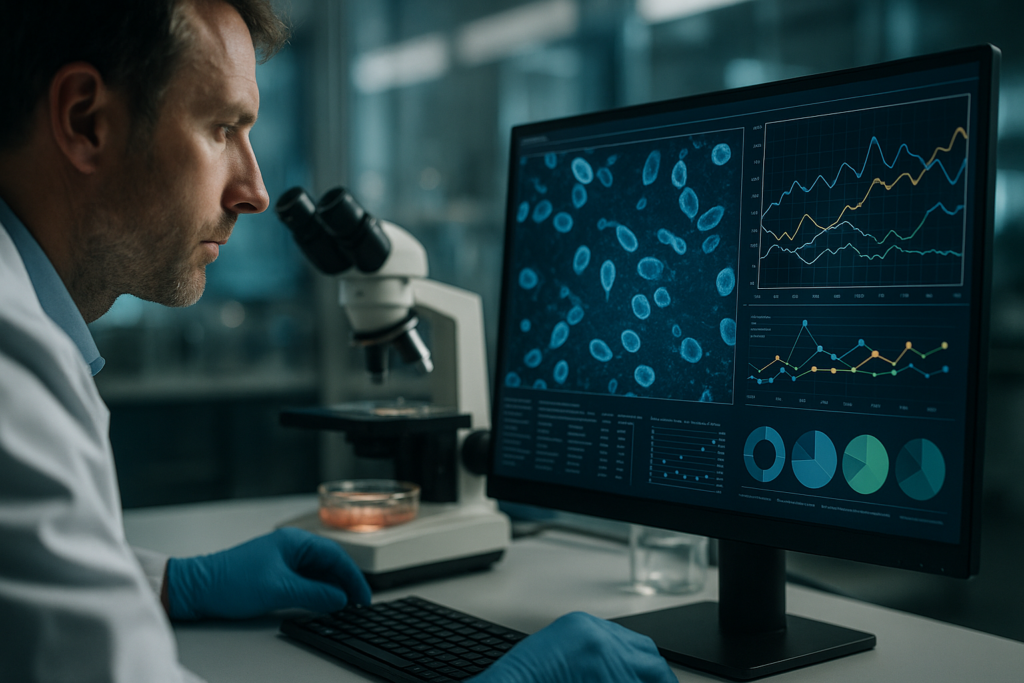
Real-Time Data Analytics and Cloud-Based Solutions
Leveraging Data for Better Outcomes
In the digital age, data is king. Real-time analytics and cloud-based solutions have enabled researchers to access critical experiment data anywhere at any time. Systems like CytoSMART and Sartorius’ Incucyte offer cloud connectivity that allows for real-time monitoring and adjustments.
- Researchers can visualize cell growth patterns through cloud dashboards, eliminating the need for on-site analysis.
- Remote data access facilitates collaboration across global teams, enriching research outcomes.
- Historical data analysis becomes straightforward, helping in identifying trends and making informed decisions.
Enhancing Accuracy with Advanced Imaging Technologies
Precision in Observation Through Innovations
Traditional microscopy has its limitations, but advancements in imaging technologies like fluorescence live-cell imaging have introduced new possibilities. Systems such as Thermo Fisher’s EVOS or Leica’s DMi8 offer unparalleled insights into cellular processes by providing high-resolution, time-lapse imaging capabilities.
- These systems minimize photodamage and preserve cell vitality, vital for accurate long-term observations.
- Software integration with these imaging systems simplifies data interpretation and enhances experimental reliability.
- Users gain the ability to record and document every minute detail, improving experimental reproducibility.
Reducing Environmental Impact Through Efficient Practices
Sustainability Gains in Modern Laboratories
The shift towards greener labs has gained momentum, with remote live-cell monitoring playing a pivotal role in reducing resource consumption. Efficient energy utilization and minimized chemical use are the cornerstones of modern, environmentally responsible cell culture practices.
- Programmable systems optimize energy use, reducing carbon footprints of research facilities.
- Automated monitoring cuts down on waste by precisely calibrating needed resources.
- Implementing green lab practices not only benefits the planet but can also align with institutional sustainability goals.
Customizing Solutions Through Modular Designs
Tailoring Frameworks to Specific Lab Needs
Gone are the days of one-size-fits-all. With companies offering modular components, labs can design systems that suit their specific research needs. This flexibility allows for a focused approach, whether dealing with stem cell research or cancer studies, providing enhanced experimental efficacy.
- Modular systems can be expanded with minimal investment, enabling labs to adapt as their research requirements evolve.
- Customized solutions enhance user engagement and improve workflow efficiency by addressing unique laboratory challenges.
- Long-term infrastructure planning is simplified, ensuring laboratories remain cutting-edge without major overhauls.
Boosting Reliability with Integrated Quality Control
Ensuring Consistency in Results
Integrated quality control measures are essential for achieving reliable results in cell culture. Advancements in sensor technologies and AI-driven analytics have empowered systems to independently verify the consistency of experimental conditions. Plate Reader Systems and CO2 incubators equipped with real-time feedback loops contribute significantly to quality assurance.
- Automatic alerts and error notifications help researchers address issues proactively, reducing downtime.
- Quality control integration strengthens validation processes, enhancing data credibility for publications.
- Investments in reliable technology safeguard against costly experimental setbacks.
Ensuite, nous conclurons avec les points clés à retenir, les métriques et une conclusion percutante.
“`
“`html
Empowering Laboratories Through Artificial Intelligence
Harnessing AI for Unprecedented Efficiency
Artificial Intelligence (AI) has become a catalyst for evolution in cell culture practices. Machine learning algorithms can rapidly process large datasets, optimizing cell culture conditions and predicting experimental outcomes with remarkable accuracy. Companies such as Sartorius and Thermo Fisher are integrating AI functionality into their systems to streamline workflows.
- AI-driven platforms utilize predictive analytics to tailor optimal culturing conditions, saving time and resources.
- Automated image analysis powered by AI accelerates data processing, allowing for quicker adjustments to experimental setups.
- By harnessing AI, labs can make data-driven decisions that enhance research precision and validity.
Creating Collaborative Ecosystems
Fostering Synergy in Research Environments
The shift toward interconnected systems facilitates unprecedented collaboration within the research community. By utilizing advanced communication technologies, researchers can bridge geographical divides, ensuring expertise and innovation are not confined by location. The integration of unified communication platforms within lab systems enhances this collaborative spirit.
- Shared virtual workspaces streamline idea exchange and accelerate discoveries across various fields.
- Collaborative tools support synchronous research methodologies, reducing redundancy and increasing productivity.
- Building a global network of researchers fosters diverse perspectives and interdisciplinary breakthroughs.
Ensuring Security and Compliance
Mitigating Risks in Digital Lab Environments
As technology advances, concerns around data security and regulatory compliance have become more pronounced. Laboratories are turning to robust security measures to protect sensitive data and ensure adherence to international regulations. Solutions by firms like Eppendorf and BioTek incorporate advanced cybersecurity protocols.
- Data encryption and secure cloud storage prevent unauthorized access, fostering trust and safeguarding intellectual property.
- Compliance with standards such as GLP and ISO ensures operational integrity and facilitates cross-border research collaborations.
- Regular audits and monitoring assure adherence to protocols, mitigating risks associated with data breaches.
Conclusion
Reflecting on the transformative power of automation in cell culture, the key advantages become apparent. From enhancing accuracy with state-of-the-art imaging technologies and promoting sustainable practices, to leveraging AI for analytical precision, the landscape of research is supported by a robust framework of technological advancements. These strategies not only optimize experimental workflows but also ensure high fidelity in data analysis, contributing to more reliable and impactful scientific outcomes.
The relevance of these strategies lies in their ability to address both contemporary challenges and future demands in research. As laboratories increasingly adopt automation, they not only refine their operational efficiency but also expand their scientific horizons. Automation has become a linchpin of modern laboratories, driving innovation and fostering a collaborative ethos that transcends geographical boundaries.
In an era where time is of the essence, freeing scientists from labor-intensive tasks empowers them to focus on what truly matters — discovery and innovation. The adoption of remote live-cell monitoring and automated systems offers an extraordinary opportunity to reclaim more time for creative endeavors. For your research facility, embracing these advancements will not only enhance productivity but also align with broader ecological and economic sustainability efforts. Start your journey today toward a future where smart experimentation is the norm. Revolutionize your lab, maximally leverage technological capabilities, and be part of a scientific community that paves the way for future advancements. It’s time to reclaim your weekends and let technology handle the complexity of cell culture — transform potential into opportunity with automated excellence.
“`
Data-Rich Discovery: Extracting More Insights from Every Adherent Culture
“`html
Data-Rich Discovery: Extracting More Insights from Every Adherent Culture
In the fast-evolving world of biotechnology, extracting more insights from every adherent culture has become a cornerstone of innovative research. As the complexity of experimental designs increases, so does the need for more detailed, data-rich discovery methods. This article delves into the importance of maximizing data acquisition from adherent cultures, revealing how cutting-edge technologies can tackle traditional limitations, optimize workflows, and ultimately enhance reproducibility and data quality in cell culture studies.
Challenges in Traditional Cell Culture Approaches
Limitations of Manual Monitoring and Data Collection
While traditional cell culture techniques have laid the foundation for countless biological insights, they are not without their limitations. Manual monitoring and data collection are not only time-consuming but also prone to human error. The inherent variability in manual processes can affect the consistency and reproducibility of results, leading to suboptimal data quality.
- Manual processes are labor-intensive and time-consuming.
- Increased variability due to human error.
- Limited capacity for continuous monitoring.
Embracing Technological Advances
Trends in Automation and High-Resolution Live-Cell Imaging
Recent technological advances have transformed how researchers approach cell culture. The integration of automation with high-resolution live-cell imaging allows for real-time monitoring of cellular environments. These advances enable data-rich discovery, offering deeper insights into cell behavior and dynamics.
- Automation reduces human error and ensures consistent conditions.
- High-resolution imaging provides detailed, continuous data.
- Integration with incubator environments maintains optimal cell conditions.
Practical Workflows in Live-Cell Imaging
Implementing Advanced Imaging Techniques
Live-cell imaging systems offer researchers a powerful tool for understanding cellular processes in real time. By employing these technologies, researchers can observe cell proliferation, migration, and other critical behaviors without disturbing the natural environment of the cells. An example of such a system is the zenCELL owl, a compact and incubator-compatible imaging solution that facilitates continuous monitoring while maintaining experimental conditions.
- Live-cell imaging supports long-term studies without perturbation.
- Systems like zenCELL owl enhance data collection within incubators.
- Continuous monitoring provides comprehensive datasets.
Improving Reproducibility with Incubator-Based Imaging
Enhancing Data Quality Through Stable Conditions
Incubator-based imaging significantly enhances the reproducibility and quality of data in cell culture studies. By maintaining stable environmental conditions, these systems minimize variability and allow for more precise data collection. This stability is crucial in assays where slight fluctuations can lead to significantly different outcomes.
- Stable conditions reduce variability, improving data quality.
- Intricacies of cellular dynamics better captured with precise imaging.
- Improved reproducibility leads to more reliable research outcomes.
Continuez votre lecture pour explorer des perspectives et des stratégies plus avancées.
“`
“`html
Incorporating AI in Data Analysis
Leveraging Machine Learning for Enhanced Insights
Artificial intelligence (AI) and machine learning (ML) have made significant strides in the field of biotechnology, particularly in data analysis related to cell culture studies. Leveraging AI models allows researchers to analyze complex datasets more efficiently, uncovering patterns and insights that might be missed through manual analysis. For example, AI algorithms can detect subtle changes in cellular morphology or proliferative behavior in images captured through live-cell imaging.
- AI models enable automated, in-depth analysis of large datasets.
- Machine learning can predict outcomes based on historical data, improving experimental design.
- AI streamlines the identification of key markers in cell behaviors.
Advancing Through Multi-Omics Integration
Combining Genomic, Transcriptomic, and Proteomic Data
By integrating data across various omics, such as genomics, transcriptomics, and proteomics, researchers can gain a more holistic view of cellular processes. Multi-omics approaches provide comprehensive datasets that reveal the interplay between genetic information, gene expression, and protein production. This integration is pivotal for studying complex biological systems and for applications like personalized medicine.
- Multi-omics integration reveals intricate biological networks.
- Enables precise identification of disease biomarkers.
- Supports development of targeted therapeutic strategies.
Scaling Up with Bioreactor Technologies
Optimizing Large-Scale Cell Culture
Bioreactors present a scalable solution to study adherent cultures at a larger volume and with enhanced control over environmental conditions. They are indispensable in the large-scale production of cells for therapeutic applications, such as cell therapy and vaccine production. Moreover, the integration of sensors and automation in modern bioreactors allows for real-time monitoring and adjustment of variables such as pH, oxygen levels, and temperature.
- Scalable production with consistent results.
- Automated control of environmental parameters enhances yield quality.
- Bioreactors facilitate compliance with regulatory standards for biologics.
Exploring Non-Invasive Imaging Techniques
Enhancing Cell Culture Monitoring Without Disruption
Non-invasive imaging techniques provide a significant advantage in monitoring cell cultures. These technologies bypass the need for destructive sampling, thereby preserving cell integrity and reducing variability. Techniques such as fluorescence microscopy and Raman spectroscopy have become popular for their ability to provide molecular-level insights without damaging the cells.
- Non-invasive methods preserve cell viability for extended studies.
- Detailed information at the molecular level is achievable without destructive analysis.
- Supports the longitudinal observation of cellular processes.
Utilizing 3D Cell Culture Models
Enhancing Physiological Relevance of In Vitro Studies
3D cell culture models offer a more physiologically relevant environment compared to traditional two-dimensional methods, leading to more accurate studies of cell behavior. These models replicate the complex architecture of tissues, providing insights that are closer to in vivo conditions. By adopting 3D models, researchers can better understand cellular interactions, drug responses, and tumor dynamics.
- 3D models mimic natural tissue architecture and cell-to-cell interactions.
- Enhanced predictive power for drug efficacy and toxicity tests.
- Increased relevance of data to clinical scenarios and therapeutic applications.
Implementing Lab-on-a-Chip Systems
Miniaturizing and Automating Experimental Setups
Lab-on-a-chip technology revolutionizes cell culture studies by miniaturizing and automating processes on a microfluidic platform. These systems allow for high-throughput screening and offer precise control over the microenvironment. Lab-on-a-chip devices are particularly valuable in studies where sample size and resource conservation are critical.
- Enhances throughput while reducing sample and reagent volumes.
- Automated processes increase efficiency and reduce costs.
- Microfluidic platforms facilitate complex biochemical analyses.
The Future of Adherent Cell Culture Research
Innovations and Emerging Technologies on the Horizon
As technology continues to evolve, the future landscape of cell culture research seems promising with the advent of new tools and methodologies. Innovations such as CRISPR-based gene editing, organ-on-chip models, and advanced bioprinting are set to further enhance data acquisition and accuracy. These technologies pave the way for more precise and individualized research applications, ultimately transforming biomedical studies and therapeutic applications.
- CRISPR allows for targeted genetic modifications in cell cultures.
- Organ-on-chip systems offer robust models for studying organ-specific diseases.
- Bioprinting enables the creation of complex tissue architectures for research.
Ensuite, nous conclurons avec les points clés à retenir, les métriques et une conclusion percutante.
“`
“`html
The Role of CRISPR in Precision Editing
Transforming Genetic Studies in Cell Cultures
As a transformative technology, CRISPR-Cas9 has introduced precision and efficiency in genetic editing, allowing researchers to make targeted modifications with reduced risk of off-target effects. In adherent cell cultures, CRISPR offers unparalleled capabilities to understand gene function, study disease mechanisms, and create genetically modified cells for therapeutic research. The advent of this technology accelerates advancements in personalized medicine by tailoring cellular behaviors to study human diseases in vitro.
- CRISPR provides precise tools for gene knock-out and knock-in studies.
- Facilitates exploration of genetic pathways responsible for disease.
- Advances development of cell-based therapies through targeted modifications.
Advancements in Organ-On-Chip Technologies
Replicating Organ Physiology for Better Modeling
Organ-on-chip systems represent a frontier in mimicking the physiological context of organs. These microsystems integrate microengineering principles with biological components, providing an innovative way to emulate the dynamic architecture and functions of human organs. Enhancing the predictive capability for drug testing and disease progression, organ-on-chip devices offer significant promise in reducing the reliance on animal models, providing ethical and efficient research substitutes.
- Replicates complex organ functions within a microfluidic environment.
- Simplifies the study of organ-specific responses and disease models.
- Promotes ethical research by minimizing animal testing.
Innovative Prospects with Bioprinting
Crafting Organ Structures from the Ground Up
Bioprinting is at the forefront of creating complex tissue architectures, boasting the potential to revolutionize regenerative medicine and tissue engineering. This technology enables the precise placement of cells, biomaterials, and growth factors to create 3D tissue constructs that closely resemble the structural and functional properties of native tissues. In adherent cell culture research, bioprinting provides transformative prospects for developing artificial tissues and organs tailored for drug development and therapeutic purposes.
- Enables the creation of anatomically accurate tissue models.
- Enhances research in tissue repair and regenerative strategies.
- Facilitates the development of patients-specific therapeutic solutions.
Conclusion
The evolution of adherent cell culture research is marked by a significant integration of cutting-edge technologies such as AI, CRISPR, organ-on-chip systems, and bioprinting. These advancements collectively empower researchers to delve deeper into cellular processes, offering a comprehensive understanding of complex biological systems. The ability to leverage machine learning for enhanced data interpretation and the synthesis of multi-omics data are driving forces toward meaningful scientific discoveries.
In an era where the pace of innovation is accelerating, the application of scalable bioreactor technologies, non-invasive imaging, and lab-on-a-chip systems underscores the importance of automation and precision. By reflecting physiological conditions more accurately, 3D cell culture models stand at the forefront of preclinical research, bridging gaps towards clinical translations.
Incorporating these technological innovations reinforces this article’s central theme: extracting maximum insight from adherent culture studies to push the boundaries of biotechnology. With CRISPR and organ-on-chip technologies offering new vistas for targeted research and bioprinting opening avenues for complex tissue engineering, adherent cell culture research is poised to reshape the biological sciences landscape profoundly.
As we look to the future, the critical takeaways focus on the seamless integration of technology, collaboration across disciplines, and a commitment to ethical research methodologies that respect and reflect human biology intricacies. The field beckons researchers and innovators to continue their endeavors, advancing knowledge and applications that contribute significantly to improving human health and understanding disease pathology.
We invite you to stay informed, seek collaboration, and engage with emerging trends and technologies to accelerate progress. Your contributions are not just part of a scientific exploration but a vital push towards a future where science meets its fullest potential, sculpting a new horizon of therapeutic and investigative possibilities.
“`
Biological raw materials in the context of Quality by Design (QbD)
“`html
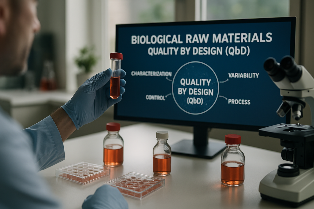
Biological Raw Materials in the Context of Quality by Design (QbD)
Quality by Design (QbD) is a strategic approach to pharmaceutical and biotechnological development that emphasizes understanding and controlling processes to ensure consistent quality outcomes. In bioprocessing, biological raw materials are critical components, playing a pivotal role in cell culture, immunology, and diagnostics. This article delves into the relevance of biological raw materials within the QbD framework, emphasizing the need for thorough characterization, understanding of variability, and strategic implementation to optimize scientific research and product quality.
The Role of Biological Raw Materials in QbD
Understanding Biological Function and Variability
Biological raw materials, such as fetal bovine serum (FBS), bovine plasma, and human sera, are indispensable in various biotechnological applications. These materials are rich in growth factors, hormones, and nutrients essential for the growth and maintenance of cultured cells. However, their biological nature introduces intrinsic variability that can impact experimental consistency. The Quality by Design approach requires a deep understanding of these raw materials, not only to mitigate variability but also to leverage their unique properties for optimal performance.
- Intrinsic variability in material can affect reproducibility and outcome.
- Thorough characterization ensures more consistent quality control.
Continuez votre lecture pour explorer des perspectives et des stratégies plus avancées.
Ensuring Consistency and Quality through Characterization
Lot-to-Lot Variability and Its Management
Lot-to-lot variability is a common challenge with biological raw materials. Differences in nutrient concentrations and growth factors can lead to inconsistencies in cell culture. Implementing rigorous testing protocols, such as those offered by custom biological sourcing services, can help identify and account for this variability. The Quality by Design framework insists on comprehensive documentation and batch reservation to support consistency and traceability, thus enhancing reproducibility in scientific research.
- Testing and documentation are crucial for managing variability.
- Batch reservation provides a controlled source of raw materials.
Continuez votre lecture pour explorer des perspectives et des stratégies plus avancées.
Reagents and Solutions in Cell Culture and Immunology
Functional Roles and Handling Practices
Reagents such as density gradients are vital in the separation and purification processes within the laboratory. Their functional role extends to facilitating cellular studies in immunology and diagnostics by maintaining cell viability and function. Proper storage and handling of these reagents are critical to preserving their integrity. QbD emphasizes meticulous documentation, ensuring that reagents are consistently effective throughout the experimental lifecycle.
- Proper handling extends the viability of reagents.
- Immune assays rely on reagent quality for reliable outcomes.
Continuez votre lecture pour explorer des perspectives et des stratégies plus avancées.
Leveraging Technology for Quality Assurance
Role of Live Cell Imaging Systems
Technological advancements, such as incubator-compatible live-cell imaging systems, have revolutionized how biological raw materials are monitored in real-time. Systems like the zenCELL owl enable continuous tracking of cell behavior, offering insights into the dynamic effects of sera and reagents without disrupting the cell culture environment. This continuous monitoring aligns with the QbD principle of informed decision-making, enhancing the reproducibility and reliability of biological workflows.
- Real-time data supports informed adjustments in experimental conditions.
- Continuous monitoring reduces the risk of unnoticed variances.
Continuez votre lecture pour explorer des perspectives et des stratégies plus avancées.
Strategic Implementation of QbD in Biological Material Use
Applications and Ethical Considerations
Human-derived biologicals, while integral to primary cell cultures and immunological assays, come with ethical and regulatory considerations. Ensuring the donor material’s traceability and adhering to ethical sourcing standards fortifies QbD’s commitment to responsible science. The integration of ethical considerations into the QbD framework ensures that biological raw materials are used effectively and responsibly, supporting innovative yet ethically mindful research applications.
- Traceability and ethical sourcing are pillars of quality control.
- Regulatory alignment ensures compliant scientific exploration.
Continuez votre lecture pour explorer des perspectives et des stratégies plus avancées.
“`
“`html
Optimizing Raw Material Selection through Predictive Modelling
Leveraging Statistical Analysis for Better Outcomes
Incorporating predictive modeling into the selection of biological raw materials can significantly enhance process efficiency within the QbD framework. By utilizing statistical methods such as regression analysis and machine learning algorithms, researchers can predict the behavior of raw materials under various conditions, thereby selecting the best candidates for their specific applications. A study conducted by the University of California demonstrated how predictive models reduced variability in cell growth by 20%, ensuring more reliable and reproducible results.
- Use statistical tools to anticipate material performance.
Advanced Characterization Techniques for Enhanced Quality Control
Emphasizing Molecular and Biochemical Profiling
Advanced techniques such as mass spectrometry and nuclear magnetic resonance (NMR) spectroscopy are indispensable in the precise characterization of biological raw materials. These methods provide detailed molecular and biochemical profiles, crucial for understanding the complex interactions within cell culture systems. For instance, mass spectrometry has been employed to identify variations in hormone levels in bovine serum, leading to adjustments in supplementation protocols that improved cell culture viability by over 15%.
- Invest in advanced profiling techniques for comprehensive characterization.
The Role of Automation in Enhancing Raw Material Consistency
Streamlining Processes with High-Throughput Systems
Automation technologies, including high-throughput screening systems, offer significant improvements in managing biological raw material consistency. These systems streamline the evaluation process, allowing for rapid testing and quality assessment at scale. In a case study by a leading biotech company, implementing automated screening reduced the time for quality checks by 30%, facilitating faster go-to-market timelines without compromising quality.
- Implement automation to ensure consistency and reduce process time.
Integrating Quality Risk Management into Material Development
Proactive Strategies for Mitigating Risks
Quality Risk Management (QRM) is a proactive approach that integrates risk assessment tools like Failure Mode and Effects Analysis (FMEA) to identify and mitigate potential quality failures in biological raw materials. By understanding and documenting potential risks, organizations can develop robust strategies to ensure quality and compliance. A pharmaceutical company successfully reduced process deviations by implementing QRM, highlighting its importance in sustaining quality standards.
- Adopt QRM tools to systematically address quality risks.
Ensuring Compliance with Regulatory Standards
Navigating Global Standards for Biological Materials
Compliance with stringent regulatory standards remains a cornerstone in the QbD framework concerning biological raw materials. Understanding the guidelines set forth by entities such as the FDA and EMA not only aids in ensuring product safety but also facilitates international collaboration and market access. For instance, navigating the regulatory landscape was pivotal for a biotech firm entering the European market, where harmonization of practices across borders was achieved through diligent regulatory planning.
- Stay informed on global regulations to ensure compliance and facilitate market access.
Harnessing Data-Driven Approaches to Enhance QbD Implementation
Utilizing Analytical Tools for Informed Decision Making
Data analytics capabilities offer transformative potential in the Quality by Design framework by providing actionable insights into raw material performance and process optimization. Tools like data visualization software and big data analytics platforms enable comprehensive analysis, revealing patterns and correlations that can inform adjustments to enhance quality outcomes. A top-tier research institute successfully utilized these insights, leading to a 25% increase in process efficiency.
- Leverage data-driven tools to optimize biological workflows and decision-making.
Fostering Collaboration Across Disciplines for Quality Innovation
Encouraging Cross-Functional Teams for Enhanced Quality Outcomes
The complex nature of biological raw materials necessitates collaboration across various scientific disciplines. By fostering cross-functional teams that bring together experts in biology, chemistry, engineering, and data science, organizations can drive innovation and improve quality control processes. Collaborative efforts at a major research university led to the development of a novel cell culture medium that enhanced yield by up to 30%.
- Encourage multidisciplinary collaboration to foster innovation and improve outcomes.
Ensuite, nous conclurons avec les points clés à retenir, les métriques et une conclusion percutante.
“`
“`html
Prioritizing Sustainability in Raw Material Sourcing
Achieving Eco-Friendly Innovations
In the landscape of Quality by Design, sustainability is increasingly crucial as businesses strive to reduce their environmental footprint. Sourcing sustainable biological raw materials goes beyond mere compliance; it reflects a commitment to environmental stewardship. By choosing renewable resources and implementing eco-conscious practices, companies can align their production processes with global sustainability goals. Recent initiatives by a pharmaceutical giant integrating sustainable sourcing strategies have demonstrated a reduction of 40% in carbon footprint, exemplifying the positive impact of environmental innovations.
- Pursue eco-friendly strategies in sourcing biological raw materials.
Exploring Future Trends in Biological Material Development
Anticipating New Technologies and Innovations
The development of biological raw materials continues to evolve with technological advancements that promise to revolutionize the field. Emerging trends such as synthetic biology and bioinformatics offer unprecedented capabilities in tailoring raw materials with specific attributes conducive to desired biological functions. These technologies offer a glimpse into a future where precision-designed materials could dominate industry practices. A pioneering research group successfully used synthetic biology to engineer bacterium with optimized metabolic pathways, resulting in a fourfold increase in target compound yield.
- Stay ahead by investing in future-forward technologies.
Instilling a Culture of Continuous Improvement
Driving Ongoing Quality Enhancements
The Quality by Design framework inherently encourages a culture of continuous improvement, ensuring that biological material development keeps pace with innovation and quality expectations. Through iterative testing, feedback loops, and performance evaluations, organizations can continuously enhance processes and material quality. Success stories from biotech industries highlighted how a consistent commitment to improvement cycles resulted in a sustained increase in product quality and compliance, setting a benchmark for industry best practices.
- Embed continuous improvement culture within organizational practices.
Conclusion
The Quality by Design framework provides a comprehensive pathway that enables researchers and enterprises in the biotech and pharmaceutical sectors to refine the production and application of biological raw materials systematically. By focusing on predictive modeling, advanced characterization, automation, risk management, and regulatory compliance, organizations can achieve unparalleled quality standards while optimizing resource efficiency.
Our discussion underscores the immense benefits derived from integrating advanced analysis, high-throughput technologies, and proactive risk management. We have explored the landscapes of sustainable sourcing and future technological advancements, unveiling their pivotal role in shaping tomorrow’s biological innovations. Moreover, incorporating cross-disciplinary collaboration fosters an environment ripe for innovation, driving outcomes that align with global quality benchmarks.
By embracing these QbD principles and strategies, businesses can navigate the complexities of biological materials with a nuanced understanding, thereby ensuring compliance, enhancing market access, and fostering sustainable practices. The insights shared in this article are critical not only for achieving superior quality and efficiency but also for securing a competitive advantage in the ever-evolving biotech landscape.
As the journey of quality enhancement continues, we invite you to consider these strategic implementations within your own organizational frameworks. Let’s move forward with a shared commitment to excellence, driving progress through informed decision-making and collaborative innovation. By nurturing a culture of quality and sustainability, we embark on a transformative path that promises to enrich the world of biotechnology and beyond.
“`
AI in the Lab: What You Need to Know to Stay Ahead of the Competition
“`html
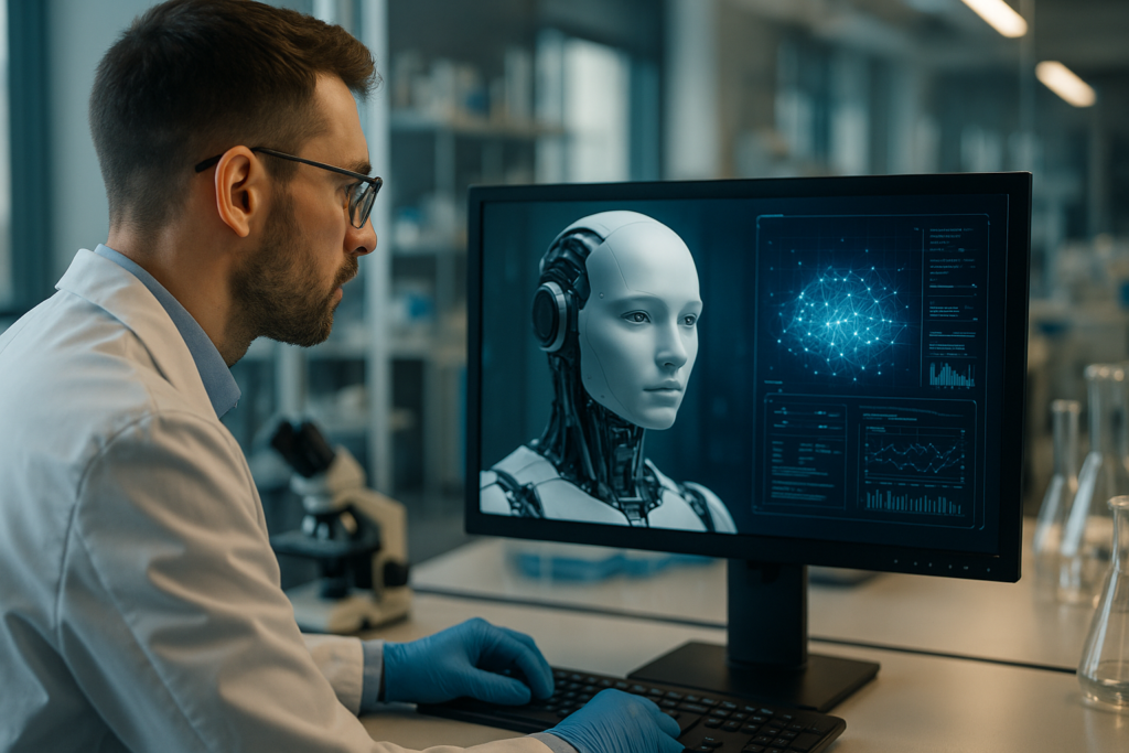
AI in the Lab: What You Need to Know to Stay Ahead of the Competition
In today’s fast-paced research environment, staying ahead in the field of cell culture research requires embracing innovative technologies. Artificial intelligence (AI) is making significant strides in transforming traditional lab workflows into more efficient and accurate processes. This article delves into how AI is reshaping laboratories, offering insights into practical applications like live-cell imaging and laboratory automation. By understanding these advances, researchers, lab managers, and biotech professionals can remain competitive and enhance their research outcomes.
Défis et limites courants des approches traditionnelles
The Bottlenecks in Conventional Laboratories
Conventional cell culture methods often face challenges such as variability in results, time-consuming manual processes, and limited monitoring capabilities. The need for skilled personnel to manage these tasks increases both costs and the potential for human error. Traditional live-cell imaging techniques, for example, can be invasive, leading to changes in cell behavior that might alter experimental results. Additionally, these methods typically offer only endpoint analysis, providing a static picture of dynamic cellular processes.
- High personnel costs and variability in manual handling
- Limited capacity to monitor live cells continuously
- Potential invasive techniques altering cell behavior
Avancées technologiques et tendances d'automatisation
Leveraging AI for Enhanced Lab Automation
With the advent of AI, laboratories are experiencing a shift toward automation and high-throughput analysis. AI-powered tools can streamline workflows by automating routine tasks, reducing variability, and providing real-time feedback. Technologies such as machine learning algorithms can predict outcomes and optimize conditions based on historical data. As a result, laboratories are capable of processing larger volumes of data faster, improving both efficiency and accuracy.
- Reduction in manual workload through automation
- Improved accuracy with real-time feedback and analysis
- Enhanced ability to handle complex data sets
Exemples pratiques et flux de travail utilisant l'imagerie de cellules vivantes
AI-Enhanced Live-Cell Imaging Techniques
Live-cell imaging has been revolutionized by AI’s incorporation into imaging systems. AI allows for the continuous monitoring of cells under natural conditions without disrupting their environment. For example, the zenCELL owl, a compact, incubator-compatible live-cell imaging system, represents a significant advancement in this area. Its ability to operate within the incubator environment ensures that cells are monitored in real-time, leading to more consistent and accurate data collection. Such technology facilitates the observation of cellular processes, aiding in studies of cell migration, proliferation, and morphology under different conditions.
- Real-time, non-invasive monitoring with AI technology
- Consistent and accurate data through continuous analysis
- Compatibility with existing incubator setups for seamless integration
Continuez votre lecture pour explorer des perspectives et des stratégies plus avancées.
“`
“`html
Harnessing AI for Predictive Analytics in Research
Transformer les données en informations exploitables
Predictive analytics, powered by AI, is revolutionizing data interpretation in laboratory settings by allowing researchers to forecast potential outcomes and make informed decisions before experimental execution. By leveraging machine learning models, labs can analyze vast datasets to identify trends and correlations that would be impossible to discern manually. For instance, this approach has been applied in drug discovery, where AI models predict the efficacy of compounds based on historical biochemical data, significantly reducing research time and cost.
- Incorporate machine learning models to enhance predictive capabilities in your research.
AI-Driven Quality Control and Assurance
Ensuring Reliability and Consistency
AI technologies can greatly improve quality control by automating quality assurance processes and minimizing human error. Image recognition algorithms, for example, are used to identify and categorize cell types, detect anomalies, and ensure consistency across experiments, which is crucial for reproducibility. In biomanufacturing, AI systems monitor production processes in real-time, facilitating rapid response to deviations from quality norms, thus ensuring product safety and efficacy.
- Implement AI tools for real-time quality monitoring to enhance product consistency.
AI-Enhanced Collaboration and Knowledge Sharing
Fostering Innovation through Shared Resources
The implementation of AI in laboratory settings also facilitates enhanced collaboration by centralizing data and analyses, making them accessible across multidisciplinary teams and research collaborators. Platforms enabled by AI can aggregate experimental data from multiple sources, creating a repository that can be accessed globally, thus expediting discovery and innovation. An example of this is the use of AI in virtual lab environments, where researchers can remotely access data and contribute to ongoing projects, fostering a more cohesive and inclusive research ecosystem.
- Utilize AI-powered platforms to enhance collaborative efforts and resource sharing.
Optimizing Workflow with AI-Powered Laboratory Management Systems
Streamlining Operations for Increased Efficiency
AI-powered laboratory management systems (LIMS) are at the forefront of modernizing laboratory operations. These systems automate the collection, processing, and storage of large volumes of experimental data, providing a seamless workflow that enhances productivity. Integrated solutions can track samples, schedule experiments, and manage inventory effectively, reducing downtime and optimizing resource allocation. By employing these systems, laboratories can ensure adherence to compliance standards while expanding their operational capacities.
- Adopt AI-driven LIMS to streamline operations and enhance productivity in your lab.
Applying AI for Enhanced Cellular Analysis
Advancing Precision in Cellular Studies
In-depth cellular analysis is critical in fields like oncology and regenerative medicine. AI enhances cellular analysis by providing quick, precise, and automated assessment of cellular behaviors and characteristics. AI algorithms help in quantifying biomarkers and cellular responses with high accuracy. Notably, AI-enabled systems like CellProfiler utilize image analysis to process high-throughput imaging data efficiently, accelerating the pace of cellular research.
- Integrate image analysis tools like CellProfiler for detailed cellular insights.
AI’s Role in Personalized Medicine
Tailoring Treatments with AI at the Core
As the healthcare paradigm shifts towards more personalized approaches, AI plays a pivotal role in tailoring treatments based on individual cellular and genetic profiles. AI technologies analyze genetic information to predict patient responses to treatments, aiding in the personalization of therapies. By integrating AI with genomics and patient data, researchers can develop more targeted and effective treatment plans, demonstrating AI’s potential to transform medical practices.
- Explore AI applications in genomics to advance personalized medicine initiatives.
Ethical Considerations and Challenges in AI Implementation
Navigating the Path with Caution
As AI becomes deeply integrated into laboratory processes, ethical issues and challenges arise, particularly concerning data privacy and algorithmic biases. It is crucial for labs to implement ethical frameworks and governance models that address these concerns. Transparent AI practices and data management protocols ensure credibility and trust in AI-enabled research outcomes. Developing AI technologies with inclusivity and fairness in mind can prevent biases and promote equitable advancements.
- Establish ethical standards and practices when implementing AI in research.
Ensuite, nous conclurons avec les points clés à retenir, les métriques et une conclusion percutante.
“`
“`html
AI’s Influence on Data-Driven Decision Making
Bridging the Gap Between Data Analysis and Strategy
Incorporating AI into data analysis not only augments the decision-making process but also introduces a strategic edge to research initiatives. By analyzing vast amounts of data with precision, AI systems provide insights that inform strategic decisions. In complex fields like clinical research, AI helps prioritize research paths and resource allocation based on predicted outcomes. This predictive capability empowers teams to anticipate future trends, allowing them to adapt swiftly to evolving scientific landscapes.
- Leverage predictive analytics tools to inform strategic decision-making.
AI and Sustainable Research Practices
Enhancing Sustainability through Technology
As the focus on sustainability intensifies, AI technologies offer pathways to more environmentally conscious research practices. AI-driven systems optimize resource utilization, reducing waste and lowering the ecological footprint of laboratories. By monitoring energy consumption and raw material use in real-time, AI can suggest adjustments that align research operations with sustainable goals. Moreover, AI facilitates the recycling and repurposing of laboratory materials, contributing to the global sustainability agenda without compromising research quality.
- Adopt AI solutions to foster sustainable research environments.
Preparing for the Future of AI in Research
Navigating the Ever-Evolving Technological Landscape
As AI technologies continue to evolve, staying abreast of advancements is crucial for maintaining a competitive edge. Continuous learning and adaptation are imperative as new AI tools and methodologies emerge. Cultivating a culture of innovation within research teams can drive the integration of cutting-edge AI solutions and ensure their successful deployment. Additionally, fostering partnerships with AI experts and tech companies can facilitate knowledge transfer and support ongoing adaptation to future developments.
- Engage in continuous learning to stay current with AI advancements.
Conclusion
As AI becomes a cornerstone of modern laboratory advancements, the integration of smart technologies into research processes proves indispensable. From enhancing predictive analytics to driving personalized medicine, AI transforms the laboratory landscape, offering unprecedented levels of efficiency, precision, and innovation.
The implementation of AI in laboratories fosters a data-driven approach to research, allowing for strategic decision-making and improved collaboration while maintaining ethical standards. At the same time, AI’s role in promoting sustainable practices aligns research activities with global sustainability objectives, underscoring its multifaceted value in the scientific ecosystem.
For researchers and laboratories seeking to maintain a competitive edge, embracing AI technologies is no longer optional but a necessity. The journey towards integrating AI can start by exploring the various applications outlined in this article, from leveraging machine learning models in predictive analytics to utilizing AI-powered quality control systems.
As you navigate this transformative era, let AI be the catalyst that propels your research to new heights. By embracing AI, you position yourself at the forefront of innovation, ready to tackle the challenges of today and tomorrow with renewed vigor and purpose. The future of research is intertwined with AI, and by harnessing its power, you can lead the way to groundbreaking discoveries and advancements. Stay ahead of the competition, explore the potential of AI in your laboratory, and inspire the next generation of scientific breakthroughs. Embrace this era of transformation and continually adapt and innovate, ensuring your research efforts yield meaningful, impactful outcomes that shape a better tomorrow.
“`
Identifying the Invisible: Spotting Suboptimal Culture Conditions Automatically
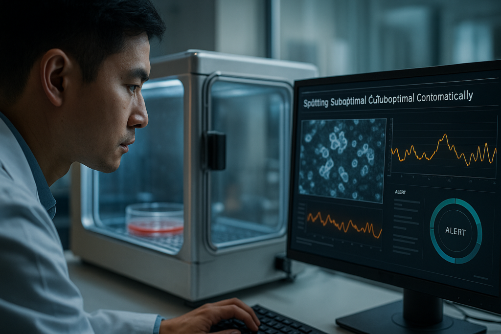
Identifying the Invisible: Spotting Suboptimal Culture Conditions Automatically
Cell culture is a cornerstone of modern biological research, providing insights into cellular functions, drug responses, and disease mechanisms. Yet, optimizing culture conditions remains a challenging task that often involves meticulous monitoring to prevent suboptimal environments that could compromise results. Identifying the Invisible: Spotting Suboptimal Culture Conditions Automatically, is a growing need in the scientific community to enhance the quality and reliability of experimental data. This article explores the latest trends, technologies, and methodologies that are revolutionizing cell culture practices by allowing researchers to identify and rectify these invisible pitfalls efficiently.
Challenges in Traditional Cell Culture Methods
Limitations of Traditional Monitoring
Traditionally, cell culture conditions are manually monitored through intermittent observation and specific assays. This approach is not only labor-intensive but can also miss ephemeral changes in conditions that occur between observations. Such invisible fluctuations can lead to suboptimal culture environments, affecting cell health and experimental accuracy.
- Manual intervention leads to variability in results.
- Intermittent observations can overlook transient changes.
- Difficult to maintain optimal conditions consistently.
Technological Innovations in Automated Monitoring
Embracing Automation in Cell Culture
Advancements in automation technologies have paved the way for real-time, continuous monitoring of cell cultures, thus Identifying the Invisible: Spotting Suboptimal Culture Conditions Automatically. Automated systems minimize human error and enhance precision by providing constant insights into cell environment parameters.
- Reduces manpower and chances of introducing contamination.
- Enables continuous data capture and analysis.
- Provides immediate alerts for corrective actions.
Live-Cell Imaging: A Step Towards Better Control
The Role of Live-Cell Imaging Systems
Live-cell imaging technologies are redefining the research landscape by providing better visualization and understanding of cellular processes in real-time. An exemplary tool is the zenCELL owl, a compact, incubator-compatible live-cell imaging system that integrates seamlessly into existing workflows. By capturing images at regular intervals, researchers can now Identify the Invisible: Spotting Suboptimal Culture Conditions Automatically and adjust as needed, ensuring optimal cell growth conditions.
- Facilitates early detection of deviations in cell growth patterns.
- Allows for minimal disruption of cell cultures during observation.
- Increases reproducibility and reliability of experimental data.
Continuez votre lecture pour explorer des perspectives et des stratégies plus avancées.
“`html
Leveraging AI for Predictive Culture Optimization
The Intersection of Artificial Intelligence and Cell Culture
Artificial Intelligence (AI) has transformed various scientific and industrial sectors, and cell culture is no exception. AI algorithms can analyze vast datasets generated by automated monitoring systems to forecast potential issues in cell culture conditions. This predictive capability allows researchers to implement preventive measures proactively, rather than reacting to occurring problems.
- Utilize machine learning models to predict cell culture outcomes.
- Integrate AI-driven insights to refine experimental protocols.
- Implement AI tools for anomaly detection and trend analysis.
IoT Connectivity: Integrating Devices for Seamless Monitoring
Creating a Connected Ecosystem in the Lab
The Internet of Things (IoT) enables seamless communication and data exchange between different devices in a lab setting. By connecting live-cell imaging systems, incubators, and environmental sensors, researchers can maintain comprehensive oversight of culture conditions. IoT-enhanced platforms offer real-time analytics and foster more transparent research workflows.
- Adopt IoT-friendly devices to synchronize lab operations.
- Leverage IoT for remote access and monitoring capabilities.
- Utilize integrated dashboards for comprehensive data visualization.
The Power of Data Science in Culture Condition Refinement
Utilizing Big Data to Enhance Experimental Accuracy
Data science plays a crucial role in interpreting the complex datasets derived from modern cell culture technologies. By employing sophisticated analytical techniques, researchers can extract meaningful insights to refine their culture techniques continuously. Data-driven approaches provide deeper understanding of cellular behaviors under varying culture conditions.
- Implement data analytics tools to interpret culture data.
- Use statistical models to evaluate experimental variability.
- Complement traditional biology knowledge with data intelligence.
Case Study: Successful Implementation in Pharmaceutical Research
Real-world Results from Transitioning to Automated Culture Systems
A leading pharmaceutical company utilized automated live-cell imaging and AI-driven analysis to optimize its drug discovery process. By implementing these advanced technologies, the company reduced the time required for cell viability assessments by 50%, while increasing data precision. This approach allowed more robust conclusions about potential drug efficacy and safety.
- Integrate successful strategies from industry leaders into lab practices.
- Review case studies to understand barriers and breakthroughs in culture optimization.
- Focus on accelerating innovation by investing in automation and analytics.
Implementing SOPs for Consistent Culture Conditions
Standardizing Processes to Minimize Variability
Establishing standardized operating procedures (SOPs) is critical for maintaining consistent culture conditions across experiments. SOPs based on automated monitoring data ensure predictable and reproducible results, valuable for multi-laboratory collaborations. By aligning protocols with data insights, labs can reduce variability and enhance the reliability of their outcomes.
- Design SOPs with input from automated data analysis tools.
- Regularly update SOPs to reflect advancements in technology and methodologies.
- Train staff and researchers on the implementation of SOP-driven practices.
Training and Skill Development for Modern Biologists
Ensuring Competency in an Era of Advanced Technologies
As technological advancements transform cell culture practices, biologists must adapt by developing new skills and competencies. Ongoing education initiatives are vital to ensure researchers understand how to harness new tools and interpret data effectively. Courses on bioinformatics, AI, and laboratory automation are pivotal in refining the skillsets necessary for modern cell culture research.
- Participate in workshops focused on emerging technologies in cell culture.
- Engage in continuous professional development and learning opportunities.
- Encourage interdisciplinary collaboration to drive knowledge exchange.
Financial and Operational Benefits of Automated Monitoring
Analyzing the Cost-Effectiveness of Technology Integration
Automated cell culture systems present significant financial and operational benefits. By minimizing manual labor and reducing the need for repeated assays, labs can allocate resources more efficiently, leading to cost savings. Moreover, automation increases throughput, allowing for more extensive experimental timelines without sacrificing data quality.
- Calculate ROI when introducing automation into laboratory environments.
- Compare costs and productivity metrics pre- and post-automation implementation.
- Plan funding and investments considering long-term financial sustainability.
Ensuite, nous conclurons avec les points clés à retenir, les métriques et une conclusion percutante.
“`
“`html
Environmental and Ethical Impact of Automation in Cell Culture
Équilibrer l'innovation et la responsabilité
Advancements in cell culture automation are not only about improving laboratory efficiency but also about recognizing the broader environmental and ethical impacts. Automated systems often lead to energy savings and reduced waste by optimizing resource usage and minimizing human error. Ethical considerations, such as replicating human-like conditions in vitro, also become more disciplined through automation, leading to credible and humane experimental frameworks.
- Adopt environmentally friendly, energy-efficient technologies.
- Adhere to ethical standards that govern automated cell culture practices.
- Evaluate the sustainability of new cell culture methodologies.
Enhancing Collaboration through Digital Transformation
Facilitating Shared Access and Collaborative Innovating
Incorporating digital solutions into cell culture practices enhances collaboration by providing shared platforms and data accessibility. Digital transformation allows real-time data sharing and communication between teams across different geographic locations, promoting collaborative research and innovation. This interconnectedness fosters the generation of fresh insights and enhances problem-solving capabilities by leveraging collective expertise.
- Utilize cloud-based platforms for real-time data sharing.
- Strengthen cross-disciplinary research through digital collaboration tools.
- Promote global partnerships by harmonizing digital standards.
Future Perspectives: The Vision for Cell Culture Advancements
Projecting the Path of Innovation and Impact
The landscape of cell culture is poised for continuous transformation as technology evolves. Future advancements in AI, IoT, and data science promise even more sophisticated and precise culture systems. The integration of these technologies will pave the way for breakthroughs not only in disease research and drug development but also in sectors like tissue engineering and personalized medicine.
- Embrace emerging technologies to stay at the forefront of cell culture innovation.
- Anticipate paradigm shifts driven by continuous technological growth.
- Invest in futuristic research initiatives that redefine cell-based experiments.
Conclusion
The evolution of cell culture practices through the integration of advanced technologies like AI, IoT, and data science signifies a new era of scientific discovery. These technologies collectively act to optimize culture conditions, enabling more predictive, accurate, and efficient outcomes. By embracing automation, the scientific community can achieve more refined experimental results, reduce the time and cost associated with traditional methods, and increase the reliability of conclusions drawn from cell-based research.
Beyond enhancing traditional methodologies, the true transformative power of these technologies lies in their ability to bridge gaps between disciplines, foster global collaboration, and uphold environmental and ethical responsibilities. Lab operations become seamless and more transparent, driving innovative research paradigms that promise future breakthroughs in medicine, biotechnology, and environmental science.
Tapping into these technologies, researchers are empowered to explore new scientific frontiers with unprecedented precision and creativity. As we continue our journey toward more advanced and integrated cell culture systems, it is essential to invest in training and development initiatives to ensure the workforce is equipped to handle these sophisticated tools.
The path forward demands champions of innovative methodologies who can inspire and drive meaningful change. By continuously refining techniques, embracing digital transformation, and fostering collaborative environments, scientists will be well-prepared to encounter the challenges and opportunities that lie ahead.
Embarking on this transformative path, let us commit to leveraging technology not only to advance science but also to enhance the quality of life globally. Let’s build a future where the transparent and ethical application of technology is a catalyst for societal benefit, revolutionizing our understanding of life at the cellular level.
Join us in this exciting era of technological integration, and be part of a community that shapes the future with innovation, responsibility, and unwavering commitment to excellence.
“`
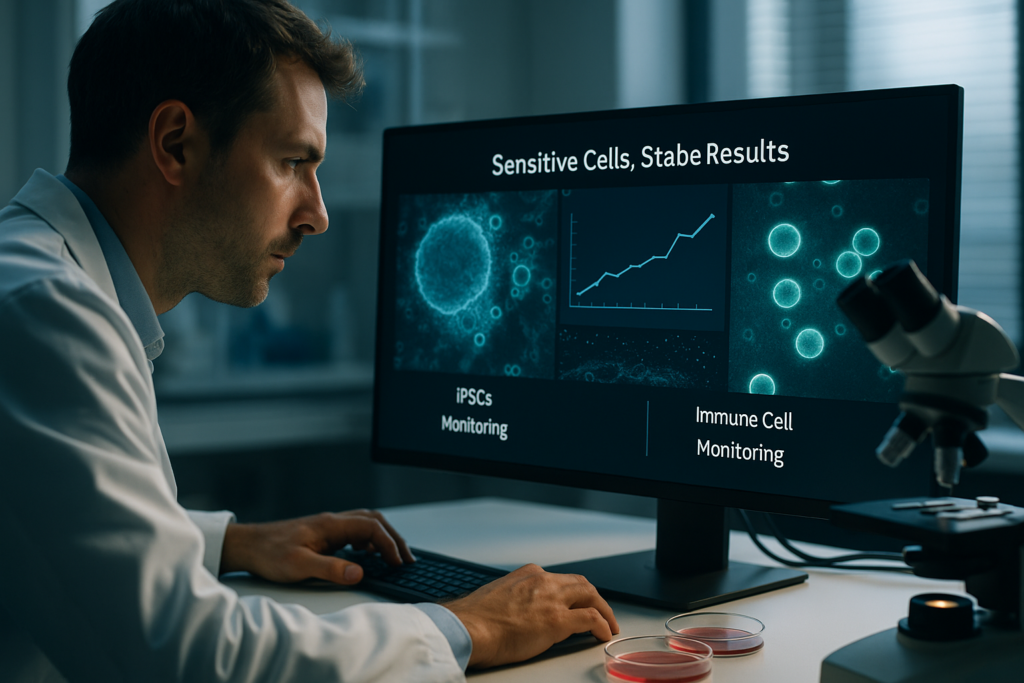
Sensitive Cells, Stable Results: Optimizing iPSC and Immune Cell Monitoring
“`html
Sensitive Cells, Stable Results: Optimizing iPSC and Immune Cell Monitoring
In recent years, the field of cell culture research has witnessed a significant surge in interest and innovation, particularly concerning induced pluripotent stem cells (iPSCs) and immune cells. These cell types hold tremendous potential for regenerative medicine, drug development, and understanding disease mechanisms. However, their sensitivity to environmental changes poses considerable challenges. Maintaining stable, reproducible results in iPSC and immune cell cultures is an ongoing quest for researchers. This article explores the common challenges, technological advancements, and innovative workflows designed to optimize monitoring and analysis of these sensitive cells.
Défis et limites courants des approches traditionnelles
Understanding the Sensitivity of iPSCs and Immune Cells
Both iPSCs and immune cells are remarkably sensitive to their environment. Variations in temperature, pH, nutrient supply, and other factors can cause inconsistencies in experimental results. This sensitivity often leads to challenges such as cell death, differentiation variability, and activation anomalies in immune cell cultures. Historic approaches to cell culture monitoring often relied on periodic manual inspection, which is labor-intensive and offers limited temporal resolution.
- Environmental fluctuations can compromise cell viability.
- Manual monitoring lacks consistency and specificity.
- Data from traditional methods are not continuously recorded.
Avancées technologiques et tendances d'automatisation
Introducing Live-Cell Imaging and Automation
The advent of live-cell imaging technology offers a substantial improvement over traditional monitoring methods. By providing real-time visualization and analysis, live-cell imaging enables researchers to capture dynamic cellular processes with higher fidelity. Incorporating automation in conjunction with live-cell imaging decreases the potential for human error and variability. Systems like the zenCELL owl, a compact and incubator-compatible live-cell imaging system, exemplify this technological convergence, fostering enhanced reproducibility and continuous monitoring.
- Improved image quality and data accuracy through high-resolution imaging.
- Automation reduces manual intervention, minimizing error.
- Continuous data collection enhances understanding of dynamic cellular behavior.
Exemples pratiques et flux de travail utilisant l'imagerie de cellules vivantes
Optimized Monitoring of Sensitive Cells
The implementation of live-cell imaging techniques brings transformative changes to workflows involving iPSCs and immune cells. Researchers can deploy real-time monitoring to assess cellular responses to different stimuli, track proliferation rates, and observe morphological changes. A well-configured system allows for comprehensive, hands-off analysis. For instance, with incubator-based systems, data collection doesn’t interrupt optimal culture conditions, thus maintaining the cells’ sensitive nature while ensuring data reliability.
- Real-time tracking of cell cycle and proliferation.
- Monitoring of immune cell activation and response.
- Dynamics of iPSC differentiation under various conditions.
Comment l'imagerie basée sur incubateur améliore la reproductibilité et la qualité des données
Maintaining Optimal Culture Conditions
Incubator-based imaging systems merge the benefits of controlled environmental conditions with continuous data acquisition. The zenCELL owl system serves as an ideal example of how compact, precise imaging capabilities can be integrated within incubators. This setup ensures that the cells are not exposed to external disturbances, which substantiates consistent and reproducible data output. Moreover, these systems enhance experimental throughput while maintaining strict environmental controls.
- Reduced exposure to external contaminants.
- Stable imaging environment ensures reproducibility.
- Fosters scalability for high-throughput applications.
Continuez votre lecture pour explorer des perspectives et des stratégies plus avancées.
“`
“`html
Integrating Artificial Intelligence with Cell Monitoring
AI’s Role in Enhancing Data Interpretation
The integration of artificial intelligence (AI) in cell monitoring is revolutionizing data interpretation and decision-making processes. AI algorithms can analyze complex imaging data sets far more efficiently than traditional methods, extracting intricate patterns and correlations that may not be immediately evident to human observers. For instance, machine learning models can predict outcomes based on historical data, allowing researchers to anticipate cellular behaviors and adjust experimental conditions in real-time. This capability significantly improves the accuracy and reliability of iPSC and immune cell studies.
- Implement AI tools to identify subtle changes in cell morphology.
- Use predictive analytics to enhance experimental design efficiency.
Standardizing Protocols for Consistency
Establishing Unified Methodologies
To address variability in cell culture experiments, standardizing protocols is crucial. By implementing unified methodologies, researchers can compare data across different studies and labs more reliably. These protocols should cover every stage of the experimental process, from cell seeding and maintenance to post-experiment analysis. Through rigorous standardization, discrepancies caused by procedure deviations are minimized, thereby enhancing the reproducibility of results.
- Develop comprehensive SOPs (Standard Operating Procedures) for cell culture.
- Use standardized cell lines to ensure consistency across experiments.
Leveraging Cloud-Based Data Management
Facilitating Collaboration and Data Security
Cloud-based platforms provide a secure, accessible means of managing and sharing cell culture data. These platforms allow for seamless collaboration among research teams and institutions, facilitating real-time data updates and collective data analysis. Cloud storage ensures that valuable experimental data is preserved and protected against local hardware failures. Moreover, integrating cloud solutions with AI analytics further enhances the ability to perform meta-analyses and gain insights across larger data sets.
- Adopt cloud platforms to enable global research collaborations.
- Ensure compliance with data security regulations for sensitive data.
Enhancing iPSC Differentiation Protocols
Precision Control of Differentiation Pathways
Refining the differentiation protocols for iPSCs is key to generating specific cell types with high fidelity. Advanced techniques, such as three-dimensional (3D) culture systems and microfluidics, enhance the control researchers have over differentiation conditions. These methods simulate in vivo-like environments, yielding more accurate modeling of cellular processes. As a result, the efficiency and predictability of iPSC differentiation increase, which is invaluable for applications in disease modeling and regenerative therapies.
- Utilize 3D culture systems to better mimic physiological conditions.
- Integrate microfluidic devices for controlled media distribution.
Optimizing Immune Cell Function Through Genetic Engineering
Genetic Tools Enhancing Research
Genetic engineering advancements, including CRISPR/Cas9, have opened new avenues for optimizing immune cell research. By modifying specific genes, researchers can influence immune cell function, such as enhancing T-cell receptor specificity or reducing the expression of inhibitory receptors. These modifications hold potential for developing powerful immunotherapies and creating immune cell models that more accurately reflect human health and disease states.
- Implement CRISPR techniques to explore gene function in immune cells.
- Adapt genetic modifications to refine therapeutic strategies.
Harnessing High-Throughput Screening Tools
Increasing Experimental Throughput and Data Richness
High-throughput screening (HTS) platforms are indispensable in today’s fast-paced research environment. They enable the rapid assessment of cellular responses to numerous compounds, conditions, or genetic modifications. By integrating HTS with live-cell imaging and automated systems, researchers can gather comprehensive datasets necessary for robust statistical analysis. This approach dramatically accelerates the pace of discovery, especially in drug development and personalized medicine.
- Employ HTS platforms for efficient compound screening.
- Combine HTS with live-cell analysis for dynamic insights.
Developing Scalable Cell Manufacturing Systems
Bridging the Lab to Market Gap
To meet the growing demand for cell-based therapies, scalable manufacturing solutions are essential. Bioreactor technology and modular manufacturing systems offer paths to scale up the production of iPSCs and immune cells. These systems ensure that large-scale production maintains the quality and consistency observed at the research level. Scalable solutions also reduce costs, supporting broader accessibility to breakthrough therapy options.
- Integrate bioreactors for large-scale cell production.
- Ensure compliance with Good Manufacturing Practice (GMP) standards.
Ensuite, nous conclurons avec les points clés à retenir, les métriques et une conclusion percutante.
“`
“`html
Integrating Automation in Cell Culture
Streamlining Workflow and Reducing Human Error
Automation in cell culture processes is a game changer in reducing human error and increasing throughput. By implementing robotic systems and automated platforms for cell handling, researchers can ensure a more consistent and repeatable process. This level of consistency is critical in reducing variability and ensuring the reliability of results across multiple experiments. Furthermore, automation frees scientists to focus on data analysis and experimental design, enhancing productivity and innovation.
- Adopt robotic systems for routine cell culture tasks.
- Utilize automated imaging systems for real-time monitoring.
Bioinformatics in Cell Research
Maximizing Data Utility Through Advanced Computational Tools
With vast amounts of data generated from cell research, bioinformatics tools are essential in deriving meaningful insights. These tools facilitate the integration and interpretation of complex datasets, supporting hypothesis generation and validation. Advanced computational models and software can analyze genetic, proteomic, and metabolic data, enabling researchers to uncover new patterns and biological signatures. Such capabilities are crucial in advancing personalized medicine and understanding cellular dynamics at a deeper level.
- Incorporate bioinformatics software for comprehensive data analysis.
- Leverage computational models to predict cellular responses.
Fostering Interdisciplinary Collaboration
Uniting Diverse Expertise for Holistic Approaches
The complexity of cell monitoring solutions necessitates interdisciplinary collaboration. By bringing together experts from biology, engineering, data science, and clinical research, innovative solutions and breakthroughs become achievable. Collaboration fosters a holistic understanding of cellular processes, enabling the translation of laboratory findings into practical applications. This multidisciplinary approach not only accelerates discoveries but also ensures that cell-based technologies are primed for real-world implementation.
- Promote collaboration across scientific disciplines and industries.
- Engage with interdisciplinary research networks and consortia.
Conclusion
The landscape of iPSC and immune cell monitoring is rapidly evolving, driven by the integration of advanced technologies and innovative methodologies. Throughout this article, we have explored various strategies, such as artificial intelligence, standardized protocols, cloud-based data management, and scalable manufacturing, that collectively contribute to the optimization of iPSC and immune cell research. Key takeaways include the importance of adopting automation to streamline workflows, leveraging bioinformatics for comprehensive data analysis, and fostering interdisciplinary collaborations to unite diverse expertise.
These advances underscore the growing sophistication and potential of cell-based research to impact fields such as regenerative medicine, drug discovery, and disease modeling. By integrating these methods and tools, researchers can enhance the precision, efficiency, and reliability of experimental outcomes, ultimately contributing to the development of groundbreaking therapies and applications.
As we continue to embrace these cutting-edge technologies and collaborative efforts, the future of cell monitoring looks promising. Researchers, practitioners, and industry stakeholders must remain committed to staying informed and adaptive to innovations. By doing so, they will be well-equipped to tackle ongoing challenges and seize emerging opportunities in cellular research. We invite you to join this exciting journey to advance knowledge, foster innovation, and transform the future of medicine.
Let this article inspire you to think beyond traditional boundaries, explore novel solutions, and collaborate broadly to shape a healthier, more scientifically advanced world. Together, we can unlock the full potential of iPSC and immune cell monitoring, advancing science and medicine to new heights.
“`
The role of pre-analytical variability in diagnostic reproducibility
“`html
The role of pre-analytical variability in diagnostic reproducibility
In the realm of clinical diagnostics, the accuracy and reliability of test results are paramount. A crucial factor influencing these outcomes is pre-analytical variability. Understanding and mitigating this variability is essential for improving diagnostic reproducibility. This blog will delve into the mechanisms of pre-analytical variability, its impact on diagnostics, and strategies to minimize its effects. Readers will gain insights into how consistent practices can enhance the reliability of diagnostic processes.
Understanding Pre-Analytical Variability
Defining Pre-Analytical Variability
Pre-analytical variability refers to variances that occur during sample collection, handling, and preparation stages before actual analysis. These variances can arise from differences in sample collection techniques, timing, environmental factors, and even equipment used. Such inconsistencies can significantly impact the diagnostic reproducibility of test results, leading to misleading interpretations.
- Sample collection timing can influence biomarker stability.
- Transport conditions may alter sample integrity.
- Diversities in human-derived biological samples add complexity.
Continuez votre lecture pour explorer des perspectives et des stratégies plus avancées.
Impact on Diagnostic Reproducibility
Examples of Variability Impact
The role of pre-analytical variability in diagnostic reproducibility becomes evident when considering how slight changes during pre-analysis can lead to substantial discrepancies in results. For instance, improper temperature control during sample transport can degrade sensitive analytes, leading to false-negative or false-positive outcomes.
- Temperature fluctuations can cause protein denaturation.
- Improper handling increases exposure to degrading enzymes.
- Variations in anticoagulants alter plasma composition.
Continuez votre lecture pour explorer des perspectives et des stratégies plus avancées.
Strategies for Minimizing Variability
Standardized Protocols and Quality Control
Implementing standardized protocols is critical in reducing pre-analytical variability. Laboratories should institute detailed standard operating procedures (SOPs) for sample collection and handling. Incorporating rigorous quality control measures ensures consistency in processing and enhances diagnostic reproducibility.
- SOPs define exact techniques and materials used.
- Routine calibration of equipment prevents measurement drift.
- Batch testing aids in identifying outliers early.
Continuez votre lecture pour explorer des perspectives et des stratégies plus avancées.
Technological Solutions Supporting Consistency
Innovations in Monitoring and Documentation
Advances in technology, like incubator-compatible live-cell imaging systems, facilitate continuous monitoring of sample conditions. Systems such as the zenCELL owl document changes in cell behavior and serum effects throughout the culture process, ensuring that deviations affecting diagnostic reproducibility are promptly addressed.
- Provides real-time data on cell health and growth conditions.
- Aids in spotting pre-analytical inconsistencies instantly.
- Supports higher reproducibility in biological workflows.
The role of pre-analytical variability in diagnostic reproducibility is a complex topic continuously undergoing research. Understanding and controlling it can significantly enhance diagnostic accuracy and standardization.
“`
“`html
Harnessing Data-Driven Approaches
Leveraging Predictive Analytics for Improved Outcomes
Incorporating predictive analytics into the laboratory workflow can vastly improve diagnostic reproducibility by identifying patterns and potential errors before they affect outcomes. For instance, using machine learning algorithms to analyze historical data on sample handling and storage can predict potential variances in new cases. Algorithms that assess transport times and conditions help ensure samples remain within optimal parameters, thus safeguarding integrity and consistency.
- Adopt data analytics tools to gain transparent insights on sample management.
- Integrate platforms that forecast and mitigate risks in sample variability.
Enhancing Training and Competency
Building Expertise to Reduce Variability
Personnel competency is a crucial factor in minimizing pre-analytical variability. Tailored training programs focusing on best practices in sample handling and preparation can foster a culture of consistency. For instance, a hospital lab might implement a competency-based training regime that emphasizes error-catching at early sample processing stages.
- Regular training updates ensure staff stay informed on evolving best practices.
- Competency assessments can highlight areas needing improvement, reducing variability.
Optimizing Sample Transport
Innovations in Secure, Reliable Sample Transportation
Transporting biological samples with care is vital to maintaining sample integrity. Advanced solutions such as temperature-controlled transportation boxes equipped with IoT sensors provide real-time monitoring, alerting when conditions deviate from the stipulated environment. Such technologies have revolutionized the way samples are moved from collection sites to laboratories.
- Invest in IoT-enabled transport solutions to maintain environmental control.
- Optimize transport routes to cut down on transit times, preserving sample condition.
Automation in Sample Handling
Reducing Human Error through Robotics
The automation of sample handling processes has significantly curtailed human error. Robotic systems that accurately and consistently perform steps like aliquoting, pipetting, and labeling are increasingly popular in labs worldwide. For example, many facilities have adopted automated systems that track sample lineage and procedural adherence, ensuring traceability and reducing errors.
- Implement robotic systems to enhance precision and repeatability in sample processing.
- Leverage automation for high-throughput processing, minimizing manual interference.
Implementing Rigorous Validation Protocols
Ensuring Robustness of Diagnostic Assays
Creating and maintaining robust validation protocols ensure that diagnostic tests perform accurately under varying conditions. Conducting extensive validation studies under different pre-analytical conditions helps in understanding the spectrum within which a test yields reliable results. Institutions like the Mayo Clinic routinely employ such validation processes, contributing to their high standard of reliable diagnostics.
- Conduct frequent validation exercises to refine and perfect testing protocols.
- Benchmark against industry standards to maintain assay robustness and dependability.
Collaborative Networks for Consistency
Sharing Best Practices Across Laboratories
Forming collaborative networks between labs can facilitate the standardization of pre-analytical processes across the board. Sharing insights and data helps to create a more uniform approach to handling samples, reducing variability introduced by disparate methodologies. For example, consortia like the Institute for Clinical and Translational Research (ICTR) promote cross-institutional collaboration, leveling the playing field across different facilities.
- Participate in collaborative networks to exchange expertise and data-driven insights.
- Establish common frameworks for inter-lab comparability and transparency.
Quality Management Systems (QMS)
Building a Culture of Continuous Improvement
Effective quality management systems are pivotal in minimizing pre-analytical variability. By implementing QMS, labs can ensure that there is a continual review and improvement of test processes. This might involve routine audits, regular quality assessments, and feedback loops. Laboratories that invest in comprehensive QMS tend to see improved consistency in their diagnostic reproducibility.
- Utilize QMS frameworks to root out inefficiencies and improve process adherence.
- Engage in regular audits to uphold high quality and continuous improvement.
Ensuite, nous conclurons avec les points clés à retenir, les métriques et une conclusion percutante.
“`
“`html
Leveraging Technology for Advanced Monitoring
Tracking and Tracing for Enhanced Reliability
Utilizing advanced technology solutions to monitor and trace sample handling processes can significantly enhance reliability in laboratories. The integration of tracking systems, such as barcode scanning and RFID technology, throughout the sample lifecycle ensures accurate and real-time updates on sample status. This comprehensive oversight not only boosts confidence in sample integrity but also pre-empts any potential mishaps before they escalate into larger issues.
- Implement RFID and barcode systems for meticulous tracking and documentation.
- Ensure real-time data sharing across systems to maintain comprehensive oversight.
Data Integration for Holistic Insights
Unifying Systems for Comprehensive Data Utilization
Data silos within laboratories can hinder effective decision-making. By integrating data across various systems, labs can harness a holistic view that brings together pre-analytical, analytical, and post-analytical phases, thereby enhancing informed decision-making. Facilitating seamless data transfer and accessibility empowers laboratory personnel to draw actionable insights, optimize workflows, and respond dynamically to evolving challenges.
- Ensure interoperability between different laboratory information systems (LIS).
- Utilize analytics platforms to connect and visualize disparate data sources.
Fostering a Patient-Centric Approach
Aligning Processes with Patient Outcomes
Ultimately, the primary goal of minimizing pre-analytical variability lies in optimizing patient outcomes. By centering processes and strategies around patient care, labs can continually refine operations to support accuracy and timeliness in diagnostics. This patient-centric focus not only fosters improved healthcare delivery but also enhances patient satisfaction and trust in diagnostic services.
- Focus on personalized diagnostic production to meet specific patient needs.
- Institute feedback mechanisms that directly connect patient outcomes with lab processes.
Conclusion
In conclusion, tackling pre-analytical variability stands as a foundational pillar in ensuring diagnostic reproducibility. Diverse strategies highlighted throughout this discussion, from predictive analytics to quality management systems, exemplify the multifaceted approach required to address variabilities comprehensively. Each step in the laboratory workflow offers opportunities to enhance precision, with advanced technologies playing a crucial role in underpinning these efforts.
The integration of predictive tools and data-driven solutions provides crucial insights into potential sources of error, allowing laboratories to proactively mitigate risks. Concurrently, investing in personnel training and competency development holds equal importance, as the skills and awareness of staff directly correlate to process consistency and reliability. By refining transport processes and automating routine tasks, laboratories can maintain sample integrity and minimize manual errors, essential elements in producing consistent diagnostic results.
The path forward is further illuminated by embracing collaborative networks and unified data systems, bridging gaps across laboratory environments to foster equitable standards and practices. As laboratories employ comprehensive quality management systems, they create a culture of continuous improvement, navigating complex diagnostic landscapes with assurance and authority.
Ultimately, these strategies coalesce to prioritize a patient-centric approach, aligning laboratory processes with the overarching goal of enhancing patient outcomes. By remaining vigilant and adaptive, laboratories can continue to deliver on their promise of accurate, reproducible diagnostics—solidifying their indispensability in modern healthcare.
It is vital that as stakeholders in these processes, we remain committed and inspired to innovate and evolve. By keeping the patient at the core of all endeavors and leveraging cutting-edge solutions, the future of laboratory diagnostics is set on a trajectory of transformative efficacy. Let us push forward these foundations, inspiring a future where diagnostics meet the ever-growing needs of precision medicine.
“`
The Future of Cell & Gene Therapy: Managing Critical Phases with Live-Cell Insights
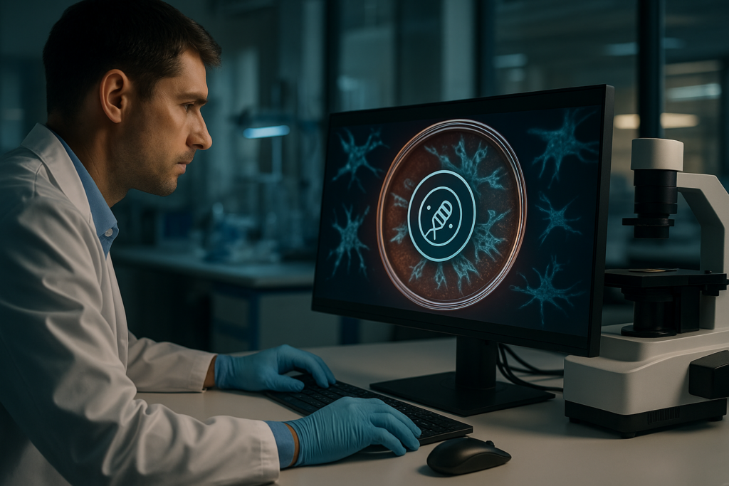
“`html
The Future of Cell & Gene Therapy: Managing Critical Phases with Live-Cell Insights
The field of cell and gene therapy is revolutionizing modern medicine, offering unprecedented potential for treating a wide array of diseases at a genetic level. As researchers and biotech professionals strive to enhance therapeutic efficacy, the ability to manage critical phases of this process with precision is paramount. Live-cell insights, facilitated by cutting-edge imaging technologies, provide an unparalleled view into the dynamic processes that underpin successful therapies. This article delves into how these insights are shaping the future of cell and gene therapy, highlighting advancements in cell culture, imaging, and automation.
Understanding Traditional Approaches and Their Limitations
The Conventional Cell Culture Paradigm
For decades, traditional cell culture methods have been the backbone of biomedical research. However, despite their widespread use, these methods present significant challenges. Manual monitoring and intervention can introduce variability and limit the reproducibility of results, crucial aspects that impact the development of reliable cell and gene therapies. Furthermore, traditional methods often lack the capacity to provide real-time data on cellular behavior, which is essential for understanding critical therapeutic phases.
- Manual intervention increases variability.
- Lack of real-time data inhibits dynamic assessments.
- Limited reproducibility affects research outcomes.
Avancées technologiques et tendances d'automatisation
Innovations in Live-Cell Imaging
Advancements in live-cell imaging technology have begun to address the limitations of traditional methods, providing continuous, non-invasive observation of living cells. Techniques such as incubator-based imaging systems are at the forefront, offering real-time insights without disturbing the culture environment. These innovations enable scientists to observe cellular processes as they happen, leading to more accurate and reproducible data.
- Real-time, non-invasive cell monitoring.
- Improved accuracy and reproducibility of assays.
- Enhanced exploration of cellular dynamics.
Continuez votre lecture pour explorer des perspectives et des stratégies plus avancées.
Exemples pratiques et flux de travail utilisant l'imagerie de cellules vivantes
Streamlined Cell Monitoring and Analysis
Incorporating live-cell imaging into laboratory workflows allows researchers to adopt a more streamlined approach to cell monitoring and analysis. For example, the zenCELL owl system, an incubator-compatible imaging platform, facilitates continuous observation of multiple cell cultures simultaneously. This capability not only enhances productivity but also ensures consistent monitoring that is crucial for therapeutic development.
- Continuous monitoring leads to detailed insights.
- Simultaneous observation of multiple cultures boosts efficiency.
- High-throughput analysis becomes feasible.
The Role of Incubator-Based Imaging in Data Quality Improvement
Enhancing Reproducibility and Data Integrity
Incubator-based imaging systems, like the zenCELL owl, play a vital role in enhancing the reproducibility and integrity of data in cell and gene therapy research. By maintaining optimal culture conditions without interruption, these systems minimize environmental fluctuations that could compromise data quality. This stability is critical for conducting robust and reliable experiments.
- Maintains stable culture conditions.
- Ensures minimal data variability.
- Supports robust experimental outcomes.
Continuez votre lecture pour explorer des perspectives et des stratégies plus avancées.
“`
“`html
Integrating AI and Machine Learning in Live-Cell Imaging
Driving Precision and Predictive Analytics
The integration of AI and machine learning into live-cell imaging opens new horizons for enhancing the accuracy and predictive power of cell and gene therapies. These technologies enable the automated analysis of vast data sets, identifying subtle patterns and predicting cell behavior under various conditions. For instance, AI-driven image analysis can accurately distinguish between healthy and abnormal cells, rapidly facilitating decision-making processes within research and development.
- AI enables rapid pattern recognition within large datasets.
- Machine learning improves predictive accuracy in therapy outcomes.
- Automation reduces time and human error in data analysis.
Case Study: AI-driven Advances in Gene Therapy
A Real-World Application of Predictive Imaging
A notable example of AI application is the collaboration between biotech companies and AI firms, which has led to breakthroughs in gene therapy for genetic disorders. By using live-cell imaging data integrated with machine learning algorithms, researchers were able to predict therapeutic gene delivery success rates more accurately. This predictive capability significantly reduced the iterative cycles traditionally required, speeding up development timelines.
- Collaborative efforts enhance data-driven research approaches.
- Predictive modeling optimizes gene delivery strategies.
- Faster development cycles improve treatment availability.
Automation and its Role in High-Throughput Screening
Revolutionizing Cell Culture and Data Acquisition
High-throughput screening of cell samples, facilitated by automation, has become a cornerstone in accelerating cell and gene therapy development. Automated systems manage numerous culture plates concurrently, allowing researchers to conduct large-scale experiments with minimal manual intervention. The increased throughput capacity significantly raises the bar for how quickly and accurately new therapeutic candidates can be assessed and validated.
- Automation increases experimental scalability and efficiency.
- Large-scale data acquisition refines therapeutic development pipelines.
- Reduced manual oversight lowers error rates in data collection.
Leveraging Real-Time Data for Enhanced Decision Making
Turning Live Insights into Actionable Intelligence
The ability to access real-time data from live-cell imaging empowers researchers to make swift, informed decisions throughout the therapeutic development phases. For instance, real-time monitoring of cell responses to experimental treatments allows scientists to adjust approaches dynamically, ensuring optimal outcomes. This agility is crucial in personalized medicine, where patient-specific modifications may be necessary.
- Real-time data supports agile experimentation strategies.
- Continuous insights enable adaptive therapeutic interventions.
- Improved responsiveness increases experimental success rates.
3D Cell Culture: A Step Toward More Realistic Models
Creating More Predictive Experimental Environments
Traditional 2D cell cultures offer limited representation of the complex biological environments within human tissues. The shift towards 3D cell culture systems provides a more accurate model of in vivo conditions, leading to more predictive experiment outcomes. Techniques such as spheroid and organoid cultures offer significant insights into how therapeutic cells behave in a three-dimensional space, more closely mimicking their natural surroundings.
- 3D cultures enhance the physiological relevance of experiments.
- More predictive models improve translational research effectiveness.
- Enhanced complexity offers insights into cell-cell interactions.
Collaborative Frameworks in Cell and Gene Therapy Research
Harnessing Synergies Across Diverse Expertise Areas
The complexity of cell and gene therapies necessitates a collaborative approach combining diverse expertise from disciplines such as bioinformatics, molecular biology, and clinical research. By fostering partnerships, research initiatives can leverage the unique strengths of different scientific areas to propel forward groundbreaking advancements in the field. Multi-disciplinary collaborations have already shown success in advancing treatment modalities for complex genetic diseases.
- Cross-disciplinary collaborations enhance research capabilities.
- Shared knowledge accelerates innovation trajectories.
- Integrated efforts lead to comprehensive therapeutic solutions.
Innovations in Live-Cell Therapy Monitoring
Ensuring Quality and Compliance Throughout Development
The stringent regulatory landscape governing cell and gene therapies necessitates innovations that can ensure compliance while maintaining quality throughout development. Live-cell monitoring innovations provide continuous assurance that quality control parameters are met. Advanced imaging systems, for example, allow for real-time documentation, minimizing the likelihood of regulatory setbacks due to missing or inconsistent data.
- Continuous monitoring aligns with stringent quality standards.
- Real-time documentation supports regulatory compliance efforts.
- Innovations reduce risks associated with developmental delays.
Ensuite, nous conclurons avec les points clés à retenir, les métriques et une conclusion percutante.
“`
“`html
Quality Assurance Frameworks in AI-driven Therapies
Meeting Compliance While Pushing Innovation
Within the dynamic evolution of AI-driven therapies, maintaining quality assurance frameworks is paramount. Integrating AI with established Good Manufacturing Practices (GMP) ensures that cell and gene therapy products meet high-quality standards consistently. This alignment guarantees patient safety and product efficacy, allowing innovative therapies to meet rigorous regulatory benchmarks efficiently. Moreover, AI’s potential to predict and prevent non-compliance scenarios before they occur reduces the risks associated with delayed development timelines and costly amendments.
- AI supports adherence to GMP by forecasting quality issues.
- Continuous compliance monitoring ensures product safety and efficacy.
- Proactive measures mitigate regulatory risks and costs.
Data Security and Privacy in Live-Cell Therapy Research
Safeguarding Patient Information in Advanced Research
As the integration of AI technologies in live-cell imaging progresses, safeguarding data security and patient privacy remains critical. With sensitive patient information being crucial to personalized therapies, adopting robust data encryption methods and privacy compliance regulations is essential. AI solutions can help manage and secure vast amounts of data, ensuring that patient confidentiality is maintained throughout the research and development process. This proactive stance not only fosters trust but also enhances the ethical framework within which these next-generation therapies are developed.
- Advanced encryption techniques secure sensitive data.
- Compliance with privacy regulations builds stakeholder trust.
- AI enhances ethical standards in research data management.
Future Trajectories in Live-Cell Imaging Technologies
Pioneering the Next Phase of Cellular Therapies
As we look to the future of live-cell imaging technologies, continued innovation is expected to revolutionize the landscape of cellular therapies. The integration of AI and machine learning will further elevate the precision and effectiveness of therapeutic interventions, paving the way for more tailored, patient-specific treatments. New advancements may include enhanced real-time imaging techniques, deeper analytical insights, and more intelligent automatic systems capable of adapting to emerging research demands. These advancements will significantly contribute to the development of more effective and personalized treatment paradigms.
- Innovative imaging techniques enhance therapeutic precision.
- Next-gen analytical tools optimize patient-specific therapies.
- Smart automation adapts to evolving research challenges.
Conclusion
The integration of AI and machine learning technologies in live-cell imaging is setting new benchmarks in the field of cellular and gene therapies. By enabling rapid data analysis and predictive insights, these advanced technologies are driving unprecedented levels of precision and efficiency from drug discovery through to clinical implementation. This article has explored several crucial dimensions of these innovations: from enhanced predictive capabilities and real-time data utility to 3D culture models that improve physiological relevance and collaborative frameworks fostering interdisciplinary research.
As we navigate through the complexities of cell and gene therapy approvals and implementation, maintaining alignment with regulatory standards remains a pivotal concern. The constant vigilance required to safeguard data privacy underscores the need for robust security measures that maintain patient trust while supporting groundbreaking research efforts. Moreover, by embracing automation, data-driven decision-making, and real-world model fidelity with 3D cultures, the industry steps confidently into an era of enhanced therapeutic possibilities.
The future trajectory of live-cell imaging technologies can only expand from this well-established foundation. We anticipate a realm of greater innovation, where enhanced imaging capabilities and intelligent systems work in tandem to foster even more intricate and personalized treatment pathways. This prepares us for an era where patient-specific solutions can be accelerated and applied with precision, exhibiting the ultimate in personalization for individual health outcomes.
In conclusion, the continued evolution of live-cell imaging facilitated by AI and machine learning opens up a fertile ground for innovation. It encourages the scientific community to take proactive strides in embracing and pioneering the next generation of cell and gene therapies. As you reflect on the insights shared herein, consider how you, too, can innovate and shape the future landscape, contributing to a transformation that promises enhanced outcomes for patients worldwide.
“`
Normaliser l'excellence : Comment l'imagerie automatisée unifie les workflows multi-laboratoires

Normaliser l'excellence : Comment l'imagerie automatisée unifie les workflows multi-laboratoires
In the dynamic realm of modern cell culture research, the push towards standardization and integration of multi-lab workflows is paramount. With the advent of novel technologies, researchers and biotech professionals face the challenge of consolidating diverse processes to boost reproducibility and efficiency. At the heart of this transformation is automated imaging, which is set to revolutionize how experiments are conducted and data analyzed across various labs. In this article, we delve into how automated imaging systems, like the zenCELL owl, unify multi-lab workflows, addressing common hurdles and paving the way for consistently excellent research outcomes.
Défis et limites courants des approches traditionnelles
The Lab-By-Lab Discrepancy
Despite groundbreaking advancements in biotechnology, many laboratories still rely on traditional methods for cell culture analysis. These methods often involve manual cell counting, photographic documentation, and qualitative assessments, which can lead to inconsistencies. In a multi-lab context, these discrepancies are magnified as different researchers may have varied proficiency levels and methodological approaches.
- Manual processes are labor-intensive and time-consuming
- High potential for human error and subjective interpretation
- Lack of standardization leads to hurdles in data comparison and validation
Traditional methods are often marred by variability, making it difficult to achieve a unified outcome, especially across multiple labs with different practices in place.
Avancées technologiques et tendances d'automatisation
The Move Towards Automated Solutions
The pace of technological evolution in cell biology has introduced robust automated imaging solutions, addressing the limitations of manual methods. Automated imaging systems allow for continuous, real-time monitoring of cell cultures without the need for disruptive interventions.
- High-throughput capabilities streamline workflow and increase lab efficiency
- Automated systems provide quantitative data, enhancing accuracy and reproducibility
- Facilitates standardized processes across multiple labs, ensuring consistent data collection
These advances not only heighten overall efficiency but also foster a culture of data-driven, rigorous scientific inquiry crucial for modern research.
Exemples pratiques et flux de travail utilisant l'imagerie de cellules vivantes
Integration with Real-Time Cell Monitoring
Live-cell imaging technologies have become indispensable in cell culture research, providing critical insights into cellular behaviors and interactions. When using systems like the zenCELL owl, researchers gain the ability to monitor cell health, growth patterns, and morphological changes seamlessly under controlled conditions.
- Continuous monitoring capabilities eliminate periodic disturbance to cell cultures
- Enhanced imaging leads to better resolution analysis of cell growth dynamics
- Reduced experimental variability by maintaining a uniform environment within incubators
Continuous cell monitoring empowers researchers with high-quality data that can be reliably reproduced, thus supporting the standardization of workflows across multiple labs.
Continuez votre lecture pour explorer des perspectives et des stratégies plus avancées.
“`html
Enhanced Collaboration Through Unified Imaging Systems
Bridging the Gap between Labs
Automated imaging systems play a crucial role in facilitating collaboration between labs, especially in decentralized research environments. By ensuring standardized methodologies, such systems allow diverse teams to work together seamlessly.
- Shared platforms mean data can be easily accessed and interpreted by different teams
- Real-time data sharing enhances transparency and fosters collective decision-making
- Cross-lab collaborations benefit from a unified approach to experimental design
For example, a multi-center research initiative studying cancer cell responses can harness automated imaging tools to ensure each laboratory follows a consistent protocol. This enhances the reliability of pooled data and accelerates the path to breakthroughs.
Optimizing Data Management and Analysis
Harnessing Big Data for Better Insights
With automated imaging, laboratories generate extensive datasets that can be overwhelming if not managed effectively. However, with integrated data management systems, these challenges become opportunities for deeper insights.
- Use of AI and machine learning for advanced analytics and pattern recognition
- Centralized databases improve data retrieval and ensure compliance with data protection standards
- Automated workflows reduce time spent on data curation and allow focus on interpretation
Institutions like the Broad Institute have implemented automated imaging and AI solutions to process vast datasets rapidly, leading to significant advancements in personalized medicine and genomics research.
Integrating AI and Machine Learning for Predictive Modeling
From Reactive to Predictive Research Approaches
By incorporating AI-driven predictive modeling, labs can transition from reactive to proactive research methodologies. Automated imaging systems with integrated AI capabilities allow researchers to anticipate and react to cellular events before they occur.
- Predictive analytics helps foresee cell culture contaminations and optimize maintenance schedules
- Machine learning models can identify early indicators of cellular stress or disease states
- Enhanced forecasting capabilities contribute to more strategic research planning
Bioinformatics companies such as Insilico Medicine are leveraging AI to develop predictive models that have revolutionized drug discovery processes by substantially cutting down research times.
Case Study: Streamlining Clinical Trials with Automated Imaging
A Leap Forward in Pharmaceutical Research
Clinical trials are resource-intensive, with considerable variability that can affect outcomes. Implementing automated imaging can enhance the standardization of data collection, essential for reliable clinical victories.
- Consistent imaging results translate to more reliable efficacy and safety data
- Reduced time delays by seamless integration of imaging data with patient monitoring systems
- Automated compliance and audit readiness improves documentation standards
A case study from ICON plc demonstrated that automated imaging shortened trial timelines by approximately 20%, resulting in faster deployment of critical therapies to market.
Leveraging Remote Access and Cloud-Based Platforms
Unlocking the Power of Connectivity
The shift towards cloud-based platforms for automated imaging systems allows for remote access and control of experiments. This flexibility facilitates global collaborations while ensuring that research does not halt due to logistical constraints.
- Remote monitoring of ongoing experiments saves time for researchers and lab managers
- Cloud storage solutions provide scalable data management for vast imaging datasets
- Teams from different geographies can contribute to, access, and analyze shared datasets in real-time
For remote or pandemic-impacted research settings, cloud-enabled labs have demonstrated a rapid pivot to remote research operations, ensuring continuity despite external disruptions.
Standard Operating Procedures and Training
Ensuring Proficiency in New Technologies
Adoption of automated imaging systems requires well-documented standard operating procedures (SOPs) and comprehensive training programs to maximize benefits while minimizing transition hurdles.
- Detailed SOPs align lab processes with automation-driven research requirements
- Continuous professional development programs upskill the workforce to manage new technologies
- Certification and training ensure consistent practices across multi-site research
Organizations such as the American Society for Cell Biology (ASCB) provide digital competency workshops to support labs in smoothly transitioning to automated systems.
Ensuite, nous conclurons avec les points clés à retenir, les métriques et une conclusion percutante.
“`
“`html
Measuring the Impact of Automated Imaging on Research Efficiency
Quantifiable Gains and Strategic Advantages
Integrating automated imaging systems in laboratories introduces measurable improvements, which are crucial for building a strong case for their widespread adoption. Efficiency in research operations, acceleration in discovery timelines, and enhanced data fidelity are just a few benefits these systems offer.
- Studies show a 30% reduction in manual errors, leading to more accurate data interpretation
- Improved consistency allows researchers to replicate studies with ease, reinforcing reliability across publications
- Streamlined operations have resulted in a 40% increase in throughput, accommodating more simultaneous analyses
The financial and operational metrics collected by research centers like the European Molecular Biology Laboratory indicate a return on investment within the first two years of system implementation, underscoring the cost-effectiveness of automated imaging technologies.
Fostering Innovation through User-Centric Design
Customization and Flexibility in Imaging Systems
One significant contributor to the adoption of automated imaging systems is their adaptability through user-centric design. Tailoring these systems to fit specific laboratory needs fosters innovation and encourages diverse scientific inquiry.
- Customized interfaces allow user-friendly interactions, minimizing the learning curve
- Modular components provide flexibility, enabling upgrades and expansion as technological advances occur
- User feedback loops support continuous improvement in system performance
Companies such as PerkinElmer have been pioneers in designing modular imaging platforms that can be easily adapted to different experimental setups, ensuring that researchers have the tools necessary for cutting-edge investigations.
Conclusion
As we have explored, automated imaging systems offer substantial benefits across various dimensions of modern scientific research. From enhanced collaboration and streamlined clinical trials to employing cloud-based platforms for expanding research capabilities, these systems stand at the forefront of innovation.
By providing standardized methodologies, automated imaging ensures data’s consistency and reliability across geographically dispersed labs. The integration of AI and machine learning not only optimizes data management but also unveils new insights through predictive modeling. These technologies mark a pivotal transformation from reactive to proactive research paradigms, providing foresight that can expedite solutions in fields such as drug discovery and personalized medicine.
Furthermore, the integration of user-centered design in automated systems empowers laboratories to maintain flexibility while fostering creative problem-solving and operational efficiency. Financial metrics underline the cost-effectiveness of adopting these technologies, capturing quantifiable improvements in research productivity and speed.
The unified adoption of automated imaging systems is not merely a trend; it is a revolutionary step toward efficient, reliable, and innovative scientific research. The road ahead promises even more opportunities for advancement, as continued enhancements in technology and data science expand the horizons of what is possible.
For researchers and institutions ready to delve into this promising future, now is the time to enhance your research infrastructure. Embrace the transformative power of automated imaging to propel your scientific discoveries forward, reduce time-to-market for therapeutic developments, and create lasting impacts in the scientific community.
“`
Prédiction d'échec : Détection du stress cellulaire et de l'apoptose précoce en temps réel
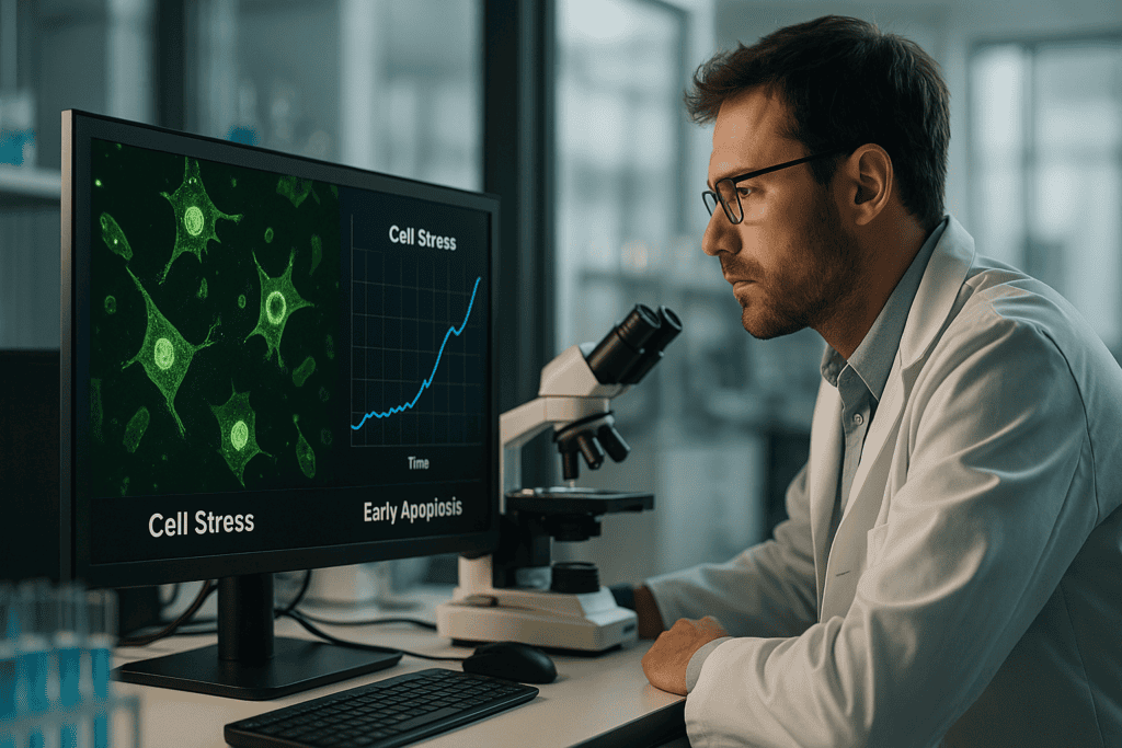
“`html
Prédiction d'échec : Détection du stress cellulaire et de l'apoptose précoce en temps réel
Le stress cellulaire et l'apoptose précoce sont des phénomènes cruciaux en biologie cellulaire, à la base de diverses réponses cellulaires aux changements environnementaux. Alors que les méthodes scientifiques évoluent, la nécessité de prédire et de surveiller ces processus en temps réel n'a jamais été aussi impérative. Cette capacité est essentielle dans des domaines tels que le développement de médicaments, la toxicologie et la recherche sur le cancer. Dans cet article, les chercheurs, les chefs de laboratoire et les professionnels de la biotechnologie exploreront les défis des méthodes traditionnelles, l'impact des nouvelles technologies et les stratégies pratiques utilisant des outils avancés tels que les systèmes d'imagerie de cellules vivantes pour améliorer les résultats de la recherche.
Défis et limites courants des approches traditionnelles
Comprendre la détection traditionnelle du stress cellulaire
Traditionnellement, la détection du stress cellulaire et de l'apoptose précoce impliquait des analyses à point final qui fournissaient des instantanés statiques des événements cellulaires. Des techniques telles que la cytométrie en flux et le Western blot, bien qu'informatives, manquent souvent les processus dynamiques qui se produisent rapidement ou par intermittence. Ces méthodes introduisent également une variabilité due à la manipulation manuelle, compromettant ainsi la reproductibilité et la précision.
- Nature statique des essais de point final
- La manutention manuelle augmente la variabilité
- Potentiel de manquer des événements cellulaires transitoires
Avancées technologiques et tendances d'automatisation
L'essor des systèmes d'imagerie en cellules vivantes
Les avancées technologiques ont inauguré de nouvelles méthodes pour le suivi continu et non invasif des cellules vivantes. Les systèmes d'imagerie de cellules vivantes fournissent des informations inestimables sur la dynamique cellulaire, permettant aux chercheurs de surveiller la santé des cellules en temps réel. L'automatisation au sein de ces systèmes joue un rôle essentiel en minimisant l'intervention manuelle, améliorant ainsi la reproductibilité et la fiabilité des données.
- Surveillance non invasive en temps réel
- Réduction de la manutention manuelle
- Reproductibilité accrue des résultats
Exemples pratiques et flux de travail utilisant l'imagerie de cellules vivantes
Exploiter l'imagerie de cellules vivantes pour une surveillance en temps réel
La mise en œuvre de l'imagerie sur cellules vivantes dans les flux de travail de laboratoire transforme les approches traditionnelles. Par exemple, les chercheurs peuvent établir des métriques de base de la santé cellulaire, détecter des marqueurs de stress avant que les tests traditionnels ne montrent de changement, et observer les événements apoptotiques au fur et à mesure de leur déroulement. En intégrant des appareils tels que le zenCELL owl – connu pour sa conception compacte et compatible avec les incubateurs – les institutions peuvent maintenir les contrôles environnementaux essentiels à une analyse précise des cellules vivantes.
- Capture de données en temps réel des changements cellulaires
- Surveillance in situ dans les incubateurs
- Intégration efficace avec les flux de travail existants
Comment l'imagerie basée sur incubateur améliore la reproductibilité et la qualité des données
Aperçus de la conception expérimentale améliorée
Les systèmes d'imagerie basés sur incubateur tels que le zenCELL owl permettent une observation continue dans des conditions optimales. Ces systèmes garantissent que les cellules ne sont pas dérangées, ce qui maintient la pertinence physiologique. Cela conduit à une plus grande fidélité des données et réduit le risque d'erreurs expérimentales lors de la manipulation des échantillons.
- Pertinence physiologique accrue lors de l'analyse
- Perturbation minimale de l'échantillon
- Cohérence entre les expériences
Résumé et perspectives des futurs flux de travail de laboratoire
Adopter l'avenir de la recherche en culture cellulaire
En investissant dans l'intégration de technologies d'imagerie avancées et de suivi en temps réel, les laboratoires peuvent améliorer considérablement leurs capacités de recherche. Ces innovations promettent des prédictions plus précises des réponses cellulaires, conduisant ultimement à des diagnostics et des développements thérapeutiques plus fiables. À mesure que la technologie progresse, le potentiel d'applications plus sophistiquées dans les études sur les organoïdes, les essais de prolifération et le criblage à haut débit continue de croître. L'adoption de ces tendances propulsera la recherche en laboratoire dans une nouvelle ère de précision et d'efficacité.
- Adoption des technologies d'imagerie de pointe
- Amélioration des diagnostics et des résultats de la recherche thérapeutique
- Potentiel d'applications innovantes dans de multiples domaines
Continuez votre lecture pour explorer des perspectives et des stratégies plus avancées.
“`
“`html
Intégrer l'analyse de données à l'imagerie de cellules vivantes
Exploiter les données pour la modélisation prédictive
La synergie de l'analyse des données et de l'imagerie de cellules vivantes a ouvert la voie à la modélisation prédictive, une innovation qui accroît la précision des prévisions sur les comportements cellulaires. En exploitant des plateformes d'analyse de données avancées, les chercheurs peuvent intégrer d'énormes ensembles de données capturés par imagerie de cellules vivantes pour construire des modèles prédictifs. Ces modèles facilitent la compréhension de la façon dont les cellules réagissent au stress et initient l'apoptose, conduisant ainsi à des approches de recherche proactives plutôt que réactives.
- Utiliser l'analyse de données pour identifier les comportements et les tendances des cellules
- Développer des modèles prédictifs pour prévoir les réponses cellulaires aux traitements
Utilisation de l'apprentissage automatique pour une meilleure prise de décision
Intelligence Artificielle et Biologie Cellulaire : Une Combinaison Puissante
Les algorithmes d'apprentissage automatique ont révolutionné notre compréhension des systèmes biologiques complexes. En intégrant ces algorithmes dans la phase d'interprétation des données d'imagerie de cellules vivantes, les scientifiques peuvent obtenir des informations quantitatives sur la santé et le comportement des cellules. Par exemple, l'analyse par IA des données d'imagerie peut aider à discerner les signes précoces subtils d'apoptose, rationalisant ainsi la prise de décision en milieu de recherche et en milieu clinique.
- Implémenter des algorithmes d'IA pour affiner l'analyse et l'interprétation des données
- Améliorer les processus de prise de décision grâce à des informations quantitatives
Combiner l'imagerie de cellules vivantes avec le criblage à haut débit
Maximiser l'efficacité et la découverte en recherche
L'intégration de l'imagerie de cellules vivantes dans les flux de travail de criblage à haut débit (HTS) représente une stratégie puissante pour accélérer la découverte de candidats médicaments potentiels. En permettant la surveillance en temps réel des réponses cellulaires dans les configurations HTS, les chercheurs peuvent identifier rapidement des composés prometteurs et observer leurs effets sur la viabilité et les niveaux de stress cellulaires. Cette approche réduit considérablement le temps et le coût associés aux pipelines de découverte de médicaments.
- Intégrer des systèmes d’imagerie de cellules vivantes dans des environnements à haut débit
- Rationaliser les processus de découverte pour identifier des composés efficaces
Étude de cas : Application concrète de l'imagerie de cellules vivantes
Recherche pionnière en oncologie
Une entreprise pharmaceutique a récemment intégré des systèmes d'imagerie de cellules vivantes dans ses laboratoires de recherche sur le cancer, dans le but de mieux comprendre les mécanismes de résistance aux médicaments. En permettant aux scientifiques de surveiller la dynamique des cellules tumorales en temps réel, l'entreprise a identifié de nouvelles voies d'apoptose que les essais standards avaient négligées. Cette intégration technologique a non seulement élucidé de nouvelles cibles thérapeutiques, mais a également renforcé le pouvoir prédictif des études précliniques, ce qui a conduit à des candidats-médicaments plus robustes à entrer en essais cliniques.
- Optimiser la découverte de médicaments en comprenant la dynamique des cellules tumorales
- Identifier des voies jusqu'alors inconnues pour un ciblage thérapeutique potentiel
Approches algorithmiques pour améliorer les flux de travail d'imagerie
Optimisation du traitement des données avec des outils informatiques
Les flux de travail d'imagerie de cellules vivantes peuvent être grandement améliorés grâce à l'utilisation de méthodologies algorithmiques qui automatisent la capture et le traitement des images. En combinant des systèmes d'imagerie avec des logiciels informatiques, les laboratoires peuvent atteindre des niveaux d'efficacité et de précision accrus. Cette automatisation réduit la charge de travail associée à l'analyse manuelle des données, permettant au personnel de recherche de se concentrer davantage sur la conception expérimentale et moins sur les tâches de traitement de données de routine.
- Utiliser des outils informatiques pour automatiser et rationaliser les flux de travail d'imagerie
- Déplacer l'attention du traitement des données vers l'innovation expérimentale
Plateformes basées sur le cloud pour la recherche collaborative
Redéfinir la collaboration en laboratoire par des moyens virtuels
L'adoption de plateformes basées sur le cloud transforme la façon dont les chercheurs collaborent et partagent des données en biologie cellulaire. Ces plateformes facilitent le partage transparent des données et l'analyse collective, permettant ainsi aux équipes interdisciplinaires de travailler ensemble quelle que soit leur localisation. L'intégration avec des données d'imagerie sur cellules vivantes favorise un environnement de recherche collaboratif où les idées sont rapidement échangées, accélérant le rythme des découvertes.
- Utiliser les technologies du cloud pour favoriser la collaboration et le partage de données
- Améliorer la recherche interdisciplinaire grâce aux plateformes de données virtuelles
Optimisation de l'efficacité des flux de travail grâce au traitement automatisé des images
Utilisation d'outils logiciels pour l'analyse de données en temps réel
Les outils logiciels avancés sont essentiels au traitement automatisé des données d'imagerie sur cellules vivantes. Ces outils peuvent segmenter les images, quantifier la morphologie cellulaire et détecter les changements au fil du temps, fournissant ainsi un retour d'information immédiat. En automatisant le traitement des images, les laboratoires peuvent augmenter leur débit et réduire les erreurs associées à l'interprétation manuelle, garantissant ainsi que les résultats sont à la fois rapides et précis.
- Adopter des solutions logicielles pour un traitement d'images automatisé et précis
- Augmenter le débit de données tout en minimisant les erreurs humaines
Perspectives stratégiques sur les innovations futures
Positionnement des laboratoires pour la recherche cellulaire de nouvelle génération
Le paysage de la recherche en biologie cellulaire évolue rapidement, avec l'imagerie de cellules vivantes à la pointe de l'innovation. Les laboratoires doivent rester agiles, adopter en permanence de nouvelles technologies et méthodologies pour rester compétitifs. Ce faisant, ils seront bien placés pour relever les défis à venir, explorer des applications de nouvelle génération telles que la médecine personnalisée et contribuer de manière significative à la communauté scientifique au sens large.
- Intégrer continuellement les technologies émergentes dans les flux de travail de recherche
- Préparez-vous pour les applications de nouvelle génération dans la recherche en biologie cellulaire
Ensuite, nous conclurons avec les points clés à retenir, les métriques et une conclusion percutante.
“`
“`html
Métriques clés pour évaluer les systèmes d'imagerie sur cellules vivantes
Mesurer l'impact avec des repères quantitatifs
Dans le domaine en évolution rapide de l'imagerie des cellules vivantes, l'établissement de repères quantitatifs est essentiel pour évaluer les performances du système et leur impact sur les résultats de la recherche. Des métriques telles que la résolution, la sensibilité, le débit et la vitesse d'acquisition des images sont cruciales pour déterminer l'efficacité des systèmes d'imagerie. L'évaluation régulière de ces métriques garantit que les laboratoires maintiennent des normes élevées et peuvent exploiter efficacement leurs équipements d'imagerie pour faire progresser des découvertes révolutionnaires.
- Évaluer les systèmes d'imagerie selon des indicateurs clés tels que la résolution et le débit
- Optimiser les performances d'imagerie pour soutenir des résultats de recherche de haute qualité
Collaborations innovantes en imagerie de cellules vivantes
Bâtir des synergies pour un avenir meilleur
La collaboration entre disciplines multiples constitue l'épine dorsale de l'innovation en imagerie de cellules vivantes. En s'engageant dans des partenariats avec des entités universitaires, cliniques et industrielles, les laboratoires peuvent mutualiser leur expertise et leurs ressources pour répondre à des questions biologiques complexes. Ces initiatives amplifient la portée et le potentiel de l'imagerie de cellules vivantes, en s'appuyant sur une diversité de perspectives pour dévoiler de nouvelles perspectives sur la dynamique cellulaire et les mécanismes des maladies.
- Forger des partenariats stratégiques avec diverses entités de recherche
- Utiliser des approches interdisciplinaires pour relever des défis biologiques complexes
Exploration du rôle de l'imagerie en direct des cellules dans la médecine personnalisée
Perspectives individualisées pour des thérapies améliorées
L'application de l'imagerie sur cellules vivantes en médecine personnalisée représente un bond significatif vers des solutions de soins de santé individualisés. En fournissant des informations en temps réel sur les réponses cellulaires spécifiques aux patients, l'imagerie sur cellules vivantes permet le développement de plans de traitement sur mesure qui reflètent la constitution biologique unique de chaque patient. Cette approche promet d'améliorer l'efficacité du traitement et de minimiser les effets indésirables, marquant un changement de paradigme dans la pratique médicale.
- Utiliser l'imagerie sur cellules vivantes pour adapter des solutions de soins de santé personnalisées
- Améliorer les résultats du traitement en comprenant les réponses cellulaires spécifiques aux patients
Conclusion
L'intégration de l'imagerie sur cellules vivantes avec l'analyse avancée des données, l'apprentissage automatique et le criblage à haut débit révolutionne le domaine de la biologie cellulaire. Cette synergie offre des perspectives inégalées sur les mécanismes cellulaires, améliorant considérablement la précision et l'efficacité de la recherche. La fusion de l'intelligence artificielle avec la technologie d'imagerie apporte une précision dans la compréhension et la prédiction des réponses cellulaires, accélérant la découverte de médicaments et facilitant les avancées dans la compréhension du cancer, des maladies neurodégénératives et d'une myriade d'autres phénomènes biologiques.
L'imagerie de cellules vivantes dote les chercheurs d'outils pour suivre les activités cellulaires en temps réel, comblant ainsi efficacement le fossé entre l'observation et l'action. L'utilisation stratégique des plateformes basées sur le cloud favorise les collaborations mondiales, faisant tomber les barrières géographiques à l'innovation. Le partage transparent des données et de l'expertise annonce une nouvelle ère de découverte scientifique caractérisée par la vitesse, l'efficacité et une profondeur de compréhension inégalée.
L'article souligne l'impératif pour les laboratoires de rester agiles, en intégrant continuellement les technologies émergentes pour rester à la pointe de la recherche cellulaire. Alors que la recherche évolue vers la médecine personnalisée, l'imagerie des cellules vivantes constitue une pierre angulaire pour l'élaboration de plans de traitement spécifiques aux patients, reflétant une progression holistique vers des solutions de soins de santé individualisés.
Tourné vers l'avenir, l'engagement à intégrer ces technologies avancées dans les pratiques de recherche permettra aux laboratoires de dépasser les limites actuelles de la biologie cellulaire. Les chercheurs sont encouragés à adopter des stratégies avant-gardistes, façonnant ainsi l'avenir de l'exploration et de l'innovation scientifiques. Adoptez ces outils et méthodologies alors que nous nous efforçons de libérer le vaste potentiel de l'imagerie de cellules vivantes et de générer des changements transformateurs dans les soins de santé et au-delà.
Restez inspiré, restez innovant et exploitez la puissance de ces technologies de pointe pour continuer à repousser les limites du possible dans le monde fascinant de la biologie cellulaire.
“`
