🫧 Application — Spheroid & Organoid Imaging

Spheroid Timelapse Imaging —
3D Cell Cultures. Live. Inside the Incubator.

Monitor spheroid growth, compaction, adhesion and outgrowth continuously — without removing 3D cultures from their environment. Automated quantification of area, diameter and roundness over time.

96h
continuous monitoring
10 min
imaging interval
24
spheroids in parallel
0
culture disruptions
zenCELL owl spheroid live imaging — 3D cell culture monitoring inside incubator
Why spheroids need continuous monitoring

3D Cell Cultures — More Relevant, More Demanding

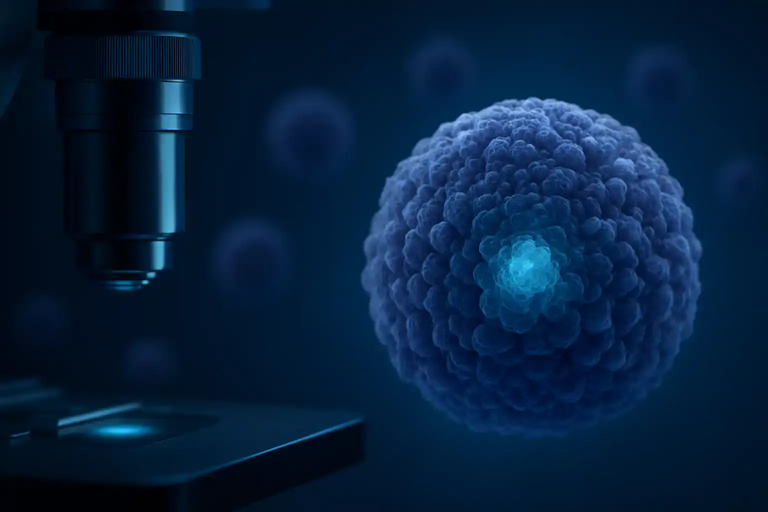
Spheroids and organoids offer a significantly better approximation of in vivo tissue environments compared to 2D cultures — but they require careful, continuous observation to capture growth dynamics, compaction and outgrowth events.

🔬
In-Incubator 3D Monitoring

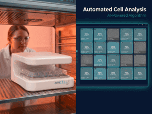
zenCELL owl images spheroids directly inside the incubator — no removal, no disruption of 3D structure, no temperature shock. Physiological conditions maintained throughout the entire experiment.

📐
Automated Morphology Quantification

Area, width, height, diameter and roundness quantified automatically using zenCELL owl and ImageJ at every imaging interval. Track structural changes over time without manual measurement.

🔄
24 Spheroids Simultaneously

Monitor up to 24 wells in parallel — different cell lines, drug treatments, or growth conditions. Compare proliferation rates and outgrowth dynamics under identical environmental conditions.

📊
Growth Kinetics Over Days

Continuous monitoring over 48–96 hours captures the complete growth profile — from initial aggregation through to full spheroid formation. Every 10 minutes, automatically.

🧬
Adhesion & Outgrowth Detection

Capture the moment spheroids adhere to treated surfaces and begin outgrowth migration. A semi-spherical 3D core with surrounding monolayer formation — documented automatically.

💊
Drug Screening on 3D Models

Apply drug treatments to spheroids and monitor effects continuously. More physiologically relevant than 2D cytotoxicity assays for predicting in vivo drug responses.

Experimental data

MCF-7 Spheroids — Two Experimental Approaches

Two parallel experiments with MCF-7 breast cancer cells demonstrate zenCELL owl's capability for both non-adhesive spheroid growth and adhesion/outgrowth monitoring.

MCF-7 spheroid growth diameter over time — zenCELL owl 96h monitoring on agarose

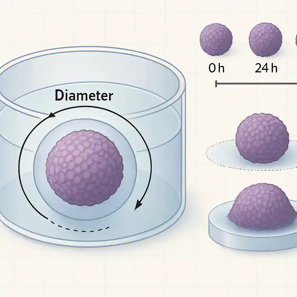
MCF-7 spheroid diameter growth over 96h on agarose-coated plates. Area, width, height and roundness quantified automatically.

Experiment 1 — Spheroid Proliferation on Non-Adhesive Surface

MCF-7 cells (6,000 cells/well) were seeded on agarose-coated 24-well plates and monitored for 96 hours at 10-minute intervals.

Result: Spheroids formed smooth, round shapes due to the non-adhesive environment. Proliferation occurred in the outer layers, increasing diameter over time. Key parameters including area, width, height and roundness were quantified automatically.

  • Platform: agarose-coated 24-well plates
  • Cell line: MCF-7 · 6,000 cells/well
  • Duration: 96 hours · interval: every 10 minutes
  • Parameters: area · diameter · width · height · roundness

Experiment 2 — Adhesion & Outgrowth on Treated Surface

7-day-old MCF-7 spheroids (6,000 cells/well) were transferred to tissue-culture treated 24-well plates and monitored for 48 hours.

Result: Cells adhered to the surface and began migrating outward. A semi-spherical 3D core with a surrounding monolayer formed within 48 hours — adhesion and outgrowth dynamics captured completely.

  • Platform: TC-treated 24-well plates
  • Spheroid age: 7 days · 6,000 cells/well
  • Duration: 48 hours · interval: every 10 minutes
  • Outcome: core + surrounding monolayer formation documented
MCF-7 spheroid adhesion and outgrowth on treated surface — zenCELL owl 48h

MCF-7 spheroid adhesion and outgrowth on TC-treated surface over 48h. 3D core with surrounding monolayer.

Results & analysis

Image-Based Analysis — All Parameters Automatically

Using zenCELL owl software and ImageJ, all key morphological parameters are monitored continuously — with no manual intervention and no environmental disruption.

zenCELL owl spheroid results — image-based analysis area roundness diameter

Results overview — spheroid expansion, adhesion dynamics and morphological parameters quantified automatically.

What Is Measured Automatically

Every 10 minutes, zenCELL owl software and ImageJ extract the following parameters from each well — no manual measurement, no environmental disruption, no bias.

  • Spheroid area (µm²) — expansion over time
  • Diameter — width and height separately
  • Roundness — structural compaction index
  • Adhesion dynamics — time to surface contact
  • Outgrowth area — monolayer expansion around 3D core
  • Morphological deviations — shape changes under drug treatment

All data exportable as CSV for statistical analysis. Timelapse videos generated automatically for each well.

🏛️

Developed in collaboration with:

University of Regensburg & Fraunhofer EMFT, Munich — with support from distribution partner Omni Life Science (ols-bio.com)

50+
peer-reviewed publications cite zenCELL owl across all applications
View all publications →
FAQ

Spheroid Imaging — Common Questions

Can zenCELL owl image spheroids inside the incubator?+
Yes — zenCELL owl sits inside the incubator and images through the transparent bottom of standard multi-well plates. Spheroids remain in their culture environment throughout the experiment. No removal, no temperature disruption, no structural damage to 3D cultures.
What morphology parameters can be quantified?+
Area, diameter, width, height and roundness are quantified automatically at every imaging interval using zenCELL owl software and ImageJ. This provides a complete kinetic profile of spheroid growth and structural changes.
Which plates are compatible for spheroid monitoring?+
Standard agarose-coated 24-well plates for non-adhesive spheroid growth, and tissue-culture treated plates for adhesion and outgrowth experiments. Any transparent-bottom multi-well plate compatible with zenCELL owl can be used.
Can I monitor drug effects on spheroids?+
Yes. Apply drug treatments and monitor spheroid morphology and growth continuously. The 24-well parallel format allows comparison of multiple drug concentrations or compounds simultaneously under identical incubator conditions.

See Spheroid Monitoring Live —
Free Remote Demo

Watch zenCELL owl monitor 3D spheroid growth inside a real incubator. On request via MS Teams.

On request via MS Teams No commitment 50+ publications Response within 24h

Developed in collaboration with:

University of Regensburg & Fraunhofer EMFT, Munich
With support from our distribution partner Omni Life Science (www.ols-bio.com)

zenCELL owl in action

See zenCELL owl
Live — 30 Min.
- Free -

Real cells. Real data. AI cell counting,
XYZ stage & full software — live inside the incubator. Twice a week via MS Teams.

Book Your Free Remote Demo

Watch zenCELL owl image live inside an incubator. Available.

Request Your Demo Slot
e.g. confluency monitoring, scratch assay, spheroids...
Live view

See zenCELL owl
live, 30 min, free online

Real cells. Real data. Real comparison. Watch AI cell counting, XYZ stage and full software live inside a real incubator — twice a week via MS Teams.

Full course of live cell imaging

Get your free live online demonstration of the zenCELL owl directly inside an incubator, available twice per week.

Request Your Demo Slot
e.g. confluency monitoring, scratch assay, spheroids...

or