“`html
Automation for Everyone: Why Complex Imaging is No Longer Just for Specialists
In the fast-paced world of modern cell culture research, the ability to consistently produce high-quality, reproducible data is crucial. With technological advancements blurring the lines between traditional research roles, automation has emerged as a powerful tool that transforms sophisticated imaging processes. Once the domain of specialized technicians, complex imaging is now accessible to a wider range of researchers and laboratory personnel. This article explores why automation is redefining the accessibility of live-cell imaging and how researchers can overcome traditional challenges efficiently.
Traditional Approaches: Challenges and Limitations
The Bottlenecks in Manual Imaging
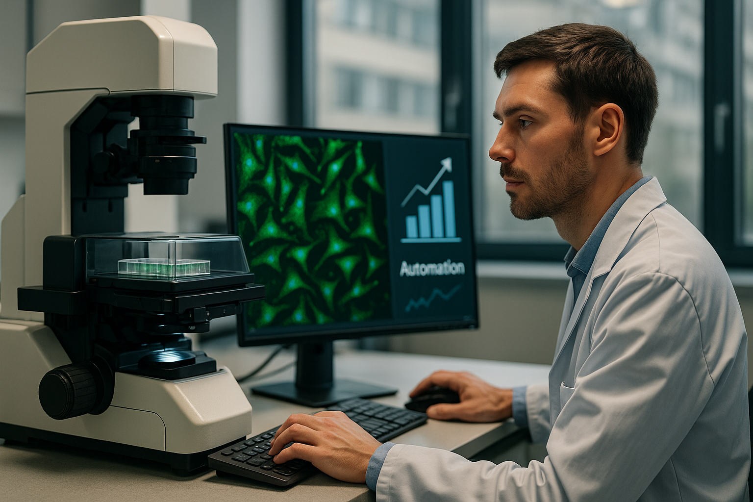
Traditionally, live-cell imaging was a labor-intensive process reliant on specialized expertise and manual operation. Researchers often faced challenges, such as the need for highly trained personnel to operate complex imaging systems, leading to bottlenecks in experimental workflows. The time-consuming nature of manual imaging not only limits throughput but also increases the risk of variability and human error, impacting data reliability.
- Requires extensive training and expertise.
- Higher chances of inconsistent results due to human error.
- Time-intensive processes reduce throughput.
Technological Advances and Automation Trends
The Rise of User-Friendly Imaging Systems
Recent technological innovations have paved the way for automation to revolutionize live-cell imaging. Advances in software and hardware design have led to the development of more intuitive imaging systems that democratize the imaging process. These systems, equipped with automated features, enable researchers to conduct complex imaging without specialized training, thus enhancing accessibility and efficiency across laboratories.
- User-friendly interfaces with automated parameters.
- Reduction in human error and increased reproducibility.
- Enhanced throughput with less manual intervention.
Practical Workflows in Live-Cell Imaging
Implementing Automated Solutions
With the advent of automation, practical workflows leveraging live-cell imaging are continually evolving. Implementing systems like the zenCELL owl, a compact, incubator-compatible imaging system, researchers can automate imaging processes seamlessly within the comfortable settings of their cell cultures. Such systems facilitate continuous monitoring, thus enabling real-time analysis and reducing disturbances to the cell environment.
- Continuous data acquisition and analysis.
- Integrated workflows for better experimental control.
- Compatibility with existing laboratory infrastructure.
End this part with a short transitional sentence like:
Continue reading to explore more advanced insights and strategies.
“`
“`html
Enhanced Data Reliability Through Automation
Minimizing Variability and Maximizing Precision
One of the most significant advantages of automation in live-cell imaging is the dramatic improvement in data reliability. Traditional manual imaging methods often suffer from variability due to human error and inconsistent handling of samples. Automated systems, on the other hand, are designed to perform tasks with high precision and repeatability, minimizing such issues. For example, the convergence of automation with real-time imaging analysis empowers researchers to capture data at regular intervals without manual intervention, ensuring that each dataset is consistent and precise.
- Automated calibration routines ensure consistent imaging conditions.
- Standardized protocols reduce variability across experiments.
- Consistent data acquisition enables more accurate comparative studies.
Reducing Operational Costs and Increasing Efficiency
Cost-effectiveness Meets Quality Output
Implementing automation in live-cell imaging not only improves data quality but also results in significant cost savings. By reducing the need for highly specialized technicians to perform routine imaging tasks, laboratories can allocate resources more efficiently. Moreover, automated systems often require less time to complete tasks, thereby increasing overall throughput and reducing operational delays.
- Lower personnel costs due to reduced need for specialized skills.
- Decreased downtime with faster processing capabilities.
- Investment in automation yields long-term savings and productivity gains.
Data Management and Integration with Automation
Simplifying Data Handling with Integrated Systems
Modern automated imaging systems often come with integrated data management solutions, simplifying the process of collecting, storing, and analyzing data. This integration is crucial for researchers handling large datasets common in cell culture studies. For instance, systems capable of interfacing with laboratory information management systems (LIMS) ensure seamless data tracking and retrieval, enhancing productivity and facilitating collaborative research efforts.
- Automated data capture minimizes manual entry errors.
- Integration with LIMS offers streamlined data management workflows.
- Facilitates real-time data sharing across research teams.
Case Study: Success with Automated Imaging Solutions
Practical Applications in Academic and Commercial Labs
Several academic and commercial laboratories have successfully integrated automated imaging solutions to overcome traditional bottlenecks. For instance, the University of California, San Diego, recently adopted incubator-compatible imaging systems across its research facilities, significantly enhancing the throughput of cell viability assays. This system allowed researchers to conduct longer-term studies without sample removal, improving the quality and reliability of the data collected.
- Increased efficiency leads to more experiments conducted annually.
- Higher accuracy results in more publishable and impactful research.
- Reduction in manual workload allows staff to focus on more value-added tasks.
Optimizing Workflow with Automation-Ready Software
Leveraging Advanced Software for Streamlined Processes
Automation-ready software plays a pivotal role in optimizing imaging workflows. By integrating advanced algorithms capable of analyzing and interpreting data autonomously, these software solutions minimize the manual effort required in data processing. Notably, software packages offering customizable templates and automated report generation can significantly streamline post-imaging workflows, saving researchers countless hours and reducing potential for user-induced errors.
- Automated analytics reduce time spent on data interpretation.
- Customizable workflows enhance flexibility in experimental designs.
- Real-time insights facilitate quicker decision-making processes.
Training and Skill Development for Automated Systems
Bridging the Gap Between Automation and User Proficiency
While automation minimizes the need for specialized manual skills, training is still essential to maximize the benefits of these advanced systems. Comprehensive training programs offered by many equipment providers focus on system operation, data interpretation, and maintenance, ensuring that lab personnel are equipped to leverage the full capabilities of automated imaging solutions. Such training often includes hands-on workshops and webinars designed to expand both theoretical and practical understanding.
- Regular training updates ensure users stay current with technological advancements.
- Collaborative learning sessions foster cross-disciplinary knowledge sharing.
- Support networks provide continuous assistance and troubleshooting resources.
Leveraging Real-Time Analysis for Timely Results
Integrating Instantaneous Feedback with Laboratory Protocols
The capability of automated systems to provide real-time analysis is transformative for experimental workflows. By harnessing dynamic monitoring capabilities, researchers can adjust experimental protocols based on instant feedback without halting operations. For example, systems capable of alerting users to changes in cell conditions allow for timely interventions, reducing the risk of losing valuable experiments due to unexpected variables.
- Real-time alerts enhance response to experimental anomalies.
- Simultaneous monitoring of multiple parameters aids comprehensive evaluation.
- Instant insights lead to more informed decision-making and research agility.
Customization and Flexibility in Automated Imaging Systems
Adapting Systems to Meet Diverse Research Needs
The adaptability of automated imaging systems to meet specific research requirements is a crucial factor in their growing adoption. Many systems offer modular designs and customizable software interfaces, allowing researchers to tailor solutions to their unique experimental needs. This flexibility ensures that laboratories can continue to meet their objectives as research demands evolve, making automation a long-term investment in scientific capability.
- Modular systems streamline scalability for expanding research projects.
- Configurable software meets diverse biological research applications.
- Continuous upgrades keep systems aligned with advancing technological frontiers.
Next, we’ll wrap up with key takeaways, metrics, and a powerful conclusion.
“`
“`html
Collaborative Research Enhanced by Automation
Pooling Expertise for Greater Scientific Breakthroughs
The integration of automated imaging systems is not only a technological advancement but also a catalyst for collaborative research efforts. By standardizing protocols and improving data consistency, these systems enable seamless collaboration between laboratories, institutions, and even cross-border research initiatives. Researchers can now easily share datasets and integrate findings, pooling expertise for more comprehensive and impactful scientific inquiries.
- Facilitates global collaboration by ensuring uniformity in data collection and analysis.
- Encourages multidisciplinary research collaborations, bringing diverse insights to complex problems.
- Paves the way for collaborative standard-setting in imaging research methodologies.
Long-term Maintenance and Support for Automated Systems
Ensuring Reliability and Longevity through Proper Care
While the initial investment in automated imaging systems may seem substantial, long-term maintenance and support strategies ensure their reliability and longevity. Manufacturers and suppliers offer comprehensive support networks that include regular system check-ups, software updates, and emergency troubleshooting services. By adhering to recommended maintenance schedules and updates, laboratories can maximize their system capabilities and minimize the risk of unforeseen downtimes.
- Routine maintenance checks prevent equipment failures and improve system uptime.
- Access to continuous technical support ensures rapid issue resolution.
- Regular software updates keep systems aligned with cutting-edge technological advancements.
Future Trends in Automated Imaging
Anticipating the Next Wave of Innovation
The future of automated imaging holds exciting possibilities, with emerging technologies poised to further enhance research capabilities. Continued advancements in machine learning, artificial intelligence, and integrated bioinformatics will likely propel automated imaging systems into new realms of efficiency and accuracy. Researchers can anticipate even greater levels of automation, where AI-driven systems offer predictive insights and autonomous experiment adaptations, further pushing the boundaries of what’s possible in life sciences research.
- AI integration brings predictive analytics into the realm of cellular imaging.
- Emerging bioinformatics tools facilitate smarter data interpretation.
- Innovative sensor technology promises higher resolution and faster data acquisition.
Conclusion
Automation stands at the forefront of revolutionizing live-cell imaging, bringing a multitude of transformative benefits to laboratories worldwide. By enhancing data reliability, reducing operational costs, simplifying data management, and facilitating real-time analysis, automated systems are reshaping how research is conducted and paving the way for new scientific breakthroughs. The adaptability and flexibility of these systems cater to diverse research needs, ensuring they remain invaluable tools in achieving and exceeding scientific objectives.
The insights presented in this article reflect the significant role automation plays in democratizing access to complex imaging techniques. By minimizing the necessity for specialized expertise, automation not only empowers seasoned researchers but also transforms laboratory newcomers into potent contributors to scientific advancement. The collective potential of standardized, high-quality data opens doors for collaborative projects, driving innovation across borders and disciplines.
As automation becomes more ingrained in laboratory practices, the role of continuous learning and adaptation cannot be overstated. By fostering a culture of knowledge sharing and skill development, research teams can stay at the cutting edge of emerging technologies. Automated imaging, supported by robust training and reliable maintenance, promises a future where research productivity and innovation are not limited by technical barriers.
In closing, the journey towards comprehensive automation in live-cell imaging is not just an upgrade of technology; it’s a paradigm shift that empowers every researcher to be at the forefront of discovery. Laboratories equipped with these advanced systems will see enhanced research outcomes and gain a competitive edge in the dynamic landscape of life sciences. Explore the future of research with automation—where every image captured is another step closer to understanding life itself in its most dynamic and glorious detail.
“`