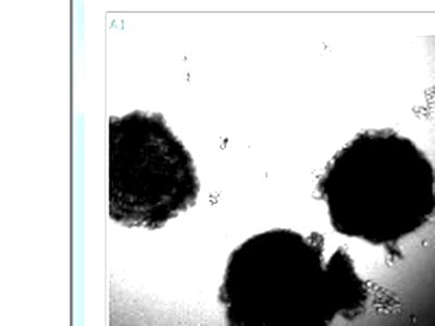
zenCELL owl permite el monitoreo continuo y no invasivo de ovocitos y embriones dentro de la incubadora de FIV. Imágenes de campo brillante sin etiquetas — desde la fertilización hasta la etapa de blastocisto — sin retirar los embriones de su entorno óptimo.
El monitoreo tradicional de FIV requiere extraer los embriones del incubador para su observación, exponiéndolos a cambios de temperatura, fluctuaciones de CO₂ y estrés mecánico. zenCELL owl elimina esto por completo. El dispositivo se coloca dentro de su incubadora de FIV y toma imágenes continuamente, 24/7, sin ninguna perturbación en el entorno de cultivo del embrión.
Los embriones nunca salen de la incubadora. La temperatura, el CO₂ y la humedad se mantienen estables durante todo el período de cultivo, desde la fecundación hasta la transferencia.
Sin etiquetas fluorescentes, sin fototoxicidad. Los embriones se visualizan en su estado natural utilizando microscopía de campo claro con luz transmitida, segura para su uso posterior.
Cada etapa de desarrollo se registra y documenta automáticamente. Además, se generan videos en lapso de tiempo para cada pocillo, lo que proporciona un registro completo del desarrollo.
La versión dedicada del software de FIV incluye herramientas de medición en tiempo real: dibuje un círculo para medir el diámetro del oocito o una línea para medir el grosor de la zona pelúcida directamente en el software.

Desde la conservación de la vida silvestre hasta la investigación clínica de FIV, zenCELL owl permite la monitorización continua de embriones en una amplia gama de especies y contextos de investigación.
Monitorear la maduración de oocitos y el desarrollo embrionario temprano en especies en peligro de extinción o raras —koala, canguro y otros marsupiales. La imagen continua no invasiva reduce el estrés por manipulación y maximiza los datos de desarrollo.
Monitoreo de embriones bovinos, equinos, porcinos y ovinos. Documentación automatizada en cámara rápida de las etapas de segmentación y desarrollo de blastocistos — para investigación, programas de cría y evaluación de la calidad embrionaria.
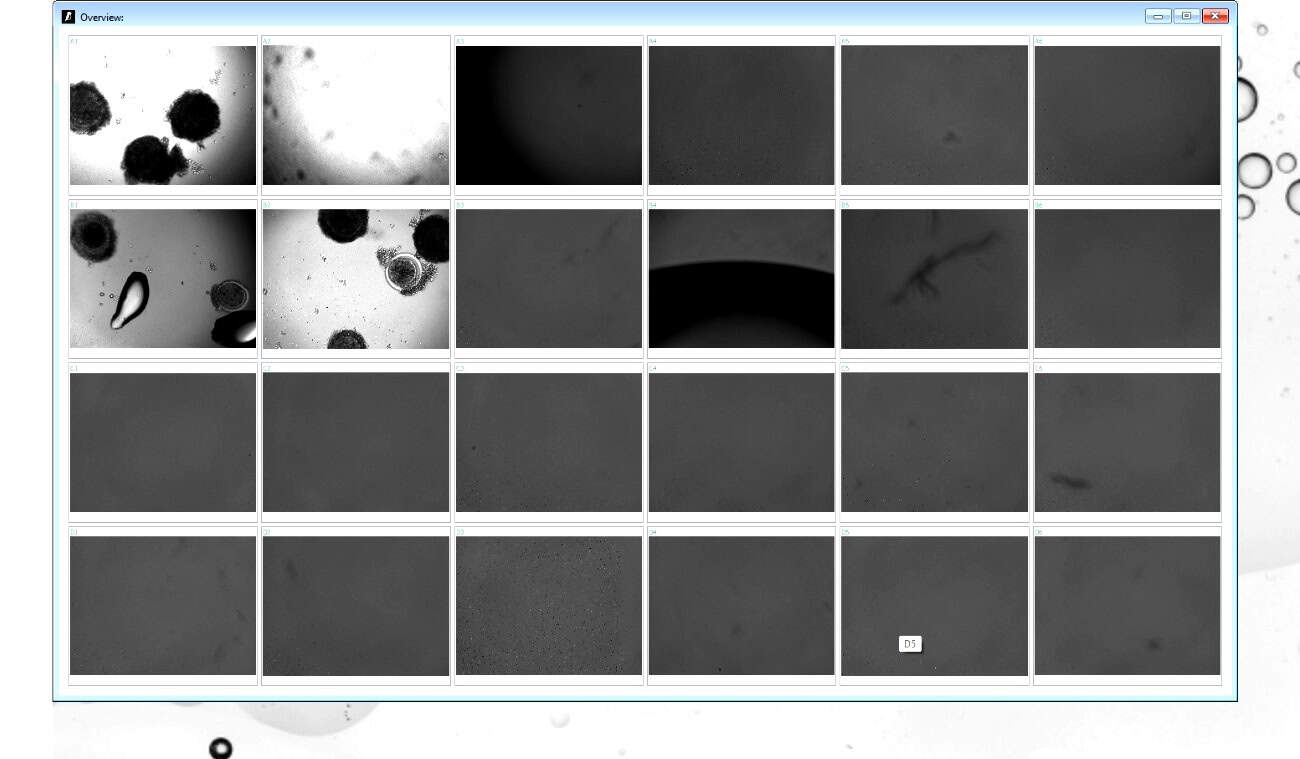
Evalúe los medios de cultivo, las condiciones de incubadora y los protocolos de FIV con datos objetivos continuos. Compare las cinéticas de desarrollo entre los grupos de tratamiento simultáneamente: 24 pocillos monitoreados en paralelo.
Observe la expansión, compactación y desprendimiento de las células del cúmulo durante la maduración in vitro. Adicionalmente, monitorice la actividad citoplasmática y la ruptura de la vesícula germinal en tiempo real.
Pruebe el efecto de diferentes formulaciones de medios de cultivo, concentraciones de oxígeno o compuestos en el desarrollo embrionario — con datos continuos objetivos de campo brillante en todos los pocillos simultáneamente.
Documentar la temporización de la primera segmentación, la compactación, la cavitación y la eclosión. Además, generar videos a intervalos y exportar datos de medición como CSV para análisis estadísticos.
El software dedicado de FIV incluye herramientas de medición en tiempo real (diámetro, distancia) y una interfaz optimizada para el monitoreo de embriones. Se instala en minutos en cualquier PC con Windows.
El dispositivo se adapta a cualquier estante de incubadora estándar — 18×18×10 cm, 1.5 kg. Un cable USB-C a su PC. Deje 2 horas para el equilibrio de temperatura antes del primer experimento.
Coloque los embriones u ovocitos en el formato de recipiente elegido. Establezca su intervalo de imagen (se recomiendan 10 minutos para FIV). Utilice la vista en vivo para ajustar el enfoque manualmente y obtener una calidad de imagen óptima.
Supervisa el desarrollo embrionario desde cualquier dispositivo, en cualquier lugar. El software funciona de forma continua sin abrir la incubadora. Los vídeos y las imágenes a intervalos se guardan automáticamente en cada punto de tiempo.
Exporta videos en cámara rápida como AVI, imágenes individuales como PNG o JPG y datos de medición como CSV. Todos los datos se almacenan localmente, sin necesidad de suscripción a la nube.
Especificaciones clave para aplicaciones de monitoreo de embriones y óvulos.
Monitoriza 24 posiciones en paralelo — ideal para investigación comparativa de FIV en múltiples grupos de tratamiento.
Campo de visión de 1.2 × 0.9 mm. Suficiente para la obtención de imágenes de ovocitos y embriones tempranos en recipientes de cultivo de FIV estándar.
Un intervalo de imagen de 10 minutos captura todos los eventos clave de desarrollo sin generar volúmenes de datos excesivos.
La versión FIV incluye herramientas de medición en tiempo real —diámetro, distancia— no disponibles en la versión estándar de cultivo celular.
Generación automática de video time-lapse por pozo. Adicionalmente, imágenes individuales exportadas como PNG o JPG.
Imágenes ininterrumpidas durante días o semanas. El PC debe permanecer activo; desactiva el modo de suspensión para experimentos largos.
Compre desde 14.000 € o alquile desde 290 €/mes. Versión de software de FIV incluida sin coste adicional.
Una conexión por cable. Windows 10/11. No requiere internet. Los datos permanecen locales en tu laboratorio.
"Aunque el ovocito no maduró completamente para hacer ICSI, es fascinante ver las células del cúmulo moviéndose y desprendiéndose con el tiempo, la actividad citoplasmática y la vesícula germinal. ¡Es un video realmente genial! ¡Gracias por el dispositivo!"
El monitoreo de embriones en FIV se refiere a la observación continua de la maduración de los ovocitos y el desarrollo embrionario temprano desde la fertilización hasta la etapa de blastocisto. El monitoreo tradicional de FIV requiere la extracción periódica de embriones de la incubadora para su observación bajo un microscopio estándar; cada extracción introduce choques térmicos, fluctuaciones de CO₂ y estrés mecánico que pueden impactar negativamente en los resultados del desarrollo.
La monitorización de embriones con lapso de tiempo utiliza imágenes continuas para capturar cada etapa del desarrollo embrionario sin interrupción de la incubadora. Además, proporciona datos cinéticos objetivos y cuantitativos sobre el momento de la escisión, la compactación y la cavitación que se correlacionan con la viabilidad embrionaria. zenCELL owl permite la monitorización con lapso de tiempo en cualquier incubadora de FIV estándar, sin requerir un sistema de incubadora dedicado para lapso de tiempo.
La FIV de vida silvestre presenta desafíos únicos: especies raras, disponibilidad limitada de oocitos y la importancia crítica de minimizar la manipulación. zenCELL owl es particularmente adecuado para la investigación de FIV de vida silvestre: monitorea hasta 24 pocillos simultáneamente, genera videos automáticos de lapso de tiempo y elimina las aperturas de la incubadora durante la observación. Además, la versión dedicada del software de FIV incluye herramientas de medición para el grosor de la zona pelúcida y la evaluación del diámetro del oocito.
zenCELL owl es compatible con placas de cultivo estándar de 24 pocillos (Ø 16mm) y placas de Petri (diámetro de 17-35mm), los formatos de recipientes más utilizados en la investigación de FIV. Para volúmenes pequeños de ovocitos, colocar las gotas en el centro del pocillo maximiza la cobertura de imagen. La función de vista en tiempo real permite el ajuste manual del enfoque para una posición óptima del embrión antes de iniciar la grabación.
Descarga nuestra nota de aplicación sobre imagenología de células vivas para FIV y ciencia reproductiva. Incluye protocolo, configuraciones de software y datos de imagen reales de experimentos de monitorización de ovocitos.
30 minutos vía MS Teams. Te mostramos muestras reales de zenCELL owl imaging en vivo, incluida la versión de software de FIV con herramientas de medición. Sin compromiso.
