“`
The Future of Cell & Gene Therapy: Managing Critical Phases with Live-Cell Insights
The field of cell and gene therapy is revolutionizing modern medicine, offering unprecedented potential for treating a wide array of diseases at a genetic level. As researchers and biotech professionals strive to enhance therapeutic efficacy, the ability to manage critical phases of this process with precision is paramount. Live-cell insights, facilitated by cutting-edge imaging technologies, provide an unparalleled view into the dynamic processes that underpin successful therapies. This article delves into how these insights are shaping the future of cell and gene therapy, highlighting advancements in cell culture, imaging, and automation.
Understanding Traditional Approaches and Their Limitations
The Conventional Cell Culture Paradigm
For decades, traditional cell culture methods have been the backbone of biomedical research. However, despite their widespread use, these methods present significant challenges. Manual monitoring and intervention can introduce variability and limit the reproducibility of results, crucial aspects that impact the development of reliable cell and gene therapies. Furthermore, traditional methods often lack the capacity to provide real-time data on cellular behavior, which is essential for understanding critical therapeutic phases.
- Manual intervention increases variability.
- Lack of real-time data inhibits dynamic assessments.
- Limited reproducibility affects research outcomes.
Avances tecnológicos y tendencias de automatización
Innovations in Live-Cell Imaging
Advancements in live-cell imaging technology have begun to address the limitations of traditional methods, providing continuous, non-invasive observation of living cells. Techniques such as incubator-based imaging systems are at the forefront, offering real-time insights without disturbing the culture environment. These innovations enable scientists to observe cellular processes as they happen, leading to more accurate and reproducible data.
- Real-time, non-invasive cell monitoring.
- Improved accuracy and reproducibility of assays.
- Enhanced exploration of cellular dynamics.
Continúe leyendo para explorar información y estrategias más avanzadas.
Ejemplos Prácticos y Flujos de Trabajo Utilizando Imágenes de Células Vivas
Streamlined Cell Monitoring and Analysis
Incorporating live-cell imaging into laboratory workflows allows researchers to adopt a more streamlined approach to cell monitoring and analysis. For example, the zenCELL owl system, an incubator-compatible imaging platform, facilitates continuous observation of multiple cell cultures simultaneously. This capability not only enhances productivity but also ensures consistent monitoring that is crucial for therapeutic development.
- Continuous monitoring leads to detailed insights.
- Simultaneous observation of multiple cultures boosts efficiency.
- High-throughput analysis becomes feasible.
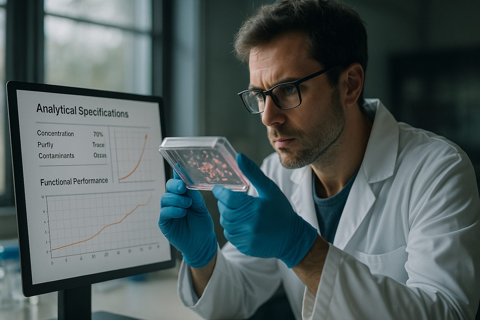
The Role of Incubator-Based Imaging in Data Quality Improvement
Enhancing Reproducibility and Data Integrity
Incubator-based imaging systems, like the zenCELL owl, play a vital role in enhancing the reproducibility and integrity of data in cell and gene therapy research. By maintaining optimal culture conditions without interruption, these systems minimize environmental fluctuations that could compromise data quality. This stability is critical for conducting robust and reliable experiments.
- Maintains stable culture conditions.
- Ensures minimal data variability.
- Supports robust experimental outcomes.
Continúe leyendo para explorar información y estrategias más avanzadas.
“`
“`
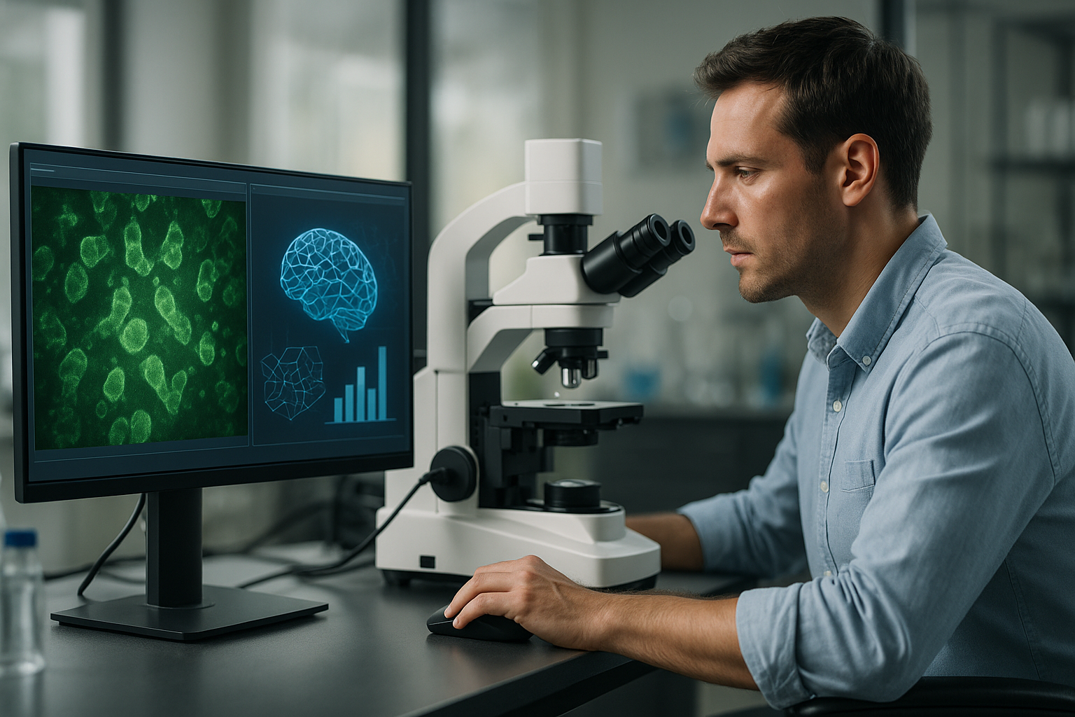
Integrating AI and Machine Learning in Live-Cell Imaging
Driving Precision and Predictive Analytics
The integration of AI and machine learning into live-cell imaging opens new horizons for enhancing the accuracy and predictive power of cell and gene therapies. These technologies enable the automated analysis of vast data sets, identifying subtle patterns and predicting cell behavior under various conditions. For instance, AI-driven image analysis can accurately distinguish between healthy and abnormal cells, rapidly facilitating decision-making processes within research and development.
- AI enables rapid pattern recognition within large datasets.
- Machine learning improves predictive accuracy in therapy outcomes.
- Automation reduces time and human error in data analysis.
Case Study: AI-driven Advances in Gene Therapy
A Real-World Application of Predictive Imaging
A notable example of AI application is the collaboration between biotech companies and AI firms, which has led to breakthroughs in gene therapy for genetic disorders. By using live-cell imaging data integrated with machine learning algorithms, researchers were able to predict therapeutic gene delivery success rates more accurately. This predictive capability significantly reduced the iterative cycles traditionally required, speeding up development timelines.
- Collaborative efforts enhance data-driven research approaches.
- Predictive modeling optimizes gene delivery strategies.
- Faster development cycles improve treatment availability.
Automation and its Role in High-Throughput Screening
Revolutionizing Cell Culture and Data Acquisition
High-throughput screening of cell samples, facilitated by automation, has become a cornerstone in accelerating cell and gene therapy development. Automated systems manage numerous culture plates concurrently, allowing researchers to conduct large-scale experiments with minimal manual intervention. The increased throughput capacity significantly raises the bar for how quickly and accurately new therapeutic candidates can be assessed and validated.
- Automation increases experimental scalability and efficiency.
- Large-scale data acquisition refines therapeutic development pipelines.
- Reduced manual oversight lowers error rates in data collection.
Leveraging Real-Time Data for Enhanced Decision Making
Turning Live Insights into Actionable Intelligence
The ability to access real-time data from live-cell imaging empowers researchers to make swift, informed decisions throughout the therapeutic development phases. For instance, real-time monitoring of cell responses to experimental treatments allows scientists to adjust approaches dynamically, ensuring optimal outcomes. This agility is crucial in personalized medicine, where patient-specific modifications may be necessary.
- Real-time data supports agile experimentation strategies.
- Continuous insights enable adaptive therapeutic interventions.
- Improved responsiveness increases experimental success rates.
3D Cell Culture: A Step Toward More Realistic Models
Creating More Predictive Experimental Environments
Traditional 2D cell cultures offer limited representation of the complex biological environments within human tissues. The shift towards 3D cell culture systems provides a more accurate model of in vivo conditions, leading to more predictive experiment outcomes. Techniques such as spheroid and organoid cultures offer significant insights into how therapeutic cells behave in a three-dimensional space, more closely mimicking their natural surroundings.
- 3D cultures enhance the physiological relevance of experiments.
- More predictive models improve translational research effectiveness.
- Enhanced complexity offers insights into cell-cell interactions.
Collaborative Frameworks in Cell and Gene Therapy Research
Harnessing Synergies Across Diverse Expertise Areas
The complexity of cell and gene therapies necessitates a collaborative approach combining diverse expertise from disciplines such as bioinformatics, molecular biology, and clinical research. By fostering partnerships, research initiatives can leverage the unique strengths of different scientific areas to propel forward groundbreaking advancements in the field. Multi-disciplinary collaborations have already shown success in advancing treatment modalities for complex genetic diseases.
- Cross-disciplinary collaborations enhance research capabilities.
- Shared knowledge accelerates innovation trajectories.
- Integrated efforts lead to comprehensive therapeutic solutions.
Innovations in Live-Cell Therapy Monitoring
Ensuring Quality and Compliance Throughout Development
The stringent regulatory landscape governing cell and gene therapies necessitates innovations that can ensure compliance while maintaining quality throughout development. Live-cell monitoring innovations provide continuous assurance that quality control parameters are met. Advanced imaging systems, for example, allow for real-time documentation, minimizing the likelihood of regulatory setbacks due to missing or inconsistent data.
- Continuous monitoring aligns with stringent quality standards.
- Real-time documentation supports regulatory compliance efforts.
- Innovations reduce risks associated with developmental delays.
A continuación, concluiremos con los puntos clave, métricas y una conclusión contundente.
“`
“`
Quality Assurance Frameworks in AI-driven Therapies
Meeting Compliance While Pushing Innovation
Within the dynamic evolution of AI-driven therapies, maintaining quality assurance frameworks is paramount. Integrating AI with established Good Manufacturing Practices (GMP) ensures that cell and gene therapy products meet high-quality standards consistently. This alignment guarantees patient safety and product efficacy, allowing innovative therapies to meet rigorous regulatory benchmarks efficiently. Moreover, AI’s potential to predict and prevent non-compliance scenarios before they occur reduces the risks associated with delayed development timelines and costly amendments.
- AI supports adherence to GMP by forecasting quality issues.
- Continuous compliance monitoring ensures product safety and efficacy.
- Proactive measures mitigate regulatory risks and costs.
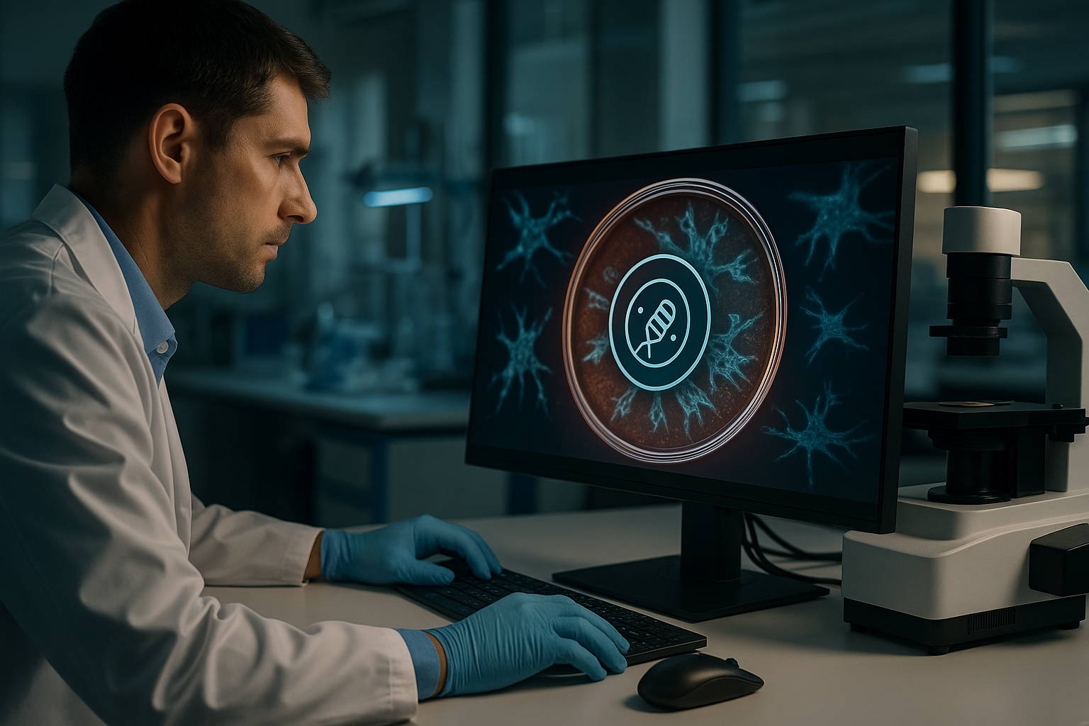
Data Security and Privacy in Live-Cell Therapy Research
Safeguarding Patient Information in Advanced Research
As the integration of AI technologies in live-cell imaging progresses, safeguarding data security and patient privacy remains critical. With sensitive patient information being crucial to personalized therapies, adopting robust data encryption methods and privacy compliance regulations is essential. AI solutions can help manage and secure vast amounts of data, ensuring that patient confidentiality is maintained throughout the research and development process. This proactive stance not only fosters trust but also enhances the ethical framework within which these next-generation therapies are developed.
- Advanced encryption techniques secure sensitive data.
- Compliance with privacy regulations builds stakeholder trust.
- AI enhances ethical standards in research data management.
Future Trajectories in Live-Cell Imaging Technologies
Pioneering the Next Phase of Cellular Therapies
As we look to the future of live-cell imaging technologies, continued innovation is expected to revolutionize the landscape of cellular therapies. The integration of AI and machine learning will further elevate the precision and effectiveness of therapeutic interventions, paving the way for more tailored, patient-specific treatments. New advancements may include enhanced real-time imaging techniques, deeper analytical insights, and more intelligent automatic systems capable of adapting to emerging research demands. These advancements will significantly contribute to the development of more effective and personalized treatment paradigms.
- Innovative imaging techniques enhance therapeutic precision.
- Next-gen analytical tools optimize patient-specific therapies.
- Smart automation adapts to evolving research challenges.
Conclusión
The integration of AI and machine learning technologies in live-cell imaging is setting new benchmarks in the field of cellular and gene therapies. By enabling rapid data analysis and predictive insights, these advanced technologies are driving unprecedented levels of precision and efficiency from drug discovery through to clinical implementation. This article has explored several crucial dimensions of these innovations: from enhanced predictive capabilities and real-time data utility to 3D culture models that improve physiological relevance and collaborative frameworks fostering interdisciplinary research.
As we navigate through the complexities of cell and gene therapy approvals and implementation, maintaining alignment with regulatory standards remains a pivotal concern. The constant vigilance required to safeguard data privacy underscores the need for robust security measures that maintain patient trust while supporting groundbreaking research efforts. Moreover, by embracing automation, data-driven decision-making, and real-world model fidelity with 3D cultures, the industry steps confidently into an era of enhanced therapeutic possibilities.
The future trajectory of live-cell imaging technologies can only expand from this well-established foundation. We anticipate a realm of greater innovation, where enhanced imaging capabilities and intelligent systems work in tandem to foster even more intricate and personalized treatment pathways. This prepares us for an era where patient-specific solutions can be accelerated and applied with precision, exhibiting the ultimate in personalization for individual health outcomes.
In conclusion, the continued evolution of live-cell imaging facilitated by AI and machine learning opens up a fertile ground for innovation. It encourages the scientific community to take proactive strides in embracing and pioneering the next generation of cell and gene therapies. As you reflect on the insights shared herein, consider how you, too, can innovate and shape the future landscape, contributing to a transformation that promises enhanced outcomes for patients worldwide.
“`